Access: a unique opportunity to use France-BioImaging technologies
France-BioImaging (FBI), offers its external users access to its state-of-the-art microbiological equipment and cutting-edge expertise.
Simply submit your request by registering through the Euro-BioImaging portal to benefit from a wide range of advanced technologies and personalized support from our specialists!
What’s your activity sector?
Choose one the options below to continue!
Users success stories
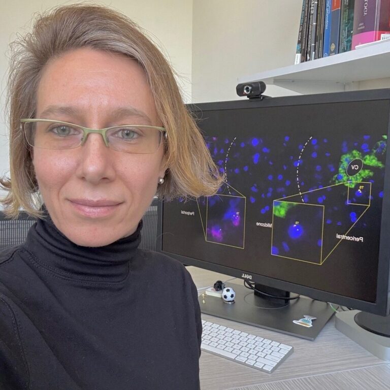
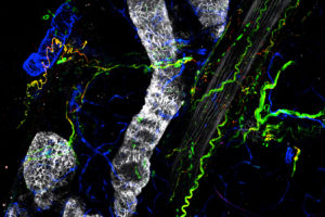
Carolina Eliscovich: Visualize the unseen
Carolina Eliscovich, group leader at Albert Einstein College of Medicine in New York, studies RNA biology with a focus on how mRNAs are spatially organized and regulated in tissues such as the liver.
Through the France-BioImaging Access Fund, she joined the Montpellier Resource Imaging facility and the Edouard Bertrand lab to apply sequential smFISH with an automated microfluidic system. Despite unexpected challenges, the collaboration enabled her to visualize, for the first time, multiple RNA species in regenerating mouse liver tissue.
She emphasizes how this unique opportunity provided unprecedented insights into gene expression heterogeneity in vivo, and highlights the generosity and expertise of the France-BioImaging teams, which made this breakthrough possible.

Hamed Abbasi: fluorescence microscopy for intraoperative use to target tumors
Hamed Abbasi, researcher at Erasmus Medical Center in Rotterdam, develops photonics-based technologies for oncological surgery, with a focus on fluorescence-guided approaches that provide surgeons with real-time tumor visualization.
Through the France-BioImaging Access Fund, he visited the PRIMACEN cell imaging platform in Normandy to perform near-infrared fluorescence lifetime imaging. Despite the technical challenges of working in this spectral range, the collaboration enabled him to generate robust proof-of-principle data.
These results were key to securing a Health~Holland grant to develop an advanced intraoperative fluorescence lifetime system, in partnership with industry. Hamed emphasizes both the cutting-edge infrastructure and the warm, supportive expertise of the PRIMACEN team, which made the collaboration scientifically decisive and personally enriching.
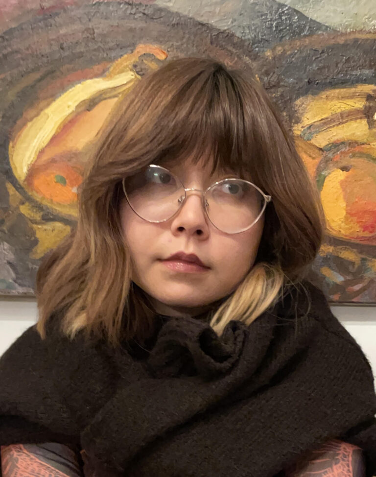
Mariia Nazarova: Understanding chromatin regulation key factors
Maria Nazarova, a PhD student at the IGBMC in Strasbourg, investigates chromatin dynamics and how enhancers interact with promoters using live-cell imaging.
Through the France-BioImaging Access Fund, she joined the MRI-CRBM platform in Montpellier to explore Lattice Light Sheet microscopy as a way to reduce phototoxicity and follow transcriptional dynamics in mouse stem cells. Despite technical limitations with time resolution, the experience offered her valuable insights into the system’s potential and the challenges of multi-color live imaging.
She highlights the warm support and expertise of the MRI team, particularly Virginie Georget, and stresses how the stay not only broadened her technical understanding but also shaped the way she designs future experiments.

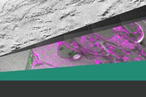
Atitheb Chaiyasitdhi: Dive into hearing mechanism
Atitheb Chaiyasitdhi, a research fellow at the University of Leicester, explores auditory mechanisms in insects, focusing on specialized sensory structures called chordotonal organs.
With support from France-BioImaging’s User Access program, he collaborated with the Imagerie-Gif platform to apply Focused Ion Beam Electron Microscopy (FIB-EM) to his study. This advanced technique enabled him to reconstruct the ultrastructure of the locust ear in 3D, achieving an unprecedented level of detail.
He highlights the responsiveness, expertise, and technical excellence of the platform, which made this remote collaboration both seamless and highly productive.
News & Events

Light microscopy reveals unexpected cardiac lymphatic remodelling
FBI CLEM WG Workshop: Cryo is far from cooling us down

Imaging unveils how symbiotic bacteria access nutrients from their insect host
2026 France-BioImaging digital calendar

Interdisciplinary access call to Structural biology, Biological imaging and Proteomics facilities
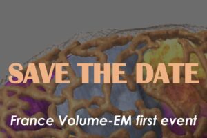
Save the date: First France Volume-EM Scientific Days
Calls
[CLOSED] France-BioImaging call for user access projects 2024
[CLOSED] Call R&D for Core Facilities 2024
[CLOSED] Africa-France Joint Initiative for Biological Imaging
Institutional partners


