Skip to content
.
  • Home
  • Services
    • Service Offer
    • Data services
      • FBI.data
      • F-BIAS
        • Open desks
      • Challenge
    • Trainings
      • FBI-AT
      • Training Portfolio
      • WG Training & Education
  • Become a User
    • How to access
    • Where to find us
      • Our Nodes
    • Fundings
    • User stories
    • Need help
    • How to acknowledge us?
  • SME & Industry
  • Opportunities
    • Fundings
    • Working Groups (WP)
    • Open Calls
    • Jobs
    • Challenge
  • News & Events
    • News
      • Newsletters
    • Our events
      • FBI Annual Meeting
      • FBI-AT
      • SFBS-2026
      • WG days
      • Sponsored events
      • Upcoming events
    • User stories
  • Community
    • Our Nodes
    • French INBS
    • International partners & cooperation
      • Euro-BioImaging
      • Global-BioImaging
      • CNRS IRN BioImage with China
      • FBI Africa-France Initiative
    • Partner communities
      • Technological Networks
      • GDR Imabio
  • About Us
    • Who are we
      • Missions & Values
      • Strategic Goals
      • Our Team
      • Governance
    • Our Nodes
    • ANR project
    • EU projects
    • CNRS IRN BioImage with China
    • Impact
    • Documents
    • Outreach
    • Contact us
Hub
Login Register

Gallery

    © David Pereira - Laboratoire Matière et Systèmes Complexes - ImagoSeine
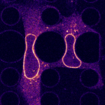
    © Théophile Déjardin – Institut Jacques Monod - ImagoSeine
    © Théophile Déjardin – Institut Jacques Monod – ImagoSeine 3t3 cells expressing fluorescent Nesprin2 going through 5µm constrictions.
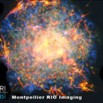
    Lionel Simmonneau, INM - Neurosphère
    Lionel Simmonneau, Institut des Neurosciences de Montpellier (INM) – Neurosphère
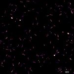
    © Patrice MASCALCHI – IINS, UMS3420 CNRS - University of Bordeaux, US4 INSERM - Bordeaux Imaging Center
    Link the dots! © Patrice MASCALCHI – IINS, UMS3420 CNRS – University of Bordeaux, US4 INSERM – Bordeaux Imaging Center DNA-Paint technique performed to image triple-spot nanorulers (DNA origami from Gattaquant company). DNA probes is covalently attached to Atto 655 dye. Images were acquired on Leica GSD.
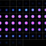
    © Mailfert Sébastien – CIML, CNRS-INSERM-AMU
    Prendre De Bonnes Resolutions © Mailfert Sébastien – CIML, CNRS-INSERM-AMU 3D confocal microscopy image of a fluorescent calibration bead to quantify the resolution of the system
    © Melina HEUZE – Institut Jacques Monod, CNRS, UMR7592 - ImagoSeine
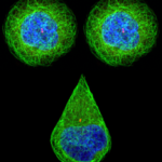
    Pattern Face © Melina HEUZE – Institut Jacques Monod, CNRS, UMR7592 – ImagoSeine MDCK cells seeded on fibronectin-coated micropatterns, fixed and stained for alpha-tubulin (green), gamma-tubulin (red) and DNA (blue).
    © Melina HEUZE - Institut Jacques Monod, CNRS, UMR7592 - ImagoSeine
    The Tracking Trip © Melina HEUZE – Institut Jacques Monod, CNRS, UMR7592 – ImagoSeine
    © Mailfert Sébastien – CIML, CNRS-INSERM-AMU
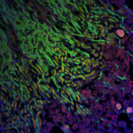
    L’Art Confocal © Mailfert Sébastien – CIML, CNRS-INSERM-AMU Mouse kidney section with AF 488 WGA in green, AF 568 Phalloidin in red, and DAPI in blue. From LifeTechnologies, F24630 FluoCells prepared slide. Confocal microscopy image obtained on a Leica SP5 ; 405nm and white light laser @488 and 568nm ; 63/1.4 oil immersion objective.
    © Noushin Mossadegh & Mailfert Sébastien - CIML, CNRS-INSERM-AMU
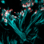
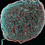
    Le bouquet de vie © Noushin Mossadegh & Mailfert Sébastien – CIML, CNRS-INSERM-AMU Coupe d’un tube séminifère de testicule de souris. On visualise les spermatozoïdes formant un « bouquet de fleurs ». Marquage en immunofluorescence des noyaux cellulaires (DAPI) représentés ici en cyan révélant la queue des spermatozoïdes. L’actine, représentée ici en rouge, révèle «la tête» des spermatozoïdes (phalloïdine marquée avec le fluorochrome Alexa-647). L’image représente 213×213μm sur 2048×2048 pixels. La coupe est d’une épaisseur de 20μm. Image de microscopie confocale sur Zeiss LSM780 ; lasers 405nm et 633nm ; objectif 40X, O.N. 1.4, immersion à huile.
    © PETREL Melina - Bordeaux Imaging Center - CNRS, Inserm, université Bordeaux Segalen
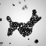
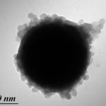
    Le caniche à sa mémère © PETREL Melina – Bordeaux Imaging Center – CNRS, Inserm, université Bordeaux Segalen Particules magnétiques encapsulées de polymère.
    © Ellen T. Arena (Pasteur)
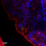
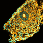
    Bioimage analysis of Shigella infection reveals targeting of colonic crypts (PNAS, 2015) © Ellen T. Arena (Pasteur)
    © Ellen T. Arena (Pasteur)
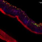
    Bioimage analysis of Shigella infection reveals targeting of colonic crypts (PNAS, 2015) © Ellen T. Arena (Pasteur)
    Dalton © Hugues Lelouard, Mailfert Sébastien & Mathieu Fallet CIML CNRS-INSERM-AMU
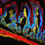
    Dalton © Hugues Lelouard, Mailfert Sébastien & Mathieu Fallet CIML CNRS-INSERM-AMU Confocal microscopy Revealing sub-population of immune cells on small intestine by 10 colors spectral imaging First Price of the FBI Image Contest 2016
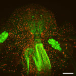
    Fly monster © Orestis Faklaris & Michael Lang – Institute Jacques Monod, CNRS UMR7592 - ImagoSeine
    Fly monster © Orestis Faklaris & Michael Lang – Institute Jacques Monod, CNRS UMR7592 – ImagoSeine Multifocal confocal microscopy (spinning disk, Spinning CSU W1) Drosophila third instar larval head, nuclear-RFP, neronal-GFP and green autofluorescence, 25x magnification, scale bar is 100 μm. Second to the FBI Image Contest 2016
    © Noushin Mossadegh & Mailfert Sébastien - CIML, CNRS-INSERM-AMU
    Le St Pierre Mediterranneen © Noushin Mossadegh & Mailfert Sébastien – CIML, CNRS-INSERM-AMU
    L'immunologie vue du ciel © Noushin Mossadegh & Mailfert Sébastien - CIML - CNRS /INSERM/AMU
    L’immunologie vue du ciel © Noushin Mossadegh & Mailfert Sébastien – Centre d’immunologie de Marseille Luminy (CIML) – CNRS /INSERM/AMU
    © Patrice MASCALCHI – IINS, UMS3420 CNRS - University of Bordeaux, US4 INSERM - Bordeaux Imaging Center
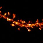
    STED image of neuron spines acquired in a fixed mouse brain slice using Atto 647N immunostaining. Image is a the result of a maximum projection of a 12 plane z-stack.
    © Noushin Mossadegh & Mailfert Sébastien - CIML, CNRS-INSERM-AMU
    © Noushin Mossadegh & Mailfert Sébastien – CIML, CNRS-INSERM-AMU Coupe d’un testicule de nouveau-né de souris. Marquage en immunofluorescence des noyaux cellulaires (DAPI).
    © Noushin Mossadegh & Mailfert Sébastien - CIML, CNRS-INSERM-AMU
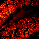
    Incandescence © Noushin Mossadegh & Mailfert Sébastien – CIML, CNRS-INSERM-AMU La chambre magmatique est en ébullition, les futures générations de spermatozoïdes se préparent à rentrer dans la vie de manière explosive. Coupe d’un tube séminifère de testicule de souris. De la périphérie à la lumière du tube, on visualise les cellules souches qui vont donner naissance aux spermatozoïdes. On distingue ces derniers grâce à leur queue. Marquage en immunofluorescence des noyaux cellulaires (DAPI). L’image représente 213×213μm sur 2048×2048 pixels. La coupe est d’une épaisseur de 20μm. Image de microscopie confocale sur Zeiss LSM780 ; lasers 405nm; objectif 40X, O.N. 1.4, immersion à huile.
    © Liu Zeng Zhen – Institut Jacques Monod - ImagoSeine
    Glowing grasp © Liu Zeng Zhen – Institut Jacques Monod – ImagoSeine Fluorescent focal adhesion proteins in a stem cell, confocal microscope Zeiss 700
    © Olga Nagy – Institute Jacques Mondod, CNRS, Drosophila Evolution Group - ImagoSeine
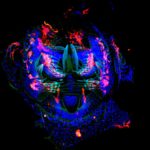
    Galactic Love © Olga Nagy – Institute Jacques Mondod, CNRS, Drosophila Evolution Group – ImagoSeine Immunostaing of Drosophila santomea pupal genitalia. Nuclei are in blue colour stained with DAPI. Neurons and glial cells are in red labeled with anti-HRP-Cy3 antibody. Hairs and chitin are in green autofluorescence. Image was taken at ImagoSeine by Zeiss LSM 710.
    © PETREL Melina - Bordeaux Imaging Center - CNRS, Inserm, université Bordeaux Segalen
    C’est l’amour © PETREL Melina – Bordeaux Imaging Center – CNRS, Inserm, université Bordeaux Segalen Algues préparées par HPF.
    © Orestis Faklaris – Institut Jacques Monod - ImagoSeine
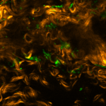
    © Orestis Faklaris – Institut Jacques Monod – ImagoSeine collagen SHG signal and autofluorescence (green) in the aorta
    © PETREL Melina - Bordeaux Imaging Center - CNRS, Inserm, université Bordeaux Segalen
    Projections solaires © PETREL Melina – Bordeaux Imaging Center – CNRS, Inserm, université Bordeaux Segalen Particules magnétiques encapsulées de polymère.
    © Patrice MASCALCHI – IINS, UMS3420 CNRS - University of Bordeaux, US4 INSERM - Bordeaux Imaging Center
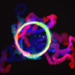
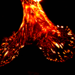
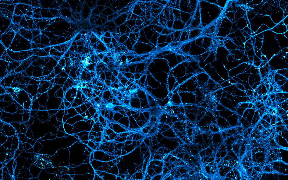
    The burning mesh © Patrice MASCALCHI – IINS, UMS3420 CNRS – University of Bordeaux, US4 INSERM – Bordeaux Imaging Center This image is about tracking of single molecules on live neurons (uPaint technique). It is the superimposition of hundred of tracks (red) and instantaneous diffusion coefficient (gradient of yellow to white).
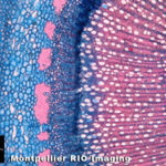
    Véronique Ripetti - DAP - Montpellier
    Véronique Ripetti, Laboratoire Développement et amélioration des Plantes (DAP), Montpellier – Tige de pommier
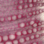
    Véronique Ripetti - DAP - Montpellier
    Véronique Ripetti, Laboratoire Développement et amélioration des Plantes (DAP), Montpellier – Tige de pommier
    Suzanne Vigneron - CRBM - Montpellier
    Suzanne Vigneron – Centre de Recherche en Biochimie Macromoléculaire (CRBM) – Montpellier -Kinétochores dans un noyau de xénope
    Steve Bourane, INM, Montpellier
    Steve Bourane, Institut des Neurosciences de Montpellier (INM), Montpellier – Colonne vertébrale de souris- MRI 2007 Image Contest 2nd prize
    Pierre Savagner, U868, CRLC, Montpellier
    Pierre Savagner, Unité Identité et plasticité tumorale (U868, CRLC), Montpellier – Glande Mammaire de souris- MRI 2006 Image contest 2nd Prize
    Michel Eybalin, INM, Montpellier
    Michel Eybalin, Institut des Neurosciences de Montpellier (INM), Montpellier – Ganglion spiral de cochlée
    Mathieu Gabut, IGMM, Montpellier
    Mathieu Gabut, Institut de Génétique Moléculaire (IGMM), Montpellier – Disque imaginal d’oeil d’embryon de Drosophile
    Willy Joly - IGH - Montpellier
    Willy Joly, Institut de Génétique Humaine (IGH), Montpellier – Embryon de drosophile
    Connect on LinkedIn Find us on X Subscribe to our newsletter
    Contact Us
    Contact Us Footer
    First
    Last

    Services Become a User Opportunities News & Events Community About Us FAQ
    Privacy Policy | Contact Us
    Credits | Designed by Yhello
    Close
    Designed by Yhello
    contact
    Yhello is a digital creation agency based in Paris, created by former scientists passionate about the web.
    Loading Comments...
    Loading login form...
    Privacy and cookies

    We use necessary cookies to run the site. Optional categories stay blocked unless you allow them.

    Your browser privacy signal is active, so analytics and marketing remain disabled.

    Privacy page

    Manage cookie preferences

    Necessary cookies are always active. Optional categories stay blocked until you allow them.
    Your browser privacy signal is active, so analytics and marketing remain disabled.

    Privacy page