Dear all, it is time to prepare the 7th MiFoBio School , 2016 !
[su_pullquote align=”right”]Pilot committee: Serge Monneret, Tristan Piolot, Laurent Héliot.
Workshops committee: Sandrine Lécart, Christine Terryn, Fabrice Cordelières[/su_pullquote]
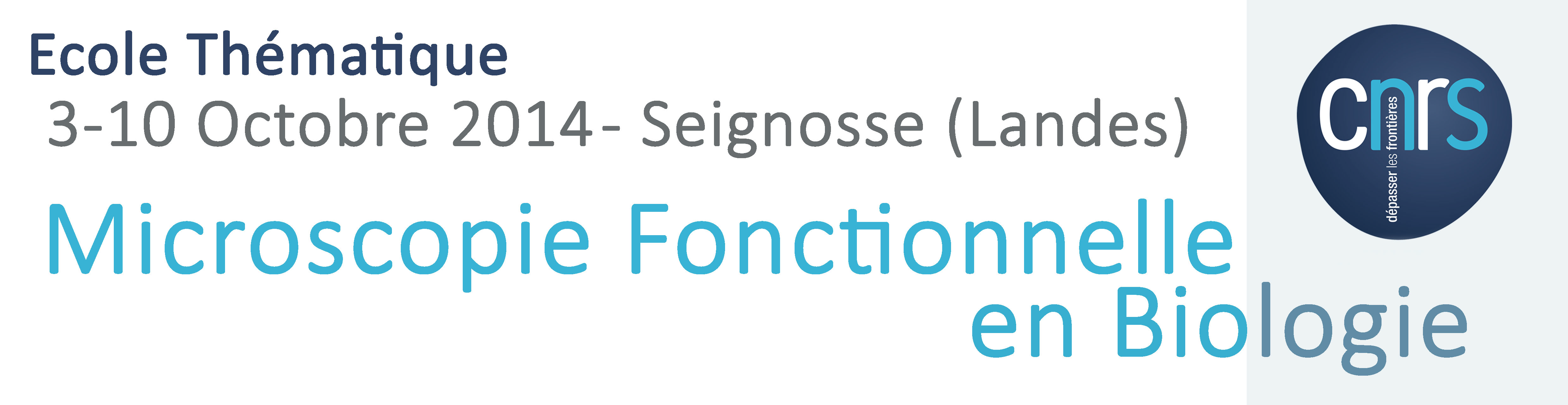
This School is organized since 2004 by the GDR2588 within the CNRS training program, with the assistance of the vocational training unit of INSERM and supports from France-BioImaging , RTmfm and ITMO BCBDE (Aviesan). This school will take place from September 30 to October 7, 2016.
The number of places is limited to 350 participants and pre-registration will be open from February 2016 to May 2016. http://gdr-miv.fr/mifobio2016
A Call for Workshops will be launched soon !
With the support of our industry partners, but also from academic laboratories we anticipate an exceptional technology park available for you to participate and propose many practical workshops.
This year four types of workshops are solicited:
- “Mastery” Workshops (AM): This is to illustrate the potential of a given technology to meet an identified biological questions; the experimental approach is mastered, the achievement being made on industrial devices, most often.
- “Innovation” Workshops (AI) : the idea is to propose experiments on new technological developments laboratories (home-made experimental setups) or original operation of a set of imaging technologies
(CLEM, multimodal microscopy, new imaging approaches, ..) - “Exploration” Workshops (AE): this is to experiment new approaches, onsite. For example exploring new types of samples by changing the type of analysis carried out or using other instrumental configurations, combine data obtained from different and complementary systems, in order to consider the question or biological hypothesis under several angles, make use of different analysis methods…We here encourage biologists to propose projects to explore, without insurance of results (in an exploratory way !) .The aim is to take advantage of the unique set of expertise and systems present during MiFoBio.
- “Theoretical approach” Workshop (AT): This year we also want to foster more theoretical approaches, including modeling. We thus request the involvement of Project Leaders for such workshops, either based on real or simulated data.
The team responsible for the implementation of workshops will very soon open a registration web portal where you can propose your workshops. This first call will be closed on January 22. If necessary, a second call will be launched at the end February on more target areas to represent all school subjects, or yet to allow the improvement of certain projects presented first call requiring changes.
So if you intend to propose a workshop, it is important to get ready with your project (title, description, equipment needs, no co-host). No more than two facilitators for workshops.
 The
The  [su_pullquote align=”right”]

 [/su_column]
 in functional microscopy. These workshops are designed to complete various imaging devices: microscopes super resolution multiphoton, small animal imaging, FLIM / FCS CARS .. provided by the industrial partners of the school or by research teams in the case of prototype systems. Several workshops computer are also introduced for the analysis and processing of data. The basic rule of the School is that industrial undertake to allow full autonomy to scientific systems available. Piloting workshops for academic and not by industrial is indeed a key to the success of these practices phases.