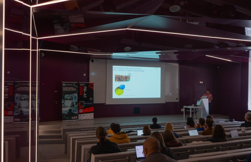
We are happy to finally announce the date of our next FBI Annual Meeting that will take place in Strasbourg.

Save the date! France-BioImaging will reunite together at the Alsace node on November 20 and 21, 2024!

This year’s edition will focus on “Live functional imaging: From chemical synthesis of the probes to instrumentation“.

Program and registration are coming soon.

As part of the scientific activities that it wishes to carry out and in continuation of its 1st seminar (Dec. 2022), the FBI “Preclinical Microscopy” Working Group is organizing its 1st webinar on February 1 from 2:00 pm to 4:00 pm. The objective of these scientific meetings is to present research projects, technologies and regulatory frameworks related to light microscopy from the organ/organoid to the living animal.

Program

2:00 pm – Welcoming & Introduction

2:15 pm – “Visualization of the ticking of circadian clock cells in freely moving mice” by Xavier Bonnefont (Institute of Functional Genomics, Montpellier).

3:00 pm – “Regulatory aspects of the use of animals for scientific purposes” by Isabelle Bardou (Cyceron, Biomedical Imaging Platform, Caen).

3:45 pm – Open discussion

Join the webinar

Cliquez ici pour rejoindre la réunion

ID de la réunion : 372 479 368 063
Code secret : tb9We5

Télécharger Teams | Rejoindre sur le web

Rejoindre avec un appareil de visioconférence

teams@vc.gustaveroussy.fr

ID de vidéoconférence : 125 390 858 6

On December 13th and 14th 2023, we have the pleasure to invite you to our Annual Meeting, to be hosted by our brand new FBI Toulouse Node at the Centre de Biologie Intégrative of the Université Toulouse 3 – Paul Sabatier.

We will be happy to celebrate yet another year of achievements and developments in bioimaging with all the members of the community.

This year, the Annual Meeting will focus on “Multiscale mechanobiology of cells and cell systems“, a topic specially selected for being one of Toulouse node’s expertise.

Mechanobiology aims to apply biophysical approaches to measure and perturb forces in complex physiological systems, and to develop in vitro systems including different types of organoids, enabling the controlled manipulation of cells and tissues and the measurement of mechanical forces to study cell mechanics and morphogenesis. 

This “mechanobiology” theme is including (i) how cells generate and transmit forces, (ii) the impact of forces on cell/tissue dynamics, (iii) the impact of extrinsic forces on cells and tissues, and (iv) the development of new tools for manipulating and measuring forces in cells and tissues in a controlled, non-invasive way.

We invite the France-BioImaging Community to present their mechanobiology-related projects during the second day of the Annual Meeting around a dedicated session with selected talks. We strongly encourage you to submit an abstract for a talk or a poster presentation during your registration!

The winner of the best talk and the best poster presentation will win their registration fees for one 2024 microscopy related event of their choice!

Core facilities will also have an opportunity to present a poster.

We look forward to meeting you there!

Gallery

Videos

Registration

Deadline: November 24, 2023

This form is currently closed for submissions.

Preliminary program

INFOS PRATIQUES:

Adresse: Centre de Biologie Intégrative, 169 Rue Marianne Grunberg-Manago, 31400 Toulouse

Télécharger le plan du campus

Comment venir?

Par la route

En venant de Bordeaux, contourner Toulouse par l’Est (direction Montpellier) ; avant le péage prendre la sortie no 23 direction Rangueil ou la no 19 direction Ramonville, puis suivre Université Paul Sabatier.

En venant de Montpellier, après le péage, prendre la sortie no 19 direction Ramonville, puis suivre Université Paul Sabatier.

En avion

Horaires aéroport Toulouse Blagnac

Une navette et le tramway relient l’aéroport et le centre ville de Toulouse (Transports en communs)

De là, il est possible de prendre le métro (lignes A ou B) pour rejoindre la station « Ramonville St Agne » (ligne B, direction Ramonville) : environ 1h de trajet

Le CBI est à 500 mètres à l’Ouest de la station et accessible à pieds ou en bus.

Il est également possible de prendre un taxi depuis l’aéroport : compter au moins 1/2h de trajet hors heures de pointe

En train

Depuis la Gare SNCF Matabiau:

Prendre le métro LIGNE A direction Basso Cambo, changer à la station “Jean Jaurès” et prendre la Ligne B pour rejoindre la station « Ramonville St Agne » : comptez environ 40 minutes depuis la gare.

Le CBI est à 500 mètres à l’Ouest de la station et accessible à pieds ou en bus.

Hôtels conseillés :

Nous vous conseillons de privilégier des hôtels en centre-ville.

Prise en charge des missions: 

Se rapprocher de votre noeud FBI (fonds mission), sauf pour les intervenants qui seront directement contactés pour la prise en charge de leur missions. 

France-BioImaging and all the French community aims to develop and promote innovative imaging technologies and methods. But microscopy images can also take an artistic, creative look and make the invisible world beautiful, allowing people to see the visual appeal of the life sciences. 

We enjoyed the diversity of the images submitted with many different microscopy techniques, models and applications represented. A big thank you to all the participants!

The National Coordination Team and the Executive Board are proud to announce the winners of the FBI Image Contest 2023:

  • 1st Place: Laurent LE, Lévêque-Fort Team, Institut des Sciences Moléculaires d’Orsay

In the blink of an eye

COS7 fixed cell. Alpha-tubulin labeled with DNA-PAINT and imaged with Atto 647N. Axial information is obtained by virtual-SAF measurement known as DONALD.

SMLM Fluorescence Microscopy with DNA-PAINT with DONALD detection

  • 2nd Place: Gonzalo QUIROGA-ARTIGAS, Team Contrôle cytoplasmique de la stabilité du génome, Centre de recherche en Biologie Cellulaire de Montpellier

“Tardigrade embryos protected by mother’s molt”

Tardigrades commonly align the time of molting with egg laying. In this image we observe a tardigrade molt covering three developing embryos (DNA in white). The microscopy technology applied was confocal microscopy, and the research aimed to investigate the synchronization of embryo development in tardigrades.

Confocal microscopy

  • 3rd Place: Hugues LELOUARD, Gorvel team, Centre d’Immunologie de Marseille Luminy

“Intestinal octopus”

Small intestine section from a LyzM-eGFP mouse containing one Peyer’s patch and stained for proliferative cells (Ki-67, yellow), Paneth cells (UEA-I, blue), epithelial cells (EpCAM, magenta), naive B cells (IgD, red), T cells (CD3, orange), helper T cells/macrophages (CD4, cyan), phagocytes (CD11c, turquoise), monocyte-derived phagocytes (GFP, green).

10-color spectral confocal microscopy

Congratulations to the winners!


Explore all the images submitted here:

As stated in the Terms & Conditions of the contest, foreign participants non-affiliated to a French institution are featured in the gallery, but were not evaluated as part of the contest.

The FBI Working Group Correlative Light-Electron Microscopy is organizing a workshop!

This workshop will take place at the Bordeaux Imaging Center (FBI Bordeaux node) from January 31st to February 2nd, 2024.

Correlative Light and Electron microscopy (CLEM) increases our capacity of biological investigation. By combining light microscopy and electron microscopy, this complementary approach takes advantages of both techniques. Light imaging provides valuable functional information thanks to its labeling power, whereas Electron microscopy excels at high resolution.

The Bordeaux Imaging Center has developed several workflows such as In-resin fluorescence and Array tomography. Both helps to determine 3D ultrastructure of a targeted area or a whole sample at different resolutions. In the framework of the FBI CLEM workshop, participants can choose one of these 2 different practicals, In-resin fluorescence and Array Tomography, in which a local specialist will show them the workflows.

Workshop 1: “In-Resin Fluorescence” – From HPF to tomography, by way of freeze-substitution and on-section fluorescence observation.

Workshop 2: “Array tomography” – From serial section to acquisition with confocal and SEM.

  • It is firstly intended to scientists with expertise in electron microscopy, who are expected to use the chosen technique.
  • 2 workshops of 4 people each (you can apply only to one workshop)
  • A practical manual will be provided, covering every step.

You want to attend this workshop? Please fill the following form before December 22nd, 2023:

The candidates whose research projects suit the workshop the best will be selected. We will get in touch with you after the Christmas break to confirm your participation at this workshop.

This form is currently closed for submissions.

Nous vous invitons au premier webinaire sur l’Initiative Commune Afrique-France pour l’Imagerie Biologique qui se déroulera le 6 décembre 2023 à 11h00 CET!

En coordination avec l’African BioImaging Consortium et Africa Microscopy Initiative et dans le cadre du programme Horizon Europe, l’Initiative Commune Afrique-France pour l’Imagerie Biologique vise à étendre et renforcer les collaborations entre collègues africains et français intéressés par l’utilisation de microscopie avancées pour leurs propres projets de recherche. C’est dans cette optique que nous avons lancé deux appels à projet : l’un favorisant l’accès aux plateformes de bioimagerie de France-BioImaging, l’autre consistant à un programme de jumelage.

Ce webinaire sera notamment l’opportunité d’en apprendre plus sur les projets sélectionnés et sur les bénéfices de cette initiative pour les scientifiques africains et français.

Webinaire

The France-BioImaging Image Contest is back for its 5th edition!

This image contest is open to all within the imaging community: core facility staff and users, R&D labs teams and co-workers, students… Submit your best microscopy images for a chance to showcase your skills, research and creativity to the French bioimaging community and beyond, allowing people to see the visual appeal of the life sciences. Images from the contest will be featured on France-BioImaging communication tools, online and in print.

France-BioImaging and all the French community aims to develop and promote innovative imaging technologies and methods. But microscopy images can also take an artistic, creative look and make the invisible world beautiful.

We are all eager to see your work !

Prizes

1 to 3 images will be awarded depending on the quantity and quality of the entries submitted. France-BioImaging will cover the registration fees for one 2024 microscopy related event of the winners’ choice (FOM, ELMI, EMC, COMULIS conference, etc.).

Important: Only French or foreign participants affiliated to a French institution can enter the contest. Foreign participants non-affiliated to a French institution can submit images and will be featured in the gallery, but will not be evaluated as part of the contest.

Submission deadline: Friday, November 10th, 2023, 23h59 UTC+2. 

Click here to consult the terms and conditions of the contest. When you are ready, submit your entry by filling the form below. You can check out last edition’s entries for inspiration. One participant can submit several entries (up to 3).

(If you have any issues when submitting your image, please contact communication@france-bioimaging.org)

This form is currently closed for submissions.

Discover last year’s submitted images on this following link: https://france-bioimaging.org/announcement/winning-images-fbi-image-contest-2022/

We are happy to announce our 8th France-BioImaging Annual Meeting! Happening on December 13th and 14th 2023, this year’s edition will be hosted by our new Toulouse node at the Centre de Biologie Intégrative (Toulouse, France).

The Annual meeting will highlight France-BioImaging’s development and perspectives. Imaging scientists and users from the infrastructure’s nodes will present their key projects and demonstrate how they have benefited from France-BioImaging and its community.

More information about the program and the registration coming soon!

France BioImaging and all the French community aims to develop and promote innovative imaging technologies and methods. But microscopy images can also take an artistic, creative look and make the invisible world beautiful, allowing people to see the visual appeal of the life sciences. 

We enjoyed the diversity of the images submitted with many different microscopy techniques, models and applications represented. A big thank you to all the participants!

The National Coordination Team and the Executive Board are proud to announce the winners of the FBI Image Contest 2022:

  • 1st Place: Carole SIRET, Van de Pavert Team, Centre d’Immunologie de Marseille-Luminy

Little Monster

The embryonic formation of lymph nodes, small organs essential for the immune response, is now known. Using light sheet microscopy, scientists were able to determine the dynamics at work in this 13.5-day-old mouse embryo. In blue, the lymphoid cells (LTi), derived from the haematogenous endothelium, a specific tissue of the embryo. They pass into the liver where they proliferate before migrating through the body to give rise to lymph nodes. The 3D information obtained thus makes it possible to follow the interactions of lymph nodes with their environment, in particular with nerve cells, in green, and blood vessels, in white. The lymphatic endothelial cells and some macrophages are visible in red.

Lightsheet Microscopy

  • 2nd Place: Magalie BENARD, Plateforme de Recherche en IMAgerie CEllulaire de Normandie (PRIMACEN), Research infrastructure HeRacLeS, Inserm US 51, CNRS UAR 2026,

“The communication link with others”

Image of a cellular interconnection between two human tumor cells whose cytoskeleton has been labeled with anti-tubulin (ATTO-647N), anti-vimentin (AlexaFluor594) antibodies and with Phalloidin probe (AlexaFluor488). Scale bar 1µm.

Confocal microscopy

  • 3rd Place: Frédéric FERCOQ, Parasites et Protistes Libres (PPL), Museum National d’Histoire Naturelle

“Sepia”

Stage 25 cuttlefish embryo (Sepia officinalis) observed under a confocal microscope.
The cuttlefish was cleared and the tissue autofluorescence was captured.

This image was produced in collaboration with Laure BONNAUD-PONTICELLI and Luis MOLINA from the BOREA laboratory.

Confocal microscopy

Congratulations to the winners!


Explore all the images submitted here:

As stated in the Terms & Conditions of the contest, foreign participants non-affiliated to a French institution are featured in the gallery, but were not evaluated as part of the contest.

In the framework of FBI-AT 2022 on Multiscale Fluorescence Imaging, which will be held in Paris from November 21st to 25th, 2022 (program available here), we have decided to open more widely the plenary lectures given by the invited researchers as well as the introductory lectures to each of the workshops.

Remote access to these lectures will be free but registration is mandatory. You will receive the connection link upon registration.

Registration form

This form is currently closed for submissions.

The France-BioImaging Image Contest is back for its 4th edition!

This image contest is open to all within the imaging community: core facility staff and users, R&D labs teams and co-workers, students… Submit your best microscopy images for a chance to showcase your skills, research and creativity to the French bioimaging community and beyond, allowing people to see the visual appeal of the life sciences. Images from the contest will be featured on France-BioImaging communication tools, online and in print.

France-BioImaging and all the French community aims to develop and promote innovative imaging technologies and methods. But microscopy images can also take an artistic, creative look and make the invisible world beautiful.

We are all eager to see your work !

Prizes

1 to 3 images will be awarded depending on the quantity and quality of the entries submitted. France-BioImaging will cover the registration fees for one 2023 microscopy related event of the winners’ choice (FOM, ELMI, EMC, COMULIS conference, etc.).

Important: Only French or foreign participants affiliated to a French institution can enter the contest. Foreign participants non-affiliated to a French institution can submit images and will be featured in the gallery, but will not be evaluated as part of the contest.

Submission deadline: Friday, November 11th, 2022, 23h59 UTC+2. 

Click here to consult the terms and conditions of the contest. When you are ready, submit your entry by filling the form below. You can check out last editions’s entries for inspiration. One participant can submit several entries (up to 3).

(If you have any issues when submitting your image, please contact communication@france-bioimaging.org)

This form is currently closed for submissions.

Our three winners have been announced! Please discover all the submitted images on this following link: https://france-bioimaging.org/announcement/winning-images-fbi-image-contest-2022/

France-BioImaging, with its partner the GDR IMABIO, organizes the 4th edition of the FBI-AT: an advanced microscopy workshop to be held in Paris from November 21st to 25th, 2022.

The aim of this France-BioImaging-Advanced Training is to train microscopy users on the most advanced imaging techniques that will allow them to perform molecular studies at the cellular level as well as in thick samples. In particular, recent developments on fluorescent probes will be highlighted. The workshop will benefit from state-of-the-art equipment available on several of the Parisian Node Imaging facilities.

This year’s edition will have plenary lectures given by experts in the microscopy development field. These seminars will be advertised as a series and will be broadcasted for a large audience.  In addition, specific techniques will be introduced.

Hands-on practicals will train attendants on these techniques every afternoon in different sites in Paris including Institut Curie, Institut Pasteur, Institut Cochin, Institut Jacques Monod, Institut de Psychiatrie et Neurosciences de Paris and ENS Paris. Access to this part of training will be restricted to selected and registered trainees.

To guaranty access to set-ups and proper training, each practical session will host only 3-4 persons. The sessions will be run in parallel.

Apply now, attendance will be limited to 25 participants! 

FBI-AT is ideal for researchers with a basic training in microscopy willing to become familiar with advanced techniques to answer their specific biological questions, or to be exposed to new developments that will allow them to tackle new questions in their project. We will consider applications from early career researchers (PhD students, post-docs), technical staff members and more senior scientists.

AT A GLANCE

The workshop contains plenary lectures and specific training sessions. Plenary lectures will be on hybrid mode and largely open.

Invited Speakers

Emmanuel Beaurepaire

Giulia Bertolin

Joerg Bewersdorf

Peter Dedecker

Claire Deo

Marie Erard

Ricardo Henriques

Christophe Leterrier

Sandrine Lévêque-Fort

Gustavo Quintas

Gaelle Recher

Jean-Baptiste Sibarita

Lothar Schermelleh

Practicals on

  • Combining micro UV-irradiation and Single Particle Tracking in living cells
  • SMLM multi-color: from sample preparation to quantification
  • FRET-based molecular tension sensors and FLIM
  • Imaging of cellular ultrastructures with expansion microscopy
  • SIM, STED or STORM ? : from sample prep to 3D imaging
  • 3D STED : Comparing flat cells vs thick samples
  • Culturing and imaging multicolour 3D live brain organoïds
  • Combining fast live 3D imaging with Z resolution preservation
  • Light sources for optogenetics
  • Non-classical genetically modified fluorescent probes for biological imaging
  • Imaging biological structures in 3D using double helix-STORM and 3D-SIM

Organizers

Florence Niedergang, Lydia Danglot, Chloe Guedj, Mickael Lelek, Pierre Bourdoncle, Audrey Salles, Xavier Baudin, Nicolas Borghi, René-Marc Mege, David Geny, Ludovic Jullien

Poster

Sponsors