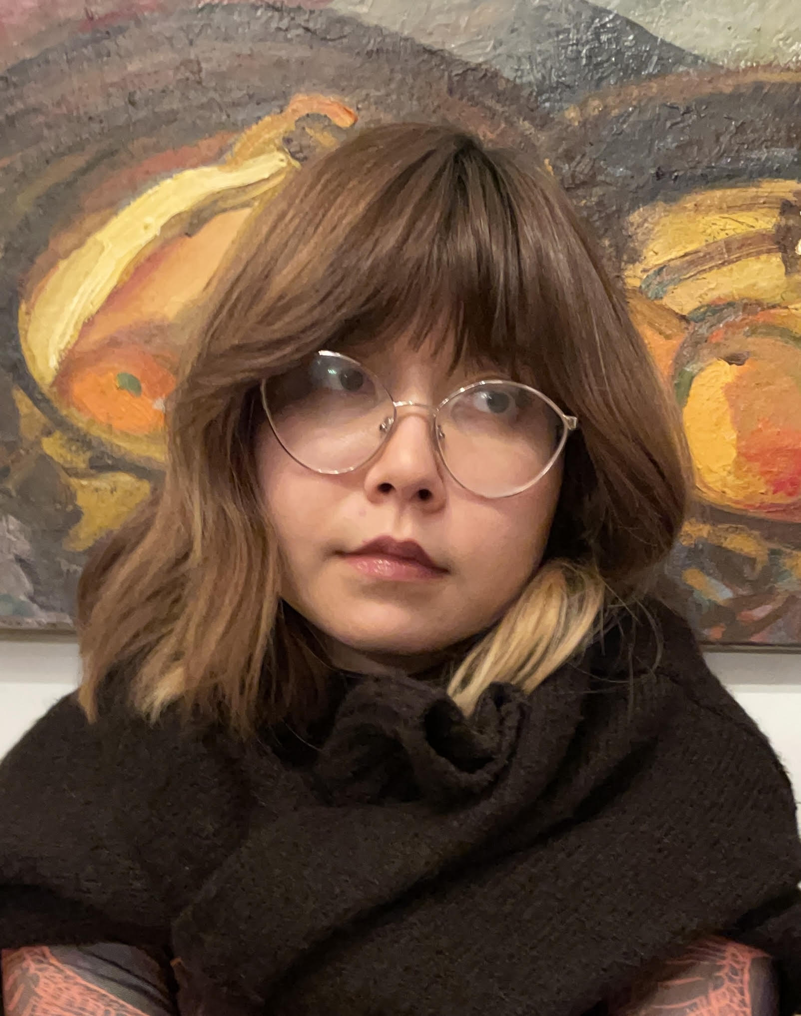
Ana Svetić is a PhD student in Nanotechnology at the University of Trieste (Italy). Her research focuses on developing a cell-derived plasma membrane model for correlative nanoscale imaging, with the aim of observing membrane nanodomains.
During a research stay abroad as part of her PhD, she visited theCBS AFM facility in Montpellier, where she used the PALM-AFM set-up to correlate AFM scanning with TIRF microscopy and investigate phase separation and nanodomains.
Through France-BioImaging User Access Support, Ana was able to acquire specific AFM tips that improved the quality of her scans and helped her obtain significant results. She also highlights the simplicity and efficiency of the access process and encourages other researchers to apply for support.
Could you introduce yourself?
Hi, my name is Ana Svetić, and I’m a biotechnologist turned biophysicist. I’m currently in the process of finishing my PhD in Nanotechnology at the University of Trieste and Elettra-Sincrotrone Trieste. My laboratory is called Nanoinnovation, and we do lots of Atomic Force Microscopy (AFM) as a supporting lab to the synchrotron.
What is your research project about?
My research project focuses on developing a cell-derived plasma membrane model, namely Giant Plasma Membrane Vesicles (GPMVs) and their patches (GPMV patches) for correlative nanoscale imaging – imaging with AFM and correlative techniques such as epifluorescence and TIRF microscopy, to ultimately observe the membrane nanodomains.
These nanodomains are, as the name suggests, nano-sized. In cells, they are just a few nanometers, very small and transient, and there hasn’t been a technique so far that allows the visualization and formation of said domains. But these ordered domains have been observed in synthetic models, namely Supported Lipid Bilayers (SLBs) and Giant Unilamellar Vesicles (GUVs).
So, my goal is to pass from more common simple synthetic membrane models (SLB and/or GUV) to a more complex one (GPMVs), in hopes to have a better, more biologically relevant model for observation of phase separation, which is responsible for the creation of previously mentioned nanodomains.
Which France-BioImaging’s facility did you visit, and which imaging technology did you use?
I was a visiting PhD student at the Centre de Biologie Structurale (CBS) – CNRS facility in Montpellier, where I’ve used the PALM-AFM set-up as a part of my PhD period abroad.
The PALM-AFM allowed me to correlate AFM scanning with TIRF (Total Internal Reflection Fluorescence), or more specifically, have the ability to measure the fluorescence intensity of the undermost layer of the GUV patches and GPMV patchesat the same time as performing an AFM scan of the same region of interest.
This ultimately allowed me to observe the phase separation and nanodomains in two ways – by seeing (TIRF) and by scanning (AFM) the surface of the membrane models.
Could you walk us through your experience accessing France-BioImaging?
I was made aware of the France-BioImaging opportunity by my mentor, Luca Costa PhD. He kindly told me about the platform, how to apply, and so I did. It was quite easy to fill out the form, and it took little-to-no-time to receive a positive reply!
I was astounded at the efficiency of the FBI and how smooth it all went, to be honest.
How did this access contribute to advancing your project?
I was already using the set-up before applying for the funding, but the funding helped me to buy and obtain AFM tips that I needed for my measurements. AFM tips that I needed were hard to get by, so I’m quite grateful for the funding of France-BioImaging. This in turn helped me get AFM scans of better quality, and at the end of the day obtain significant results for my research.
Thanks to TIRF, we were able to confirm different types of phase separations, which in turn shows up in nanodomain patterns.
AFM-TIRF image of a 20 mol% cholesterol GUV patch, same ROI. The correlative imaging demonstrates the existence of 2 phases. TIRF (left), AFM (right).
What are the next steps for your research project?
As I’ve already mentioned, I’m currently wrapping up my PhD project. Afterwards, I’m not sure, but I would like to explore more of what I’ve developed so far, and eventually localize meaningful proteins inside GPMVs and GPMV patches. This would let us know where exactly in nanodomains are proteins such as, for example, HER-2! It would be really, really cool.
What advice would you give to researchers who are considering applying for the FBI User Access Support?
Do it! Any support is meaningful and sometimes just a little bit can change the course of your research.
Meryem Aloulou is an INSERM researcher at the Toulouse Institute for Infectious and Inflammatory Diseases (INFINITy), where she leads a research programme investigating how regulatory T cells orchestrate T–B cell cooperation. She is currently exploring how the nuclear architecture of T cells contributes to the regulation of their identity.
To visualize this architecture, she needed access to an imaging technique that was not available in her laboratory. Through France-BioImaging User Access Support, she was able to visit the IBENS imaging facilityand collaborate with Samira Benadda to move her research project forward.
We had the pleasure of meeting Meryem, who shared her journey with us and presented the results she obtained thanks to this visit.
Could you introduce yourself?
I am originally from Agadir, a coastal city in southern Morocco, where my curiosity about the living world began long before I knew it had a name. What fascinated me from the start, and still does, is the paradox at the heart of immunity: the same system must defend us against pathogens without ever turning against our own tissues. That question brought me to France, where I studied immunology and completed my PhD in Immunology at Université Paris Diderot.
During my PhD, carried out in Professor Renato Monteiro’s laboratory, I investigated Fc receptors and uncovered unexpected mechanisms regulating antibody-mediated immune responses. Our work challenged the traditional view of these receptors as purely activating molecules, revealing that the very same receptors can either amplify or restrain immune responses depending on the biological context. This work highlighted the remarkable adaptability of the immune system and its ability to fine-tune inflammation.
After completing my PhD, I joined the Centre de Physiopathologie de Toulouse Purpan (CPTP) in Toulouse in the laboratory of Dr. Nicolas Fazilleau. There, I turned to the regulation of humoral immunity, where our work revealed an unexpected heterogeneity within follicular regulatory T (Tfr) cells, reshaping our understanding of their role in regulating germinal center responses.
To further broaden my expertise, I spent two years as a visiting researcher in Professor Adrian Liston’s laboratory at the University of Cambridge. I was fascinated by the way Adrian had built his laboratory around a truly interdisciplinary vision, bringing together immunologists, molecular biologists, computational biologists, mathematicians, clinicians… to tackle fundamental questions of immune regulation. Working in this environment transformed the way I approach scientific research and convinced me that the most exciting biological discoveries emerge at the interface of multiple disciplines.
Today, I am an INSERM scientist at the Toulouse Institute for Infectious and Inflammatory Diseases (INFINITy), where I have established an independent research program investigating how regulatory T cells orchestrate T–B cell cooperation in health and disease. My goal is twofold: to uncover the mechanisms that govern protective immunity, and to identify biomarkers that improve immune monitoring in vaccination, autoimmune disease, and cancer. To achieve this, I combine fundamental immunology with emerging technologies and translational research.
What is your research project about?
The nucleus is much more than a container for DNA. It is closer to a library: the way the books are arranged on the shelves determines which ones can be read. And in immune cells, we have barely started to look at the shelves
Regulatory T cells are essential for maintaining immune homeostasis. Far from being mere suppressors, they actively orchestrate interactions among T cells, B cells, and dendritic cells to ensure that effective immune responses develop while preventing excessive inflammation and autoimmunity.
Although decades of research have identified the molecular and epigenetic programs controlling regulatory T cells, we still know remarkably little about how the three-dimensional organization of the nucleus contributes to establishing and maintaining their identity.
The spatial organization of chromatin determines which genes are accessible, how they are expressed, and ultimately how a cell acquires its function. Yet this level of regulation remains largely unexplored in regulatory T cells.
To investigate this, we use two complementary populations as experimental models: CD4⁺ and CD8⁺ regulatory T cells. Our previous work revealed important molecular differences between them, leading us to hypothesize that they may also possess distinct nuclear architectures.
Ultimately, we aim to determine whether nuclear architecture represents an additional layer of immune-cell organization that shapes immune-cell function.
You received financial support to access a France-BioImaging facility. Which facility did you visit, and which imaging technology did you use?
I first heard about France-BioImaging through our imaging facility, and the User Access call arrived exactly when our project needed a technology we did not have in Toulouse.
Through the France-BioImaging User Access program, I obtained access to the imaging facility at the Institut de Biologie de l’École Normale Supérieure (IBENS) in Paris.
The project is carried out in close collaboration with Dr. Samira Bennada, Head of the IBENS Imaging Facility, whose expertise in expansion microscopy and quantitative imaging has been instrumental in shaping both experimental design and imaging strategy.
Expansion microscopy relies on a counterintuitive idea: rather than relying solely on a more powerful microscope, the biological specimen itself is physically enlarged. The sample is embedded in a swellable polymer, where molecular labels are anchored before controlled expansion physically separates them while preserving their relative organization.
In our protocol, the sample is physically expanded before imaging, increasing the effective imaging resolution and allowing quantitative visualization of nuclear structures that were previously below the diffraction limit (Figure 1).
Figure 1. Expansion microscopy enhances the visualization of nuclear organization in primary regulatory T cells. DAPI-stained CD4⁺ regulatory T-cell nucleus before expansion (left) and after expansion (middle and right), imaged using a 63× objective. Physical expansion increases the effective imaging resolution from approximately 250 nm to ~30 nm, allowing subnuclear structures that cannot be resolved by conventional fluorescence microscopy to become distinguishable. Right: maximum-intensity projection. Scale bars, 5 µm.
Our project combines expansion microscopy with two complementary imaging modalities: the NSPARC detector for high-resolution confocal imaging of chromatin-associated markers and nuclear compartments, and spinning-disk SoRa microscopy for rapid three-dimensional super-resolution imaging.
Together, these approaches provide a multiscale quantitative imaging pipeline adapted to investigating the nuclear architecture of rare primary regulatory T cells.
Why was this technology relevant or essential for your research project?
Current molecular and genomic approaches have transformed our understanding of regulatory T-cell biology by identifying the genes, transcription factors, and epigenetic programs that define these cells. However, they provide little information about how these molecular components are spatially organized within the nucleus.
Yet, the three-dimensional organization of chromatin is increasingly recognized as a fundamental regulator of gene expression. To understand how regulatory T cells acquire and maintain their identity, we need to investigate not only which molecular players are present, but also how they are arranged in space.
Expansion microscopy is uniquely suited to address this challenge because it enables nanoscale imaging while preserving the native spatial organization of the cell. By physically expanding the specimen, it allows us to visualize and quantitatively analyze subnuclear structures that would otherwise remain below the diffraction limit.
Combined with high-resolution quantitative imaging, this approach allows us to map the spatial organization of chromatin, nuclear compartments, and lineage-defining transcription factors directly in primary regulatory T cells.
Rather than replacing transcriptomic or epigenomic approaches, expansion microscopy complements them by adding a spatial dimension that sequencing alone cannot provide. Ultimately, this integrated strategy will allow us to determine whether distinct nuclear architectures contribute to regulatory T-cell specialization, stability, and function.
Could you walk us through your experience accessing France-BioImaging?
The collaboration started long before the first images were acquired. Together with Samira, we discussed the biological hypothesis, selected the most appropriate nuclear markers, optimized sample preparation, and designed the imaging workflow best suited to answer our biological question.
I still remember seeing the first successfully expanded regulatory T-cell nucleus appear on the screen. It was not yet a biological discovery, but it was the first tangible demonstration that a question we had previously been unable to address had become experimentally accessible. For the first time, we could begin to explore the internal nuclear landscape of these rare primary immune cells at a scale that had previously been beyond our reach.
For me, this illustrates one of the greatest strengths of France-BioImaging. It is not simply a network providing access to sophisticated microscopes: it is a network of scientists who help transform an ambitious biological question into a robust imaging strategy.
As an immunologist, working closely with imaging experts has been extremely enriching. This interdisciplinary dialogue has broadened the way I think about my research and opened new perspectives at the interface between immunology, quantitative imaging, and spatial biology.
How did this access contribute to advancing your project? Which results did you obtain?
The project is still ongoing, but the impact of France-BioImaging is already tangible.
Beyond providing access to technologies unavailable at our institute, the collaboration has enabled us to establish a robust multiscale imaging pipelinespecifically adapted to primary regulatory T cells. Together with the IBENS team, we optimized sample preparation, expansion protocols, image acquisition, and quantitative image analysis.
We are nowgenerating the first datasets that will allow us to quantitatively compare the nuclear organization of different regulatory T-cell populations.
Most importantly, this access has transformed what was initially a conceptual hypothesis into a biologically testable question, and has established a technological framework that will support many future projects in our laboratory.
What are the next steps or future perspectives for your research project?
Our immediate objective is to determine whether distinct nuclear architectures are associated with different regulatory T-cell states. Beyond describing nuclear organization, we aim to identify structural features that distinguish specialized Treg populations.
A particularly exciting question is whether nuclear architecture merely reflects cellular state or actively contributes to maintaining the regulatory lineage. We hypothesize that specific features of three-dimensional genome organization provide a structural framework supporting the transcriptional and epigenetic networks underlying Treg stability.
To address this, we will generate multidimensional spatial maps of the Treg nucleus by combining chromatin organization with the spatial distribution of lineage-defining transcription factors and histone modifications. Integrating this nuclear spatial imaging approach with transcriptomic, DNA methylation, and functional analyses will reveal how nuclear organization contributes to Treg stability and function.
More broadly, we envision nuclear architecture as a new dimension of immune-cell characterization, complementing molecular biomarkers and providing innovative ways to assess Treg stability.
Ultimately, this work demonstrates how advanced imaging can bridge structural cell biology and immunology by bringing spatial biology to the nuclear level, opening new perspectives for precision immunology.
What advice would you give to researchers who are considering applying for the France-BioImaging User Access Support?
I would strongly encourage researchers to apply. France-BioImaging offers much more than outstanding imaging technologies: it gives you access to an exceptional community of imaging scientists, and it fosters genuine scientific collaborations.
My advice: start with a clear biological question, not with a specific technology. The facility scientists are true scientific partners who can help refine the experimental design, identify the most appropriate imaging approaches, and develop robust quantitative workflows.
For me, the greatest strength of France-BioImaging is precisely this combination of technological excellence, scientific expertise, and collaborative spirit. It enables researchers to tackle ambitious biological questions that would be difficult to address within a single laboratory.
If I had the opportunity to apply again, I would do it without hesitation.
Like Meryem, you can benefit from France-BioImaging User Access Support to access cutting-edge imaging technologies available across the infrastructure!
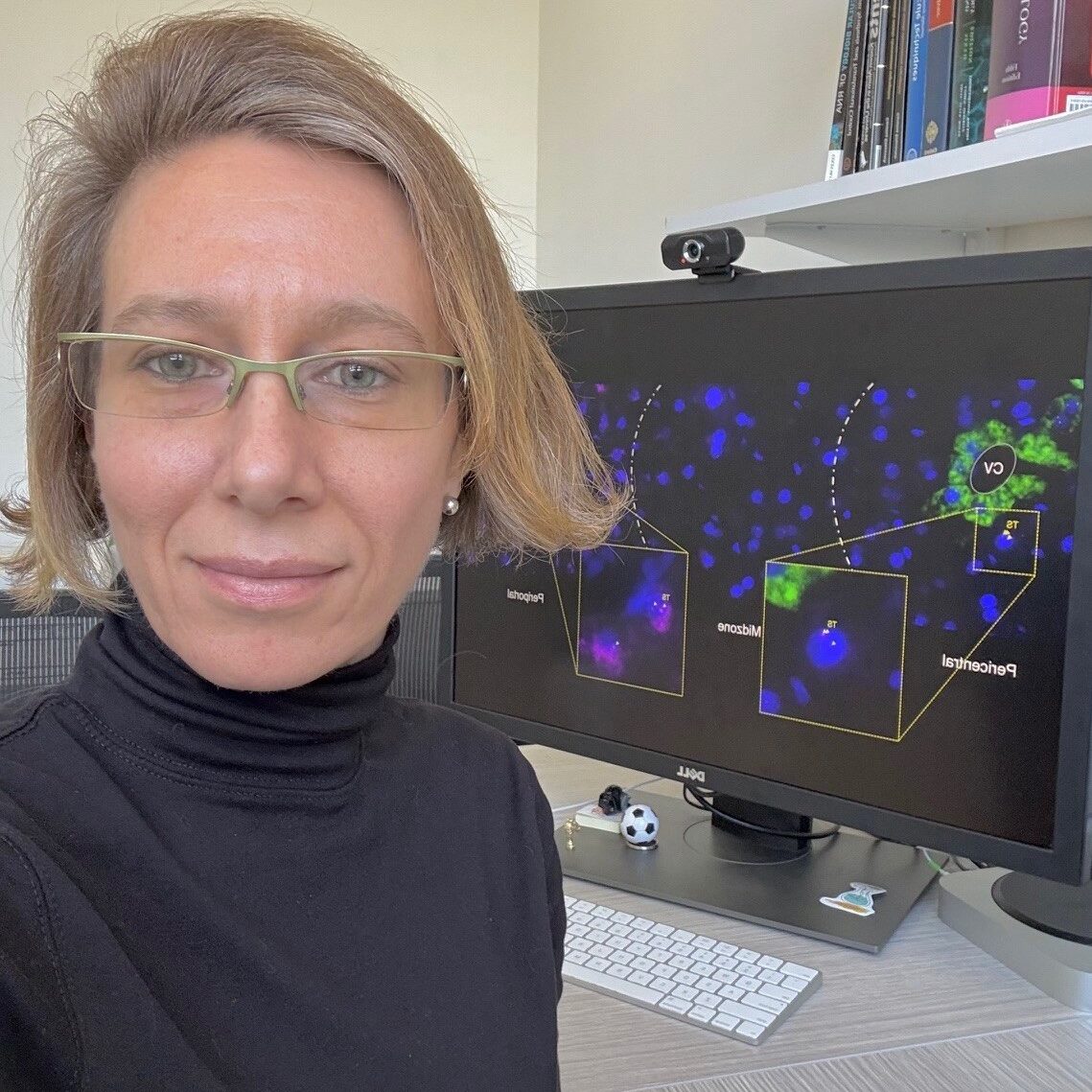
We are delighted to introduce Carolina Eliscovich, one of the awardees of the “FBI Call for User Access Projects 2024.” Carolina was granted access to one of the France-BioImaging facilities to advance her research using cutting-edge imaging technologies.
In this interview, she shares insights into her scientific journey and her current project, which explores how mRNAs are spatially organized and regulated in the liver of a living organism using advanced RNA imaging techniques.
To start, could you tell us a bit about yourself? What has been your academic journey so far, and what is your current role or area of work?
I am originally from Buenos Aires, a cosmopolitan and multicultural city in Argentina; and it is in this city located in the southern region of planet Earth where my academic journey starts inspired by the legacy of multiple local Nobel Laureates in Biomedical Sciences surrounded by yerba mate drinking and fútbol. Encouraged by my high school teachers to follow my curiosity to understand how living things function, I studied and graduated in molecular biology at the Faculty of Exact and Natural Sciences of the University of Buenos Aires.
Shortly after that, I moved to Europe to continue my scientific formation and completed a PhD in Biological Sciences at thePompeu Fabra Universityand Centre for Genomic Regulation in the vibrant city of Barcelona, Spain. My PhD thesis work, under the mentorship of Dr. Raúl Méndez, focused on the study of how maternal mRNAs encoding for proteins required for spindle formation and chromosome segregation are locally translated by the RNA-binding protein that promotes cytoplasmic polyadenylation to ensure proper meiosis progression in Xenopus oocytes.
I subsequently crossed the Atlantic Ocean back again but this time I headed to the North, to the greatest New York City in the USA, where I joined, as a postdoctoral fellow, the laboratory of Dr. Robert H. Singer at Albert Einstein College of Medicine. During my training in his lab, I acquired unique professional expertise in RNA biology by using single-cell and single-molecule fluorescence microscopy technology. We developed a robust methodology, named super registration, using single-molecule fluorescence in situ hybridization in combination with immunofluorescence (smFISH-IF) to determine the physical interaction between individual mRNA molecules and their binding protein(s) within single neuronal cells using standard wide-field microscopy. As a result of a successful collaboration with other colleagues in the lab, we also developed a novel imaging technology to visualize and quantitate the mechanism of translation of single transcripts in real time in living cells.
I have always been interested in the cell biology of mRNAs since my first days at the lab bench; and now my lab also at Einstein expands on the study of these versatile macromolecules by watching them within cells in tissues. Most of the knowledge we have about RNA regulation comes from studies in cells in culture, but little is known about how RNAs behave in the native context of the tissue under physiologic conditions, and this is the knowledge we would love to contribute to the field.
What are you currently working on in your research? What is the main topic or challenge you’re exploring?
I recently became interested in the biology of the (mouse) liver; and my current research program at Einstein focuses on the development of RNA imaging technology using fluorescent-labeled probes to visualize the spatial organization of mRNAs in the healthy and regenerating liver. The architecture of the liver is unique and what I find fascinating the most is that gene expression of the hepatocytes-the dominant cell type in this organ- are spatially regulated depending on their distance to the vasculature.
This organ is also remarkably resilient and capable of regenerating itself upon a variety of injuries or surgical removal of a significant portion of the organ mass. This process has biological and clinical relevance because it is the foundation of liver transplants and, unfortunately, organ transplantation is the only treatment for liver failure. What we found is that during this incredible regeneration response, hepatic gene expression is dynamically reprogramed, and therefore, it is an ideal model system to study mechanisms of mRNA regulation in time and space in a living organism.
A current ongoing challenge we face in our studies is to examine individual cells not only at tissue-scale but also with single molecule sensitivity while preserving tissue morphology. Also, and importantly, high-throughput and multiplexing has been always challenging in imaging-based technologies due to a limited number of different fluorophores that can be simultaneously detected because of spectrally overlapping wavelengths. Luckily, we were able to overcome these difficulties thanks to the France-BioImaging available technology!
At what point did you come across France-BioImaging, and what made you want to use its services or connect with the infrastructure?
The France-Bioimaging infrastructure came along in my research thanks to Dr. Edouard Bertrand (Institute of Human Genetics, Montpellier, France). I had the privilege of working with Edouard at Einstein while he was doing a sabbatical year in the Singer lab a few years ago. Later, he introduced me to the mission of the infrastructure and how the state-of-the-art imaging technologies and resources were bringing to all investigators across France that could not access the equipment otherwise.
I remember that I felt a bit jealous of the platform; my lab was located overseas, how would I get access to the France-BioImaging facilities that I needed?
And the answer came last year when France-BioImaging launched for the first time the call for external users (both national and international). It was a great opportunity to finally gain access to the platform and get training on the technology that was missing in our studies: to visualize, simultaneously, multiple individual mRNA molecule species in the intact mouse liver tissue during regeneration. And I submitted my application.
Could you walk us through your experience accessing France-BioImaging? Which facility did you work with, how did the process go, and what stood out to you during your time there?
The experience at the France-BioImaging facility was incredible!
My application included access to sequential FISH methodology developed by Dr. Davide Normanno, postdoc in the Bertrand lab, thus, I visited the Bertrand laboratory and theMontpellier Resource Imaging (MRI) facility in the Institute of Human Genetics in Montpellier for two weeks. Before my arrival to Montpellier, we worked virtually on the design of the fluorescent probes, protocols and details on sequential FISH methodology and equipment. Everything was planned before my arrival.
But life is certainly filled with unexpected events and on my very first days in Montpellier, Davide went suddenly on a medical emergency that required surgery and was not able to be in the lab the subsequent days*; and it was thanks to the collegiality and generosity of the investigators at the France-Bioimaging infrastructure that I was lucky to meet and count on Drs. Marcelo Nollmann and Jean-Bernard Fiche at the Center for Structural Biochemistry, just across the street, who rapidly jumped in and helped to finish the training on the sophisticated automated microscope with a microfluidic perfusion system to perform sequential FISH. We were running against the clock and against all odds our sequential FISH pilot experiment on liver tissue worked beautifully and we were able to visualize, for the first time, multiple mRNAs within hepatocytes in the intact liver tissue during regeneration, something we could not have done without accessing France-BioImaging. Thanks to all!**
I will never forget how I felt while looking at the first images we took; it was one of those unique moments in the lab where we are fortunate to see the unseen.
*Davide recovered well, and he is currently healthy and working in the lab.
**I am deeply thankful to Edouard, Davide, all the people in the Bertrand lab, Marcelo and Jean-Bernard for their kindness and generosity during my visit. I could not have done anything without them!
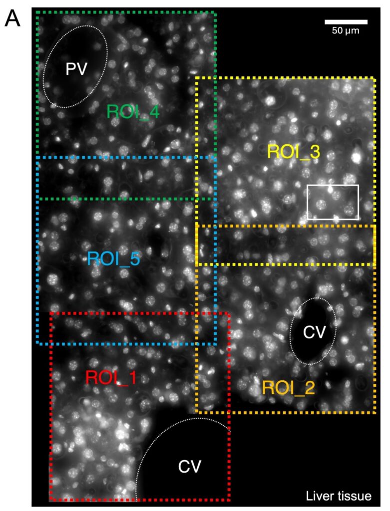
Panoramic view of a hepatic lobule showing DAPI-stained nuclei (white). The central vein (CV) and the Portal vein (PV) are indicated. Region of interests (ROI, indicated in colors) were imaged sequentially and then stitched to reconstruct the liver tissue morphology. Scale bar, 50 μm.
What did microscopy bring to your project specifically? Were there insights or results you couldn’t have obtained otherwise?
My lab is, in essence, an RNA imaging lab. Single-cell and single-molecule microscopy allow us to use the power of quiet observation and understand what every single cell is doing in its preserved microenvironment within the tissue, giving us insight about the heterogeneity present in a biological sample that is usually masked in other methodologies.
Even though we perform standard smFISH (up to 2-3 different RNA species) in liver tissue in our lab in a routinely manner, we could not have been able to advance the method to image multiple RNA species (multiplexing; up to 16 different RNA species) and in a high-throughput way (by the use of an automated microfluidic system) without accessing to the France-BioImaging facility at the MRI-IGH in Montpellier.
Sequential smFISH imaging technology applied to liver tissue allows us to study the simultaneous expression of multiple mRNAs within the same hepatocyte giving us unprecedented gene expression information of a regenerative liver cell in vivo.
The analysis of these images will give us understanding about mechanisms that the liver may employ to segregate different functions along the hepatic lobule and shed a light on the co-expression of genes that have been always thought not to be expressed at the same time in the same cell. This will allow us to explore in depth the relationship between tissue morphology and the molecular state of the cells and, therefore, cell function in vivo.
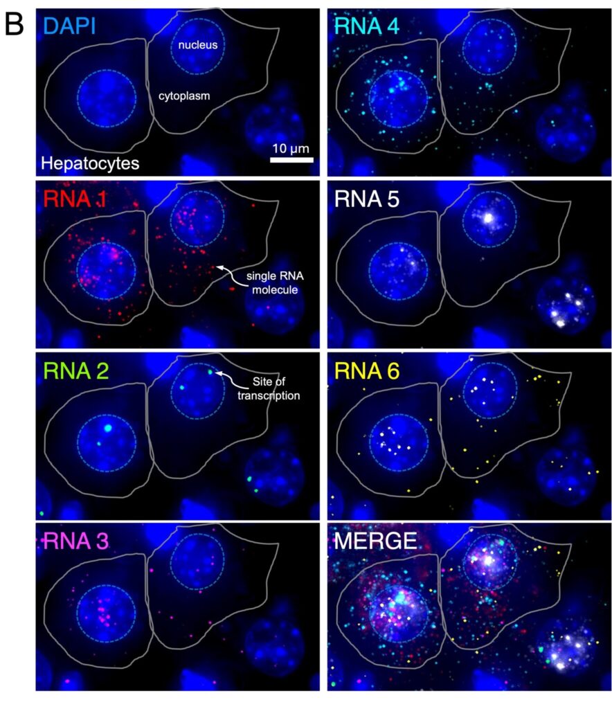
Magnification of the white box area inside ROI_3 in panel (A). Only six different mRNAs are shown in different colors to illustrate the method. DAPI-stained nuclei (blue). Single RNA molecules and sites of transcription can be visualized in the cytoplasm and nucleus of the cells, respectively. Merge image is also shown. Scale bar, 10 μm.
Looking back, would you encourage other researchers to use France-BioImaging’s platforms and access program? What would you say to someone considering it?
I would encourage everyone in the scientific community from students and postdocs to principal investigators to look for opportunities to access the France-BioImaging facilities. The first thing I would say to someone considering this is that they will never regret the experience because it is a great chance to learn and test the feasibility of a new imaging technology and/or methods applied directly to their specific research question, something that could take a lot of time (and funds) if they do not have the technology already established in their home institutions.
The France-BioImaging facility nodes include a great variety of state-of-the-art microscopes and imaging technologies available that I am sure each researcher can find the proper match for their scientific needs.
Finally, this access program would eventually help them to establish collaborations with the experts in the host lab/institution and even lead to the opportunity to reimplement the technology in their home labs or institutions where it would serve as the foundation for future directions of research in the upcoming years.
If I had the opportunity to apply to the France-BioImaging call for user access program again, I would do it without a doubt!
Discover Hamed Abbasi’s story, a researcher at the Center for Optical Diagnostics and Therapy at Erasmus Medical Center, who benefited from the “FBI Access Fund” in 2024. Thanks to this program, Hamed was able to visit PRIMACEN, a cell imaging platform that is part of the Normandie Node. This collaboration enabled him to gather robust data and secure a grant to develop an advanced fluorescence lifetime system for intraoperative use, supporting surgeons with real-time visual guidance to identify and delineate tumors.
To start, could you tell us a bit about yourself?
As far back as I can remember, I’ve been fascinated by optics. My late father was an optician, and as a teenager, I spent my summers working with him. That’s where I first learned the fundamentals of optics, which later inspired me to pursue the subject academically. I earned a Bachelor’s degree in Engineering Physics with a minor in Lasers and Optics, followed by a Master’s in Photonics, where I developed a laser-induced fluorescence spectroscopy system for non-destructive quality assessment of agricultural products as part of my thesis.
After graduation, I spent several years in industry as an Optical System Designer and Developer, working on spectrometers, hyperspectral imaging systems, colorimeters, and tunable lasers. During that time, I also worked part-time as a lecturer at an opticianry school, training industry professionals looking to deepen their understanding of optics.
Eventually, I decided to pursue a PhD and was admitted to the Biomedical Engineering program at the University of Basel in Switzerland. My doctoral research focused on developing a closed-loop optical feedback system to monitor a laser osteotomy setup using Laser-Induced Breakdown Spectroscopy (LIBS). I graduated magna cum laude in 2020.
Shortly after, I joined the Center for Optical Diagnostics and Therapy at Erasmus Medical Center in Rotterdam, the largest hospital in the Netherlands, where I still work in the Department of Otorhinolaryngology, Head and Neck Surgery. Within our department, I have developed several innovative photonics-based technologies to address unmet clinical needs in oncological surgery, including approaches based on Raman spectroscopy and fluorescence molecular imaging.
What are you currently working on in your research?
The central theme of my current research is photonics-guided surgery. Among the many optical techniques available, we focus primarily on fluorescence molecular imaging. This intraoperative imaging method provides surgeons with real-time visual guidance to identify and delineate tumors.
Within our fluorescence-guided surgery group, we have conducted multiple clinical trials using this innovative approach, applying various tumor-targeted fluorescent tracers. In these procedures, patients receive a tracer before surgery, and during the operation, dedicated imaging systems are used to visualize the tumor in real time.
We are continually working to improve both the sensitivity and specificity of our systems, which remains a significant challenge, as enhancing one often compromises the other.
At what point did you come across France-BioImaging, and what made you want to use its services or connect with the infrastructure?
Before learning about France-BioImaging specifically, I was already familiar with Euro-BioImaging, since our internal imaging center (Erasmus Optical Imaging Centre Core Facility in Rotterdam) is one of its nodes. Our microscopy center is also part of NL-BioImaging, the national imaging infrastructure in the Netherlands. I was aware that similar national infrastructures exist across Europe, but I didn’t know much about France-BioImaging until it announced a call for external users.
I came across the announcement on LinkedIn, and it immediately caught my attention. I visited the France-BioImaging website and began exploring the various facilities. After reaching out to a few of them, I eventually found the center that had the specific microscope I needed: the PRIMACEN/Cell Imaging platform in Normandy.
Could you walk us through your experience accessing France-BioImaging?
I contacted the PRIMACEN facility by email, and we quickly arranged an online meeting. That’s when I had the opportunity to speak with Ludovic Galas and Damien Schapman. During the meeting, I explained what I was looking for, and they confirmed that they had exactly the instrument I needed: a fluorescence lifetime microscope with sensitivity in the near-infrared range. It was perfect for my project.
Right after our meeting, I prepared a proposal and submitted it to the France-BioImaging user access call. Once my proposal was accepted, I reconnected with Ludovic and Damien to organize two one-week visits.
I visited the facility twice and had an excellent experience. The team at PRIMACEN was incredibly welcoming and supportive. It was not only a great opportunity to access cutting-edge infrastructure but also to meet and work with wonderful people.
What did microscopy bring to your project specifically?
Performing fluorescence lifetime imaging in the near-infrared (NIR) region is quite challenging for several reasons. First, detector sensitivity in the NIR range is generally lower than in the visible spectrum. Similarly, NIR fluorophores might have lower brightness compared to many visible ones. And finally, fluorescence lifetimes in the NIR are significantly shorter than those in the visible range. All of this makes it difficult to obtain robust and reliable data.
Despite these challenges, I was able to collect strong proof-of-principle results that supported my hypothesis. I remember leaving Normandy with a big smile on my face, almost certain that this data would help me secure a grant to develop an advanced NIR fluorescence lifetime imager for clinical use, and Bingo, it did! Just a few months later, together with our external industry partner and my colleagues at Erasmus Medical Center, Dominic Robinson and Stijn Keereweer, we were awarded a grant from Health~Holland.
Fluorescence lifetime imaging adds an additional layer of information to conventional intensity-based fluorescence imaging by measuring the temporal decay dynamics of the emitted fluorescence signal.
The project focuses on developing an advanced fluorescence lifetime system for intraoperative use, in close collaboration with our consortium partner Single Quantum, a Delft-based company known for developing the world’s fastest and most sensitive light sensors, with unmatched precision.
Without the microscopy data from PRIMACEN, it would have been much more difficult to convince the grant committee. That proof-of-principle experiment made all the difference.
Looking back, would you encourage other researchers to use France-BioImaging’s platforms and access program?
Absolutely. I would strongly encourage any researcher in need of advanced microscopy to consider using France-BioImaging’s platforms. The infrastructure is state-of-the-art, and the people are incredibly helpful and welcoming.
And don’t worry, if you receive an email from them and see “FBI”, it’s not the Federal Bureau of Investigation! It’s just the friendly acronym for France-BioImaging. My experience was both scientifically rewarding and personally enjoyable, and I wouldn’t hesitate to recommend it to others.
In this new user story, meet Maria Nazarova, a PhD student at IGBMC in Strasbourg. For her research on chromatin regulation mechanisms, she benefited from the FBI Access Fund to access cutting-edge technology available at the MRI-CRBM platform in Montpellier. But as is often the case in science, not everything went according to plan.
Read her story to find out more!
To start, could you tell us a bit about yourself? What has been your academic journey so far, and what is your current role or area of work?
I have always been interested in molecular biology, and I’ve been lucky to explore it from several angles starting with plant genetics, moving through cancer-related non-coding RNAs and immunoglobulin locus rearrangements, and finally arriving at my current favorite topic:
Chromatin dynamics and its role in crucial processes like transcription, replication, and repair
I’m now a 3rd-year PhD student in the Tom Sexton Lab at the IGBMC in Strasbourg, where I study how enhancers communicate with promoters using live-cell imaging.
What are you currently working on in your research? What is the main topic or challenge you’re exploring?
Even though enhancers are key chromatin regulatory elements in both normal cell function and disease, we still know surprisingly little about how they find, choose, and activate their target genes.
It seems these mechanisms aren’t universal, but instead depend on many variables such as gene type and function, genomic distance, nearby regulatory regions, and the local epigenetic environment.
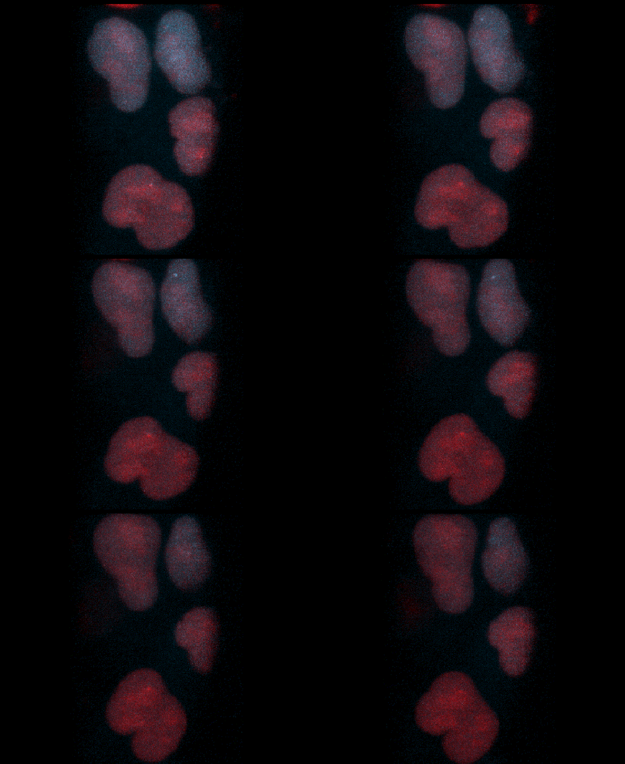
Mouse ESC with Sox2 promoter labelled (red) and Sox2 real-time expression (MS2/MCP).
My goal is to uncover some of those specific rules by modulating these factors in mouse stem cells and tracking how enhancer and promoter dynamics change during transcription under different conditions.
At what point did you come across France-BioImaging, and what made you want to use its services or connect with the infrastructure?
One of the main limitations of live-cell imaging is phototoxicity, which is especially important in my project because the cell line I use has three endogenous labels. Light Sheet technology seemed like the ideal solution for this.
To follow the dynamics of enhancer and promoter over long periods and across multiple transcriptional cycles without harming the cells, I need a system that can provide strong signal at low laser intensity.
Mounia Lagha, our collaborator from the Montpellier campus, shared the opportunity to apply for the France-BioImaging program, which I did and was fortunate to be selected.
Could you walk us through your experience accessing France-BioImaging? Which facility did you work with, how did the process go, and what stood out to you during your time there?
I spent two weeks at the Montpellier Ressources Imagerie(MRI) facility(MRI-CRBM, ed.), working with the Lattice Light Sheet microscope.
During the first week, I focused on learning the system — first with fixed samples provided by the team, and then moving to my own cells. Since my chromatin labels appear as single bright-ish spots on a background signal, it took some time to adjust, especially because I’m used to spinning disk microscopy.
The light sheet setup offers a very different perspective — both literally in terms of imaging geometry and in terms of how the data looks and behaves.
The second week was fully dedicated to data generation, though it came with some technical challenges.
The main issue was time resolution. Because I was working with three fluorophores, and the system had only one camera and needed to sequentially switch lasers, I could only get around 6 seconds per frame compared with the 1-1.5 seconds I’m used to with spinning disk.
That’s a big difference when you’re trying to follow the subtle, local dynamics of enhancer-promoter interactions in real time. So unfortunately, I wasn’t able to collect data I could use for quantitative analysis.
However, the experience itself was extremely valuable, and I’m very grateful for the opportunity. The MRI team was welcoming and supportive throughout my stay.
I am especially thankful to Virginie Georget, who guided me through the imaging process and was deeply committed to helping me get the most out of the system. Her knowledge, patience, and willingness to adapt the setup to my experiment made a big difference.
Even though the data didn’t turn out as hoped, I came back with a much deeper understanding of microscopy techniques in general and a lot of ideas for how to better design future imaging experiments.
What did microscopy bring to your project specifically? Were there insights or results you couldn’t have obtained otherwise?
Microscopy, especially live-cell imaging, is absolutely essential for my project. It allows me to directly follow the spatial and temporal behavior of enhancer-promoter pairs inside the nucleus, in real time.
Even though the data from the Lattice Light Sheet setup couldn’t be used in the end, it pushed me to think more deeply about the technical needs of my project, and it gave me first-hand experience with a powerful imaging technology that could still be extremely useful under different experimental conditions.
Looking back, would you encourage other researchers to use France-BioImaging’s platforms and access program? What would you say to someone considering it?
Yes, absolutely. The experience is not only useful for data generation, but also incredibly enriching from a learning and technical development perspective. It gives you access to cutting-edge technologies and expertise that you might not have in your home institution.
My advice would be to plan ahead as much as possible, communicate clearly with the hosting team about your needs and expectations, and if your project involves live imaging, try to negotiate at least three weeks, especially if you’re planning to use a new system. Two weeks pass very quickly, and having more time makes a huge difference.
Loading Comments...
Loading login form...
Privacy and cookies
We use necessary cookies to run the site. Optional categories stay blocked unless you allow them.
Your browser privacy signal is active, so analytics and marketing remain disabled.