Service
The Normandie Node is composed of one imaging facility so called PRIMACEN (HeRacLeS, US 51-UAR 2026, with user service, research and development activities), and six highly visible R&D teams (SFR IRIB, NORVEGE, INC3M, SCALE and BB@C) expert in microscopy techniques, controls and tools. Mainly located in Rouen and distributed to Caen and Le Havre, the Normandie node is offering high level technical and innovative methodological expertise in multi-scale imaging (TEM, STED, FLIM, TIRF, 2P, LSM…) at the interface between physiology, biology, chemistry, bioimage analysis, from the molecule to the small animal/plant. The Normandie Node has expertise in vascular sciences, microalgal biosciences and intercellular communication. Moreover, we have founded the International master program in cell imaging (IMAC) where students are intensively trained on PRIMACEN equipment and have the opportunity to go abroad including Finland thanks to a tight cooperation.
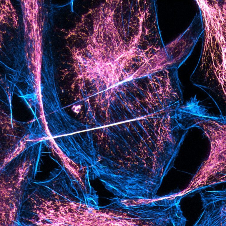
Located in Rouen, PRIMACEN, the cell imaging facility of Normandy (1000 m2) is integrated since 2022 in HeRacLeS (Univ. Rouen Normandy, Inserm US51, CNRS UAR2026). To achieve its service offering and R&D objectives, PRIMACEN proposes a continuum with 3 pillars thanks to complementary skills in cell biology-histology, instrumentation-quality control and data processing. In tight connection with animal (rodent/marine models) and plant facilities, the first transversal pillar involves living and fixed sample preparation including labeling, for advanced light and electron microscopies. Then, appropriate resolutions are proposed to users and collaborators through multi-scale and multi-modal imaging including LSM and 2P, WF/confocal micro/macroscopy, TIRFM, FLIM-STED imaging, cryo-FLIM/confocal and TEM/cryoTEM microscopies. To finally help users in their scientific projects, image processing and analysis (3D modeling, ImageJ macros, AI…) are available on highly performing working stations.
Bioorganic Chemistry team (15 people) develops bioconjugation, surface modification, multi-step synthesis, to create biocompatible chemical tools to explore the complexity of living mechanisms. The team’s expertise ranges from organic synthesis to photophysics; aiming at the development and tailoring of 1) new organic fluorophores (either push-pull or ESIPT based with high Stokes shifts and solvatochromism), with a recognized know-how in cyanins, coumarins, rhodamins, indazoles or BODIPY fluorescent scaffolds, notably their NIR derivatives, for application in conventional, two photon and STED microscopy, 2) biodetection (proteins, DNA) techniques based on profluorescent or chemiluminescent probes, and 3) new chemobiology tools such as bioconjugation reactions (and click-chemistry type tools), chemical or photoreactive cross-linkers, photolabile groups. The team collaborates with industrial companies through Carnot I2C and with international collaborators through XL-Chem program.
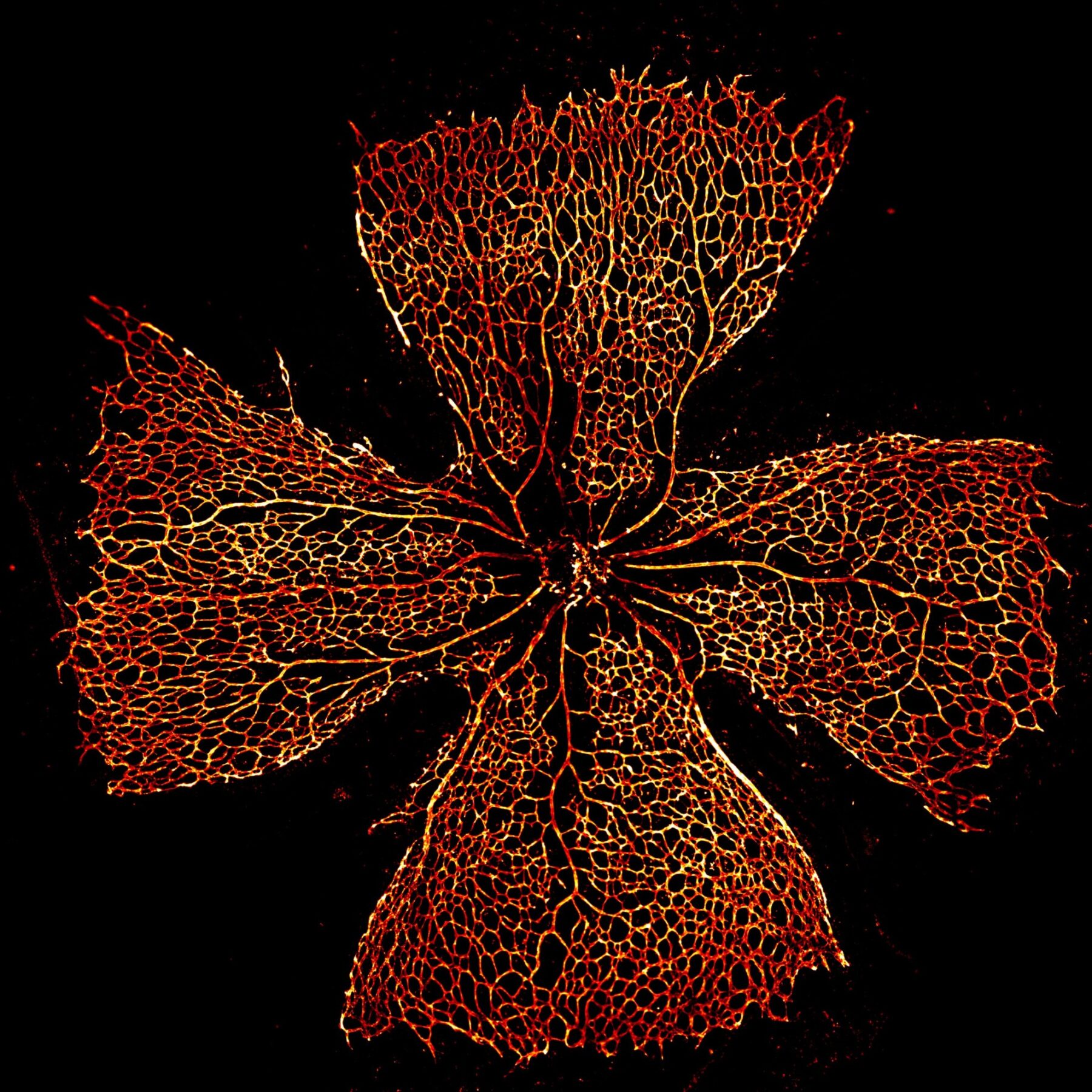
Team 4 of the Inserm research unit 1245 aims to characterize the contribution of neurovascular dysfunction in the pathophysiology of neonatal brain lesions while keeping in mind brain immaturity. Thereby, research projects paid attention to molecular, cellular and integrated processes leading to angiogenesis defects and neurodevelopmental consequences such as vessel-associated migration. The team’s research activity is deeply committed into translational research validated by patents and clinical protocols with the objective to develop diagnosis tools and neuroprotection strategies. To reach these objectives, the research team developed mouse perinatal models of white matter injury, in utero gain and loss of functions, and environmental models of fetal toxicity such as FASD. Endothelial and neuro-vascular dysfunctions are apprehended in the developing brains and retinas by imaging approaches (light sheet, FLIM-STED, time-lapse migration, in situ zymography, OCT) on ex vivo tissues (brain organotypic slices, cultured retinas). Image processing and analysis through notably machine learning is developed in partnership with several members of the Normandy FBI node.
The GlycoMEV team (research axis 3) aims in understanding the biosynthesis and secretion of glycoproteins with a special focus on N-glycosylation in microalgae models. As in all Eukaryotes, N-glycosylation occurs in the endoplasmic reticulum and the Golgi apparatus, where numerous enzymes act step-by-step to build and transfer oligosaccharides named N-glycans on secreted proteins. To decipher the sequence and regulation of these events, it has recently developed cryopreparation methods and imaging technologies (i.e. TEM) enabling the characterization the microalgae cell morphology and to analyse the subcellular localisation of distinct molecular actors. Furthermore, the team has acquired expertise in molecular biology techniques specific to these models allowing the generation of strains expressing various proteins fused to fluorescent reporters. They have also developed strategies based on confocal/FLIM imaging to characterize the targeting mechanisms responsible for the localization of glycoenzymes in the Golgi apparatus
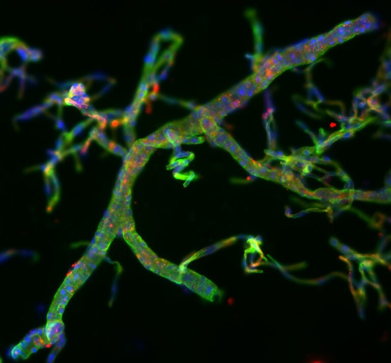
The “tPA and neurovascular disorders” team within the UMR-S U1237 – INSERM UNICAEN and GIS Blood and Brain @ Caen-Normandie Institute” (BB@C – INSERM – UNICAEN- CHU CAEN) is focused on a better understanding of the physiopathology of thrombosis/ischemic neurovascular disorders by developing and applying advanced methods (from molecular to multimodal cell and in vivo imaging). Mainly dedicated to live-cell imaging, we will provide access to dynamic imaging, super-resolution microscopy (STED). We developed innovative neuroimaging tools such as biocompatible nanoparticles, multimodal probes to detect vesicles trafficking, autophagy, neuronal activity, inflammation in the field of vascular disorders. Additionally, we used multiphoton imaging to observe in vivo vascular events including inflammation, diapedesis of immune cells within the parenchyma as well as microthrombosis. Functional ultrasound localization microscopy enables us to assess brain-wide neurovascular activity at a microscopic scale.
SEBIO is a research unit specializing in aquatic ecotoxicology with a significant experience in the development and validation of effect-based tools for monitoring and predicting environmental impact of chemicals. As experts in marine biology, SEBIO develops in vivo whole aquatic organism imaging approaches aimed at achieving breakthrough in methods and physiological data in seawater shrimps, bivalves and fish larvae. By using light-sheet microscopy, multi-modal imaging and 3D-reconstruction, these projects will decipher fine structures and detailed homeostatic regulations, in particular related to vascular anatomy, immune cell migration and blood/hemolymph tissue perfusion, regarding infection disease, intertidal cycle, osmotic stress and blood withdrawal for laboratory sampling. In relation with INERIS, technological transfers to European environmental agencies and water monitoring programs will rely on a network of reference laboratories, research centers and organizations for environmental risk assessment.
The translational Cardiovascular Research team develops fluorescence-based 3D imaging techniques and intravital microscopy approaches, in order to apply them for solving key questions related to the molecular and cellular functions, notably related to inflammatory responses in hearts and vessels. The researchers and technicians of the team, in collaboration with PRIMACEN, have implemented and developed set-ups for macroconfocal lymphangiography, vascular thrombosis evaluations as well as light sheet imaging, which are regularly optimized for new applications and projects. The team implemented in 2016 the first cardiac lymphangiography in rodents. Later, the team developed approaches for light sheet 3D imaging. These protocols are accessible to the community through their integration into the platform PRIMACEN (IBISA).
|
|
|
|
|