Oh oh oh, we’ve got many news and upcoming events to share with you! Here is a quick rundown (and by the way, have a great end of the year 2023!).
Congratulations to the France-BioImaging Image Contest 2023 winners!
We are happy to announce the winners of the France-BioImaging Image Contest 2023. Congratulations! France-BioImaging will cover the registration fees for one 2024 microscopy related event of the winners’ choice!
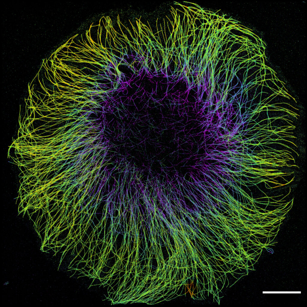
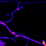
1st Place: “In the blink of an eye”, Laurent LE, Lévêque-Fort Team, Institut des Sciences Moléculaires d’Orsay
COS7 fixed cell. Alpha-tubulin labeled with DNA-PAINT and imaged with Atto 647N. Axial information is obtained by virtual-SAF measurement known as DONALD. SMLM Fluorescence Microscopy with DNA-PAINT with DONALD detection
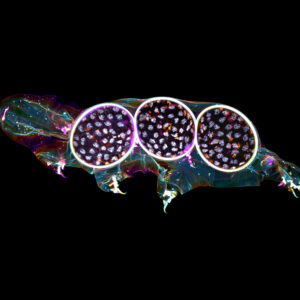
2nd Place: “Tardigrade embryos protected by mother’s molt”, Gonzalo QUIROGA-ARTIGAS, Team Contrôle cytoplasmique de la stabilité du génome, Centre de recherche en Biologie Cellulaire de Montpellier
Tardigrades commonly align the time of molting with egg laying. In this image we observe a tardigrade molt covering three developing embryos (DNA in white). The microscopy technology applied was confocal microscopy, and the research aimed to investigate the synchronization of embryo development in tardigrades. Confocal microscopy
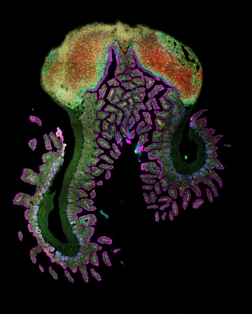
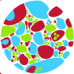
3rd Place: “Intestinal Octopus”, Hugues Lelouard, Gorvel team, Centre d’Immunologie de Marseille Luminy
Small intestine section from a LyzM-eGFP mouse containing one Peyer’s patch and stained for proliferative cells (Ki-67, yellow), Paneth cells (UEA-I, blue), epithelial cells (EpCAM, magenta), naive B cells (IgD, red), T cells (CD3, orange), helper T cells/macrophages (CD4, cyan), phagocytes (CD11c, turquoise), monocyte-derived phagocytes (GFP, green). 10 colors spectral confocal microscopy
A big thanks to all the participants! We enjoyed the diversity of the images submitted with many different microscopy techniques, models and applications represented.
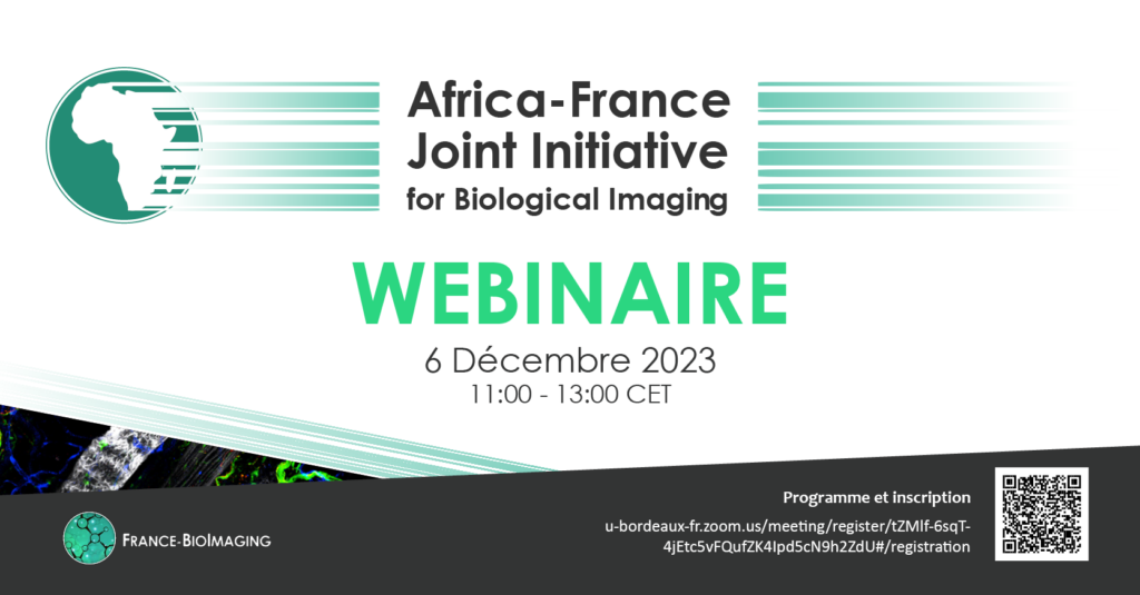
Our first webinar about the Africa-France Joint Initiative for Biological Imaging was a success!
At the beginning of December, we organized a webinar about the Africa-France Joint Initiative for Biological Imaging.
This webinar was, first, the chance to learn about the initiative and the two calls for project (now closed but some of the projects have started!). Moreover, we talked about the vision, the objectives and the opportunities that are already existing in terms of training, data analysis and international projects.
More than 40 participants, coming from 13 different countries, attended our event. We hope that this webinar (and in a bigger picture, the initiative) is just the beginning of multiple collaborations reinforcing the global bioimaging community.
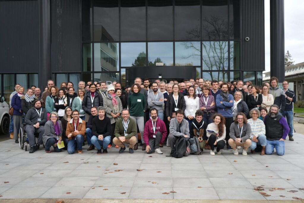
As we discussed about the future of the infrastructure and the collaborations between France-BioImaging and the national and european bioimaging ecosystem, this event was a very inspiring moment bringing together the community.
The Annual Meeting was the occasion to have updates on training, tech transfer and data projects led by the infrastructure, thanks to our mission officers. Ongoing and future projects from France-BioImaging calls have also been presented. A great success for our R&D transfer projects!
We had the pleasure to hear several wonderful presentations about imaging applied to mechanobiology. We talked, among other topics, about Brillouin microscopy, FAST-RIM, morphogenesis, compressive stress, Compression Force Microscopy and the use of organoids to study impact of cell heterogeneity.
Finally, Tatiana Merle, Laetitia Pieruccioni and Juri Aparicio Arias were awarded prizes for the best oral presentation, the best scientific poster and the best team poster respectively. Congratulations on the quality of their presentations, which have earned them free entry registration at one of 2024 microscopy events.
Overall, a very empowering meeting on the crucial role of core facilities!
Thanks to all the imaging scientists who participated to this event and a special thank you to the local organizers!
You missed it? No worries, we will display the recordings on our website in January. Stay tuned!
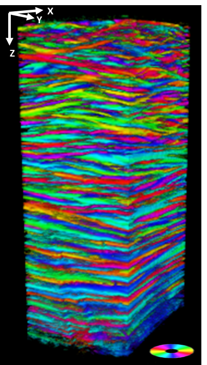
Imaging the orientation of biological structures in depth: P-SHG microscopy
Three-dimensional reconstruction of the lamellae structure of a human cornea. Colors indicate the direction of the collagen lamellae in the imaging plane, as shown in the inset color wheel. The image size is 250 x 250 x 600 µm3. The anterior part (side outside the eye) of the cornea is at the top of the image.
A key property of the human cornea is to maintain its curvature and consequently its refraction capability. Although we know that it is related to its stacked collagen lamellae structure, the distribution, size, and orientation of these lamellae along the depth of the cornea are poorly characterized up to now. A team from the Laboratory for Optics and Biosciences (LOB) has optimized a recent technology which combines Second Harmonic Generation microscopy and polarimetry (P-SHG) to image the lamellar microstructure of human corneas and more!
Acquire the structure in depth
Imaging the cornea is essential to understand how visual acuity works. This part of the eye is characterized by its transparency and refractive power but also by its unique mechanical properties. To answer the many questions of the cornea structure, the SHG microscopy is the perfect technique. This technology is based on the sample capacity to generate second harmonic light, which has half the wavelength of the light entering the material. However, the SHG is working on well-aligned assemblies of non-centrosymmetric molecules which fits perfectly with the collagen!
Apart from being specific to this kind of macromolecules, the SHG microscopy offers multiple advantages. First of all, as it does not involve the excitation of molecules, molecules do not suffer from phototoxicity or photobleaching effects. Moreover, no markers are necessary which makes this type of microscopy noninvasive. Finally, SHG microscopy allows the visualization of in-depth structure of thick samples. As a matter of fact, it is, nowadays, the gold standard technique for in situ visualization of collagen 3D organization in unstained biological tissues.
Add polarimetry and get orientation information
P-SHG first offers all the advantages of usual SHG microscopy: 3D optical imaging in depth and high specificity and sensitivity to collagen without any labeling. In this study, scientists took advantage of the light polarization to reveal the direction of the collagen fibrils that make up the lamellae of the cornea in their SHG microscopy acquisition. This recent technology is called: polarization-resolved SHG microscopy (P-SHG). The main novelty of this study was to implement P-SHG in depth to analyze intact human corneas along their full thickness (up to 600 µm).
By browsing this website, you indicate that you accept the terms and conditions, and agree to comply with them. If you do not accept these terms and conditions, please do not use this website.