Imaging the orientation of biological structures in depth: P-SHG microscopy
A key property of the human cornea is to maintain its curvature and consequently its refraction capability. Although we know that it is related to its stacked collagen lamellae structure, the distribution, size, and orientation of these lamellae along the depth of the cornea are poorly characterized up to now. A team from the Laboratory for Optics and Biosciences (LOB) has optimized a recent technology which combines Second Harmonic Generation microscopy and polarimetry (P-SHG) to image the lamellar microstructure of human corneas and more!
Acquire the structure in depth
Imaging the cornea is essential to understand how visual acuity works. This part of the eye is characterized by its transparency and refractive power but also by its unique mechanical properties. To answer the many questions of the cornea structure, the Second Harmonic Generation microscopy is the perfect technique. This technology is based on the sample capacity to generate second harmonic light, which has half the wavelength of the light entering the material. However, the SHG is working on well-aligned assemblies of non-centrosymmetric molecules which fits perfectly with the collagen!
Apart from being specific to this kind of macromolecules, the SHG microscopy offers multiple advantages. First of all, as it does not involve the excitation of molecules, molecules do not suffer from phototoxicity or photobleaching effects. Moreover, no markers are necessary which makes this type of microscopy noninvasive. Finally, Second Harmonic Generation microscopy allows the visualization of in-depth structure of thick samples. As a matter of fact, it is, nowadays, the gold standard technique for in situ visualization of collagen 3D organization in unstained biological tissues.
Add polarimetry and get orientation information
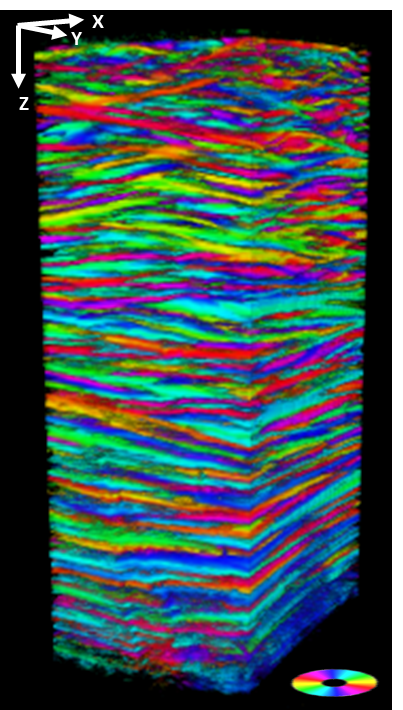
P-SHG first offers all the advantages of usual SHG microscopy: 3D optical imaging in depth and high specificity and sensitivity to collagen without any labeling. In this study, scientists took advantage of the light polarization to reveal the direction of the collagen fibrils that make up the lamellae of the cornea in their SHG microscopy acquisition. This recent technology is called: polarization-resolved SHG microscopy (P-SHG). The main novelty of this study was to implement P-SHG in depth to analyze intact human corneas along their full thickness (up to 600 µm). 3D reconstructions of P-SHG data show in a unique way the stacking of collagen lamellae with different orientations all along the thickness of the cornea. Here, imaging helped confirm that these lamellae are roughly organized parallel to the cornea surface, with different collagen orientations in sequential lamellae, and provided new information about the variation of these orientations along the depth of the cornea.
Aside from being the first quantitative characterization of the lamellar structure of the human cornea continuously along its entire thickness with micrometric resolution, this imaging technique could be a huge step forward in vivo diagnosis as it uses the detection of the reflected signal. Furthermore, this study opens the way to promising new characterizations of the cornea, such as mapping the size and distribution of lamellae as a function of depth, but also as a function of position (center or periphery of the tissue). This information will feed into mechanical modelling of corneal behavior during variations in intraocular pressure or healing processes. Finally, the study of pathological tissues will clarify the role of the corneal defective structure in certain diseases.
These results show the unique potential of P-SHG microscopy for imaging of collagen distribution in thick dense tissues. And of course, this approach is readily applicable to more than just cornea! It may be used for instance to decipher the structure of collagen in fibrotic pathologies or in other proteins that exhibit SHG, namely myosin and tubulin, or in starch and cellulose in plants. This shows the unique potential of P-SHG microscopy for imaging thick collagen-rich tissues.
Get access to one of our services!
Polarization SHG microscopy is not available in open access but we are open to collaborations!
You need classic SHG microscopy or another imaging technology or expertise that France-BioImaging provides? To get open access, please login via Euro-BioImaging website! You just have to choose the technology you want to use, then submit your proposal. All applications will be processed by the Euro-BioImaging Hub in close relation with France-BioImaging. And of course, all scientists regardless of their affiliation, area of expertise or field of activity can benefit from open access services! Users whose projects will be validated by Euro-BioImaging will benefit from a waiver for the access cost on France-BioImaging core facilities (https://france-bioimaging.org/access/).
Sources: https://www.inp.cnrs.fr/fr/cnrsinfo/cartographier-la-structure-de-la-cornee-humaine
https://www.ip-paris.fr/en/news/unveiling-structure-human-cornea
Raoux, C., Chessel, A., Mahou, P. et al. Unveiling the lamellar structure of the human cornea over its full thickness using polarization-resolved SHG microscopy. Light Sci Appl 12, 190 (2023). https://doi.org/10.1038/s41377-023-01224-0