We’ve got many news and upcoming events to share with you! Here is a quick rundown.
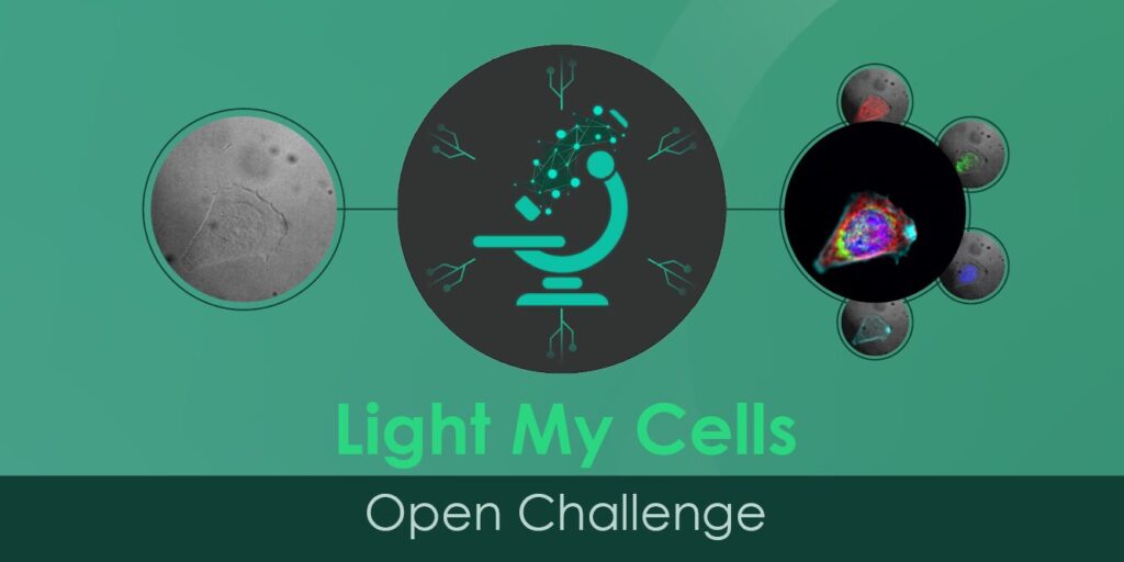
The first FBI challenge has just started! Welcome to the Light My Cells challenge
In order to answer image data analysis demands, France-BioImaging is launching its first data machine learning competition! The idea? To develop powerful methods that will then end up in creating standards & benchmarks in the field of bioimaging.
The FBI challenge is hinged on a double contribution: from core facilities engineers and from data scientists. The first group will acquire a large number of images to build a dataset, that will later be used by the algorithms. These images will be produced by microscopy engineers & technicians from FBI’s platforms. As for the second contribution, this is where the challenge starts! The challenge will be later published to have a maximum of data scientists to work on the algorithms that best fulfill the analysis task.
The first challenge is now open! We need experts to work on algorithms that will predict the fluorescence image of cell culture from a bright field, phase contrast and/or DIC microscopy label-free image.
If you are interested in creating the next generation of tools that will help the bioimaging community, check lightmycells.grand-challenge.org !
Travel Grant for the next GBI EoE in Okazaki, Japan
The next Global BioImaging Exchange of Experience will be held in Okazaki, Japan! And GBI is offering travel grants for those who wish to disseminate their work at a global scale!
Under-served regions must have fair and equitable opportunities to attend the Global BioImaging events. Therefore, the selection process for travel grants will follow a scheme that maximizes diversity and ensures equitable access globally. The selection approach will maximize regional and national diversity among grantees.
Anyone around the Globe is welcome to apply!! In addition to regional diversity and further promoting the subsequent dissemination of knowledge to a broader base, national and institutional diversity will be considered. The number of grantees from the same country will be limited, and only one application may be selected from the same institution. Lastly, efforts will be made to ensure equal gender representation.
How imaging allowed to rethink theories about skull structure and body temperature?
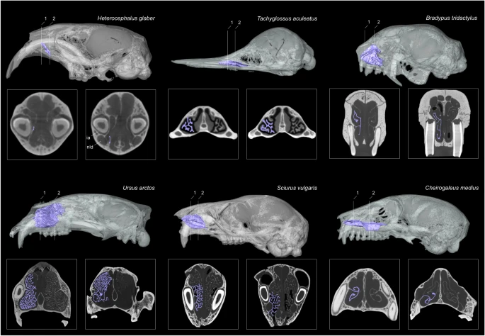
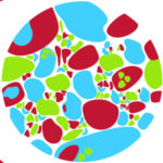
Detailed view of the maxilloturbinal in selected mammalian species with peculiar thermal and metabolic conditions or that undergo different forms of heterothermy (https://doi.org/10.1038/s41467-023-39994-1)
Most mammals can maintain a relatively constant and high body temperature. Scientists from the Institut des Sciences de l’Evolution de Montpellier (ISEM) investigate the possible correlation between the maxilloturbinal in the anterior nasal cavity and the body temperature maintenance by using Micro Tomography (or MicroCT) at the MRI core facility (FBI Montpellier node). This technique was essential in this study as it rebuilt the hypothesis around body temperature maintenance. Here is what they found.
Micro-CT is a 3D imaging technique using X-rays to see inside biological material, at a small animal or body part level. Slice by slice, this technology scans the object in a series of 2D images that are reconstructed in a 3D model. Micro CT is, thus, non-destructive.
Scientists needed to compare the structure of the maxilloturbinal in order to take conclusion. This is when Micro Tomography was very useful. They scanned 424 individuals from 310 mammal species using high-resolution X-ray micro-computed tomography, with approximatively half of the samples imaged at MRI, part of our Montpellier node. Using the obtained comparative 3D µCT dataset, they explored the anatomical diversity of the maxilloturbinal based on relative surface area, morphology and complexity. They specifically tested the relationship between multiple parameters such as the size-corrected basal metabolic rate (cBMR), the relative surface area of the maxilloturbinal (Maxillo RSA) or body temperature.
And the results surprisingly showed that there is no evidence to relate the origin of endothermy and the development of some turbinal bones!
So, what could be linked with the thermoregulation of mammals?
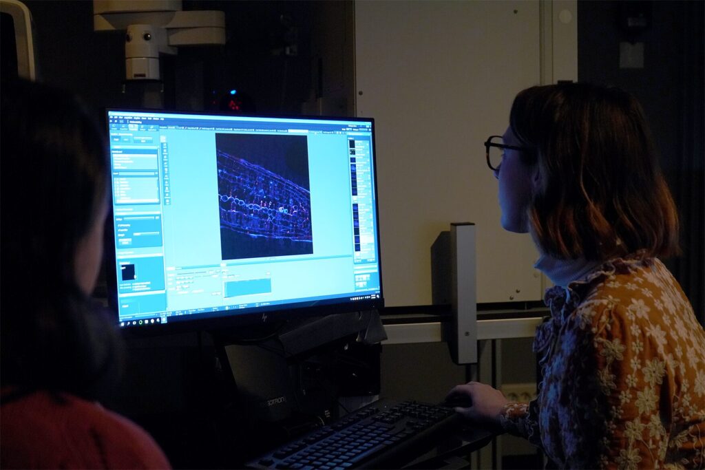
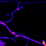
As the FBI Correlative Light-Electron Microscopy workshop happened last week at the Bordeaux Imaging Center (FBI Bordeaux node), what a better occasion to highlight a correlative microscopy technique: The Array Tomography.
By combining light microscopy and electron microscopy, this complementary approach takes advantages of both techniques. In fact, light imaging provides valuable functional information thanks to its labeling power, whereas electron microscopy excels at high resolution.
Array tomography is a versatile microscopy method that offers opportunities to explore cell and tissues in three dimensions. This technique is well suited to image large tissue volumes of your sample with fine structural and molecular details.
Different modes are available, each having its own specificity and benefits:
The fluorescence microscopy AT mode (FM-AT) delivers volumetric resolution and molecular marker multiplexing highly superior to traditional fluorescence microscopies.
The electron microscopy AT mode (EM-AT) captures three-dimensional ultrastructure at size scales that would require prohibitive effort using traditional serial-section EM methods.
And of course, you can combine both modes in a unique one FM/EM-AT with three-dimensional light and electron images acquired in perfect volumetric data.
Moreover, even though fields of applications are numerous, these attributes establish AT as an ideal choice for the most demanding analyses of diverse cellular architectures within mature and developing tissues such as brain tissue (neuroscientists, this technique is for you!).
Three microscopy techniques to understand a HIV viral assembly mechanism
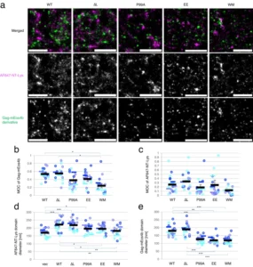
Colocalization between Gag-mEos4b curvature mutants and AF647-NT-Lys and domain analysis in PALM/dSTORM
HIV type 1 virus has a lipid envelope enriched with host cell sphingomyelin and cholesterol. In order to understand the mechanism of this enrichment, the FBI Alsace node (Laboratoire de Bioimagerie et Pathologies from Université de Strasbourg and the Imaging Center PIQ-QuESt) has participated in a study recently published in Nature Communications about HIV-1 virus assembly. Indeed, they have investigated the interplay between the HIV-1 Gag protein and the host cell lipids at the plasma membrane. This work has greatly benefited from the use of a great combination of different quantitative (FLIM-FRET and FRAP) and super-resolution (PALM/STORM) custom-made microscopes with specific probes.
Understanding the interaction between cancer and dendritic calls with the new Cryo-EM Glacios
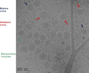
Cryo-EM image of endogeneous retrovirus-like particles released by from murine cancer cells
PICT, imaging core facility at Institut Curie, has acquired in 2023 a new Cryo-TEM Glacios from Thermo Fisher (partly purchased thanks to a France-BIoImaging grant). This powerful electron microscope will allow scientists to image and study proteins at an unprecedented resolution.
Using this powerful microscope, the Team of C. Thery (Institut Curie, U 932) has published a study on extra-cellular vesicles (EVs) released by murine tumor cells. Tumor‐derived EVs have been proposed to induce immune priming of antigen presenting cells or to be immuno‐suppressive agents. Their study revealed distinct subpopulations including endogenous retrovirus-derived components (VLP) and call for systematic re‐evaluation of the respective proportions and functions of non‐viral EVs and VLPs produced by murine tumors and their contribution to tumor progression.
By browsing this website, you indicate that you accept the terms and conditions, and agree to comply with them. If you do not accept these terms and conditions, please do not use this website.