Three microscopy techniques to understand a HIV viral assembly mechanism
HIV type 1 virus has a lipid envelope enriched with host cell sphingomyelin and cholesterol. In order to understand the mechanism of this enrichment, the FBI Alsace node (Laboratoire de Bioimagerie et Pathologies from Université de Strasbourg and the Imaging Center PIQ-QuESt) has participated in a study recently published in Nature Communications about HIV-1 virus assembly. Indeed, they have investigated the interplay between the HIV-1 Gag protein and the host cell lipids at the plasma membrane. This work has greatly benefited from the use of a great combination of different quantitative (FLIM-FRET and FRAP) and super-resolution (PALM/STORM) custom-made microscopes with specific probes.
Using FRAP to characterize mobile and immobile molecules
The Fluorescence recovery after photobleaching (FRAP) quantifies the two-dimensional lateral diffusion of fluorescently labeled molecules of interest. This technique is very useful in biological studies of cell membrane diffusion and protein binding as it not only reports on the diffusion rates of mobile fractions of molecules but also provides information about the proportion of immobile molecules.
In our case, FRAP experiments indicated that the expression of Gag significantly decreased the mobile fraction of sphingomyelin (SM)-rich domains. Besides, the technique showed that cholesterol (Chol)-rich domains were intrinsically immobile, even in the absence of Gag. It is speculated that the association of Gag with SM-rich domains restricts the lateral diffusion of the lipid domains, resulting in an increase of the immobile fraction in FRAP measurements.
Using PALM/dSTORM to localize molecules at high resolution
The Photo-activated localization microscopy (PALM) is a widefield fluorescence microscopy imaging method that provides images with a resolution beyond the diffraction limit. By collecting a large number of images each containing just a few active isolated fluorophores, the collection of these images allows to stochastically activate each fluorophore and thus to obtain a global image of the sample with high resolution.
The Stochastic optical reconstruction microscopy (STORM) works on the activated state of a photo-switchable molecule that leads to the consecutive emission of sufficient photons to enable precise localization before it enters a dark state or becomes deactivated by photobleaching.
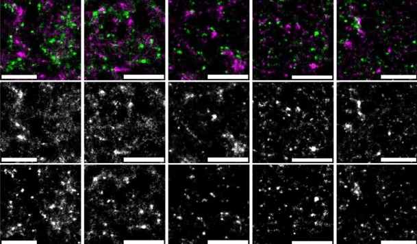
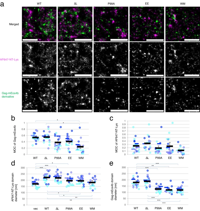
Coupling these two techniques, scientists next investigated at high resolution the localization of Gag and SM-rich or Chol-rich domains, both labeled with specific fluorescently labeled lipid binding proteins.
PALM/dSTORM visualized domains of different sizes labeled with the two lipid binding proteins, showing that the expression of Gag induced the formation of larger SM-rich domains but not the formation of larger Chol-rich domains. The main hypothesis is that the formation of large lipid domains may be due to the coalescence of smaller lipid domains.
Using FLIM-FRET to identify molecule proximity and interaction
And last but not least, the Fluorescence-lifetime imaging microscopy (FLIM) is an imaging technique for producing an image based on differences in the fluorescence-lifetime rather than its intensity. By quantifying variations in the exponential decay rate of the fluorescence from a fluorescent sample (fluorescence-lifetime) it is possible to report on molecule proximity. Since the fluorescence-lifetime is insensitive to changes in fluorophore intensity or concentration, it is the most quantitatively precise technique to report on fluorescence resonance energy transfer (FRET).
FRET is a mechanism describing energy transfer between two light-sensitive molecules (chromophores). A donor chromophore, initially in its electronic excited state, may transfer energy to an acceptor chromophore through non-radiative dipole-dipole coupling. FRET is extremely sensitive to small changes in distance and therefore an excellent reporter on molecule proximity and interaction.
In this third and final part, to better understand the possible effect of Gag on the lipid distribution in the plasma membrane, scientists investigated by two-photon FLIM-FRET the interaction of Chol-rich lipid domains with SM-rich lipid domains and its dependence on Gag multimerization. These last results showed that Gag multimerization induces SM-rich and Chol-rich domains to be in close proximity and that membrane curvature affects the apposition of SM-rich and Chol-rich domains.
A great example of application on how a combination of high-end technologies in microscopy can help you understand multiple aspects of a biological mechanism. So, what are you waiting for? Dive into the true potential of microscopy!
Get access to one of our services!
You need FRAP, two photon FLIM-FRET, PALM/dSTORM at France-BioImaging? To get open access, please login via Euro-BioImaging website! You just have to choose the technology you want to use, then submit your proposal. All applications will be processed by the Euro-BioImaging Hub in close relation with France-BioImaging. And of course, all scientists regardless of their affiliation, area of expertise or field of activity can benefit from open access services! Users whose projects will be validated by Euro-BioImaging will benefit from a waiver for the access cost on France-BioImaging core facilities (https://france-bioimaging.org/access/).
Tomishige, N., Bin Nasim, M., Murate, M. et al. HIV-1 Gag targeting to the plasma membrane reorganizes sphingomyelin-rich and cholesterol-rich lipid domains. Nat Commun 14, 7353 (2023). https://doi.org/10.1038/s41467-023-42994-w
Technical information from www.eurobioimaging.eu