Array Tomography: the best of both worlds
As the FBI Correlative Light-Electron Microscopy workshop is currently happening at the Bordeaux Imaging Center (FBI Bordeaux node), what a better occasion to highlight a correlative microscopy technique: The Array Tomography.
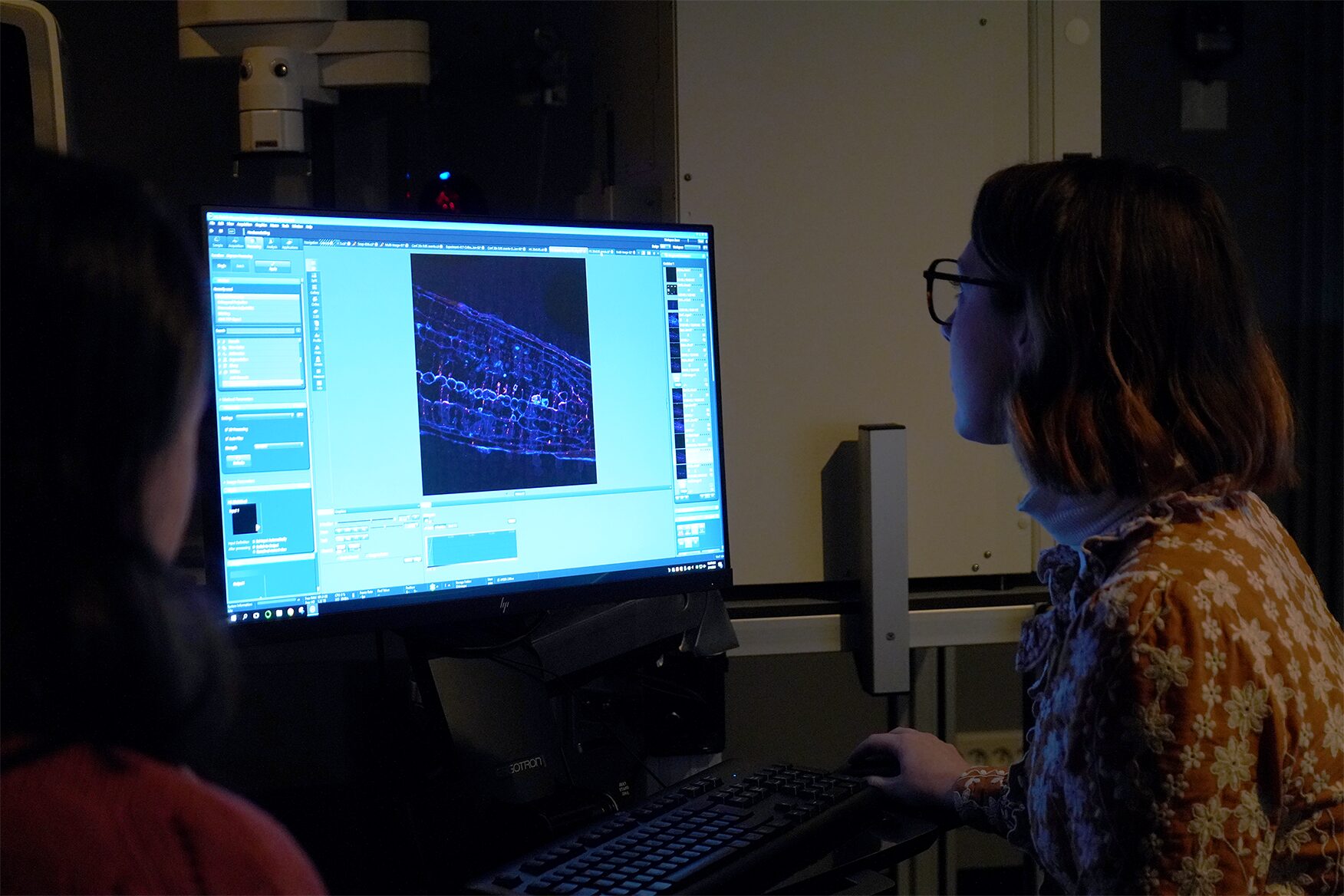
Correlative Light and Electron microscopy (CLEM) increases our capacity of biological investigation. By combining light microscopy and electron microscopy, this complementary approach takes advantages of both techniques. In fact, light imaging provides valuable functional information thanks to its labeling power, whereas Electron microscopy excels at high resolution.
Where array tomography (AT) is special is that this technique is based on serial ultramicrotomy (cutting many sections less than a micrometer in thickness) of the sample, section collection onto support, and serial scanning EM (SEM) imaging.
An array of microscopy modes
Array tomography is a versatile microscopy method that offers opportunities to explore cell and tissues in three dimensions. This technique is well suited to image large tissue volumes of your sample with fine structural and molecular details.
Different modes are available, each having its own specificity and benefits:
- The fluorescence microscopy AT mode (FM-AT) delivers volumetric resolution and molecular marker multiplexing highly superior to traditional fluorescence microscopies.
- The electron microscopy AT mode (EM-AT) captures three-dimensional ultrastructure at size scales that would require prohibitive effort using traditional serial-section EM methods.
- And of course, you can combine both modes in a unique one FM/EM-AT with three-dimensional light and electron images acquired in perfect volumetric data.
Why you should consider this technique next time?
The use of FM-AT should be considered for volumetric fluorescence imaging of fixed tissue specimens whenever there is need for very high resolution, high-order molecular multiplexing and/or rigorously depth-independent quantification of fluorescence signal intensities. Use of EM-AT offers perhaps the most convenient approach to volumetric electron microscopy available. Moreover, even though fields of applications are numerous, these attributes establish AT as an ideal choice for the most demanding analyses of diverse cellular architectures within mature and developing tissues such as brain tissue (neuroscientists, this technique is for you!).
Finally, while originally developed for EM, physical cutting of ultrathin sections was found to improve the axial resolution and provide accessibility to the sample for molecular labeling, beneficial for both, EM and light microscopy. Need another argument? The same or adjacent sections can be imaged with different modalities in correlative or even conjugate microscopy!
Get access to one of our services!
You need Array Tomography or another imaging technology or expertise that France-BioImaging provides? To get open access, please login via Euro-BioImaging website! You just have to choose the technology you want to use, then submit your proposal. All applications will be processed by the Euro-BioImaging Hub in close relation with France-BioImaging. And of course, all scientists regardless of their affiliation, area of expertise or field of activity can benefit from open access services! Users whose projects will be validated by Euro-BioImaging will benefit from a waiver for the access cost on France-BioImaging core facilities (https://france-bioimaging.org/access/).
Technical information: https://www.arraytomography.org/
Smith SJ. Q&A: Array tomography. BMC Biol. 2018 Sep 6;16(1):98. doi: 10.1186/s12915-018-0560-1