France-BioImaging wishes you a happy new year 2023, full of successes and astonishing images of cells and living organisms, for which we welcome you on our microscopes and facilities.
Here are the first news of 2023!
ISIDORe user access funds for research projects related to infectious diseases
ISIDORe TNA Calls are still OPEN!We are pleased to remind you that ISIDORe funding have been extended to other pathogens. Researchers interested in the following infectious diseases can apply for funding to access imaging technologies and services from Euro-BioImaging portfolio, including France-BioImaging facilities:
Respiratory Pathogens: Zoonotic betaCoronaviruses, Influenza A, Y. pestis, M. tuberculosis / bovis, C. burnetii.
Vector-borne pathogens and their vectors: Rift valley fever virus, Zika virus, Chikungunya virus, West Nile virus, Dengue virus, Japanese encephalitis virus, Yellow fever virus, Tick-borne encephalitis virus, Plasmodium, B. burgdorferi.
Risk Group 4 Pathogens: Ebola virus, Marburg virus, Nipah virus, Hendra virus, Lassa virus, New World arenaviruses, CCHF virus.
If you are currently working or plan to work on one of these infectious diseases in the coming months, please contact us so that we can discuss your project and help you apply for support from this fund!
We strongly encourage you to get in touch with us as soon as possible.
Useful links:
Full announcement and access funding opportunities are available here
For more information on the project and to apply, visit the ISIDORe website.
Unsure whether or not your work would be eligible? Contact the Euro-BioImaging Scientific Project Managers for advice: info@eurobioimaging.eu
Congratulations to the France-BioImaging Image Contest 2022 winners!
We are happy to announce the winners of the France-BioImaging Image Contest 2022. Congratulations! France-BioImaging will cover the registration fees for one 2023 microscopy related event of the winners’ choice!
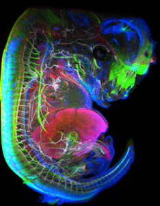
13.5-day-old mouse embryo. Lymphoid cells (blue) pass into the liver to proliferate before migrating through the body to give rise to lymph nodes. Nerve cells (green), blood vessels (white), lymphatic endothelial cells & some macrophages (red).
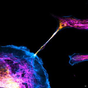
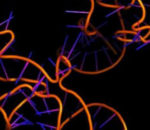
Image of a cellular interconnection between two human tumor cells whose cytoskeleton has been labeled with anti-tubulin, anti-vimentin antibodies and with Phalloidin probe. Imaged with confocal microscopy.
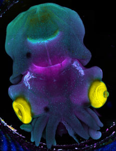
Stage 25 cuttlefish embryo (Sepia officinalis) observed under a confocal microscope. The cuttlefish was cleared and the tissue autofluorescence was captured.
A big thanks to all the participants! We enjoyed the diversity of the images submitted with many different microscopy techniques, models and applications represented.
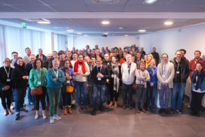
The France-BioImaging Annual Meeting: 10 years of operation and scientific advances!
As we discussed about the future of the infrastructure and the collaborations between France-BioImaging and the national and international bioimaging ecosystem, this event was a very inspiring moment bringing together the community.
The Annual Meeting was the occasion to have updates on training, tech transfer and data projects led by the infrastructure, thanks to our mission officers. Ongoing and future projects from France-BioImaging calls have also been presented. A great success for our R&D transfer projects!
Overall, a very empowering meeting on the crucial role of core facilities!
Thank to all the imaging scientists, R&D teams and users for contributing to these 10 years of operation and scientific advances!
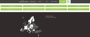
Looking for a specific imaging technology?Euro-BioImaging, the European bioimaging research infrastructure, worked on a quick and easy way to find the imaging technique & expertise you need. Simply, answer to a few questions to get in touch with facilities providing the technology you want. The map also displays all the services by country.
And of course, all the services from France-BioImaging are available on EuBI website. This is a great upgrade to facilitate project application!
By browsing this website, you indicate that you accept the terms and conditions, and agree to comply with them. If you do not accept these terms and conditions, please do not use this website.