
Multi-modal imaging reveals a new viral transmission mechanism
A recent study1 led by researchers from Karim Majzoub’s team (IGMM – Institut de Génétique Moléculaire de Montpellier) reveals an unexpected mechanism of deltavirus transmission, in which deltaviruses can be physically incorporated into helper virus particles. This work highlights how the combination of advanced imaging approaches was essential to identify, characterize, and validate this “viral Trojan Horse” mechanism.
Structural evidence of virus-virus association
Negative-stain electron microscopy first revealed the presence of atypical “double particles” within viral preparations. These structures, observed in superinfected cells producing both deltavirus and helper virus particles, displayed a characteristic morphology consisting of a main virion associated with a smaller satellite-like structure. These observations suggested a physical association between the two viral entities, but did not allow identification of their composition.
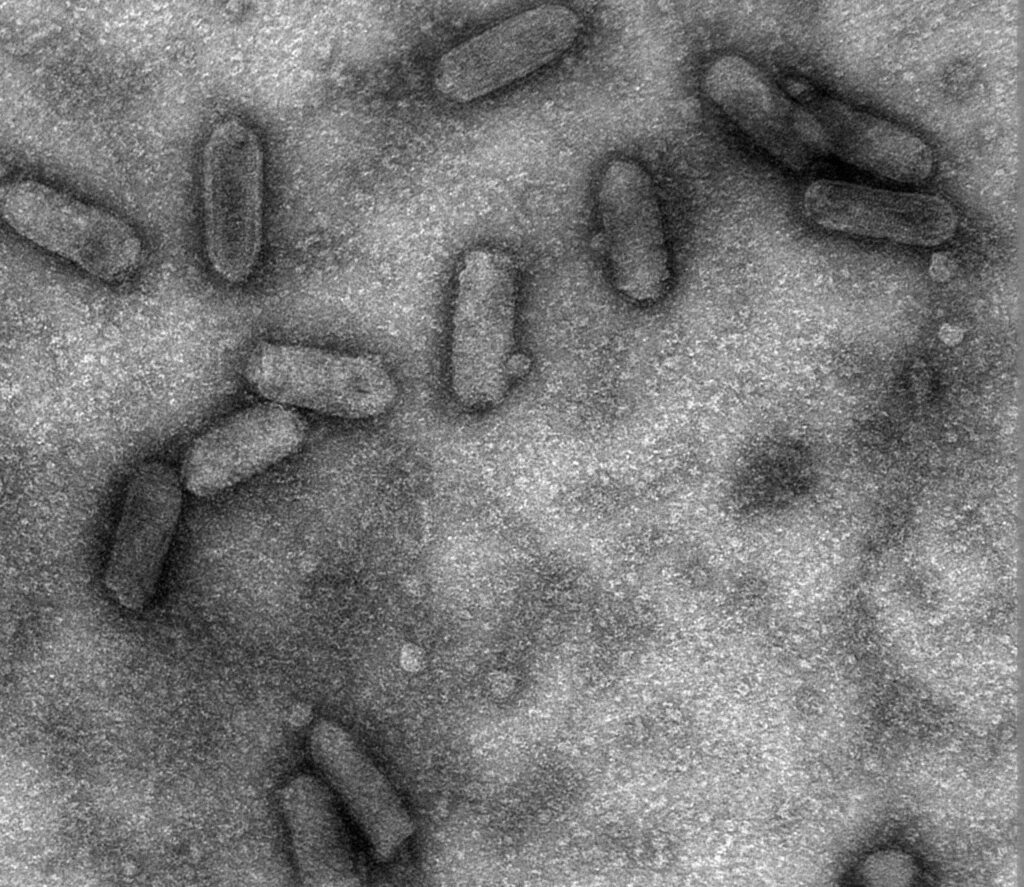

Cryo-electron microscopy provided further insight by enabling visualization of these particles in near-native conditions. In some cases, the observed structures appeared to share a continuous membrane, supporting the hypothesis that deltaviruses could be incorporated within helper virus particles rather than simply interacting at their surface.
Atomic force microscopy (AFM) was used as an independent validation approach. By imaging non-fixed samples in a BSL-3 environment, AFM confirmed the presence of similar particle morphologies, including protruding structures consistent with those observed by electron microscopy. This step was essential to exclude fixation-related artifacts and to establish the physical reality of the virus-virus association.

Multi-modal imaging for composition analysis
To determine the molecular composition of these particles, fluorescence-based approaches were applied. Super-resolution microscopy (STED) enabled the co-localization of deltavirus proteins (RDAg) and helper virus proteins within the same structures, using specific antibodies.

Isolated viral particles of VSV (shown in gold) and deltaviruses (in cyan), produced by cells, were deposited on glass coverslips and imaged using super-resolution STED microscopy, followed by deconvolution. Higher-magnification views reveal that some VSV particles exhibit a cyan signal at their periphery, indicating the presence of deltavirus within the VSV particle, consistent with a hitchhiking mechanism.
These findings were further supported by immunofluorescence and RNA FISH, which confirmed the presence of both viral proteins and deltavirus genomic RNA. In parallel, immunogold labeling combined with electron microscopy provided ultrastructural evidence linking deltavirus proteins to the observed particles.
Beyond structural characterization, these results support a model in which deltaviruses can be transmitted within helper virus particles. By exploiting the entry pathways and tropism of their helper viruses, this mechanism may facilitate infection of a broader range of cell types, tissues, and potentially hosts. In some cases, such as with HSV-1, deltavirus infectivity appears to depend on this association, highlighting the functional relevance of this “Trojan Horse” mechanism.
Ongoing work aims to further investigate this mechanism using live-cell imaging approaches, with a focus on the intracellular trafficking of deltavirus-containing particles during superinfection.
1 Joe McKellar, Aurélien Fouillen, et al., Deltaviruses spread through a viral Trojan Horse, Cell, Volume 189, Issue 9, 2026, Pages 2748-2761.e17, ISSN 0092-8674, https://doi.org/10.1016/j.cell.2026.01.037
 Marine Beraud • May 12, 2026