F-BIAS: A distributed national network for bioimage analysis
▽ Scroll down
Category: Announcement
F-BIAS (France-BioImaging Analysts) is a France-BioImaging support initiative that brings together bioimage analysts across France to deliver national-level image analysis services for the life science community. This distributed network addresses two major challenges: reducing the isolation of analysts, who are often the sole experts in their institutions, and expanding access to high-level image analysis expertise.
Responding to the challenges of bioimage analysis
With the rapid growth of imaging technologies and the increasing complexity of image data and analysis tools (deep learning, AI, custom pipelines…), many researchers lack local support to analyze their datasets. F-BIAS was created to fill this gap by federating analysts embedded within imaging core facilities that are part of France-BioImaging. Launched in 2021, the network now includes about 20 members.
Fig.1 The geographical repartition of F-BIAS members and their host structure (here in 2024).
A network designed by and for analysts
F-BIAS operates as a collaborative and horizontal structure. Each analyst, employed within a host institution, dedicates part of their time to the network. Monthly meetings, an active chat system, and annual hackathons foster knowledge sharing, peer support, and collaborative tool development. This model enhances both motivation and skill development across the team.
Image analysis services for the scientific community
F-BIAS offers two main services for researchers in France:
Consultations (Open Desks):Free one-hour online sessions held every two months. Analysts advise users on tools and workflows, demonstrate solutions, or provide quick analysis prototypes. Over 35 sessions have been conducted since 2022, with highly positive feedback.
Collaborative Projects: When a consultation is not sufficient, users can request longer-term collaborations. These projects involve tailored image analysis developments and are carried out by a dedicated analyst. They are billed at a subsidized rate and may include technical mentorship between analysts.
Fig.2 Overview of the bioimage analysis services provided by F-BIAS.
A flexible infrastructure complementing local platforms
As a virtual and distributed structure, F-BIAS complements local imaging core facilities. It pools expertise, tests and disseminates tools, and supports researchers without local access to analysts. In return, host platforms benefit from the enhanced skills of their F-BIAS-affiliated analysts. The network also serves as a relay for other France-BioImaging initiatives, such as FBI.Data or the development of tools like Icy, BioImageIT, and MorphoNet.
A reproducible model
F-BIAS’s success lies in its strong scientific value for members and the strategic support of France-BioImaging. Its flexible model is transferable to other countries. It demonstrates the power of distributed approaches to provide high-quality, accessible image analysis services at the national level.
We are pleased to introduce Ludovic Galas, head of the Normandie node, which has recently joined Euro-BioImaging as part of the French node. In this interview, Ludovic shares his background and presents the unique strengths of the Normandie node, from its state-of-the-art imaging platforms to its scientific expertises. He also reflects on the significance of integrating into the France-BioImaging and Euro-BioImaging communities and how this connection enhances visibility, fosters collaboration and provides new opportunities for users at both national and international levels.
Could you introduce yourself and your role within the Normandie node?
Attached to the Inserm Health TechnoIogies Institute, I am a cell biologist (PhD, HDR) with a research engineer position (IR HC HEB) and former international scientific experience in the Netherlands, the United States and Japan.
I am also author and co-author of more than a hundred publications in various fields due to facility activities, awarded from the French Society of Neuroendocrinology (2003) and from Inserm (Prix Innovation, 2017), co-founder of the international master program in Cell Imaging (University of Rouen Normandie, 2004), and reviewer for international journals and national « equipment » calls. I joined in 2025 the IBiSA scientific committee and the 2026 INBS roadmap working group (MESRI).
In 2022, I was appointed as director of HeRacLeS (Inserm US 51, CNRS UAR 2026, University of Rouen Normandie) with 7 facilities or services including the cell imaging platform of Normandie so called PRIMACENof which I am the scientific leader. I am also the head of the Normandie node of France-BioImaging managing together with Isabelle Bardou (PhD, HDR, University of Caen Normandie), applications to calls, organizing meetings and seminars of the node and finally defining the scientific and technological node signatures but also the strategies for new equipment and associated human resource profiles. I also identify needs from Normandie node users that can be found in other FBI nodes.
Which platforms and R&D teams compose your node?
At that time, the Normandie node is composed of a single platform so called PRIMACEN which offers both advanced light and electron microscopy approaches. Thanks to expertise of human resources, we propose a full workflow from living and fixed sample preparation and labelling, image acquisition and image processing and analyses.
6 complementary R&D teams are also integrated in the Normandie node including a chemobiology CNRS team (UMR 6064-Rouen, Dr Xavier Franck), 3 Inserm teams in vascular sciences (UMR 1096-Rouen, Pr Jérémy Bellien and Dr Ebba Brakenhielm ; UMR 1237-Caen, Pr Denis Vivien ; UMR 1245-Rouen, Pr Gaël Nicolas and Dr Bruno Gonzalez), an ecotoxicology INERIS team (UMR-I 02-Le Havre, Prs Céline Boulangé-Lecomte and Frank Le Foll) and a team in microalgae biosciences (UR 4358-Rouen).
Historically, PRIMACEN and R&D teams have several common publications facilitating exchanges and collaborations within the Normandie node.
Which are the main application domains of your node?
Within the Normandie node, the first main application domain is « vascular sciences » including neurovascular dysfunction in the pathophysiology of neonatal brain, physiopathology of thrombosis/ischemic neurovascular disorders, inflammatory responses in hearts and vessels, vascular anatomy, immune cell migration and blood/hemolymph tissue perfusion in marine invertebrate models.
The second main application is related to the biosynthesis and secretion of glycoproteins with a special focus on N-glycosylation in microalgae models used as cell factories for biotherapies.
Finally, two more transversal domains are the development of new fluorescent probes and the investigation of intercellular, including cell-to-cell, communication modalities.
Can you share a scientific or technical success achieved within your node?
As a recent technical success, US 51 PRIMACEN platform (Rouen) has recently published a series of papers describing the combination of FLIM, confocal microscopy and STED nanoscopy for multi-labelling experiments in living samples (Bénard M et al.: Int J Mol Sci. 2021; Life Sci Alliance, 2024; Bio Protoc. 2025). Indeed, cell-to-cell communication via tunneling nanotubes (TNTs) is a challenging topic with a growing interest. Several innovative tools that use red/near-infrared dye labeling and employ lifetime-based imaging strategies were proposed to investigate the dynamics of TNTs in a living mesothelial H28 cell.
In a recent scientific advance, UMR 1237 PhIND (Caen) recently demonstrated that during aging, central nervous system-associated macrophages(CAMs; i.e., resident immune cells located along the brain vasculature at the interface between the bloodstream and the parenchyma) become key coordinators of the neuroimmune responses following stroke. Moreover, CAMs ensure a long-term fine-tuning of the immune responses triggered by stroke (Levard et al., Nat Neurosci 2024).
What are your perspectives following your node’s integration into France-BioImaging?
Following integration of the Normandie node into France-BioImaging, Damien Schapman, Christophe Chamot and Magalie Bénard (PhD) have contributed respectively to integration working group, training mission and Africa-France joint initiative. Marc Ropitaux, Sophie Bernard and Philippe Chan are involved in the organization of CLEM working group days (Rouen, 2026). We also stimulate the internship of Rouen master’s students in other nodes including Paris-Centre (2024), Bordeaux (2025) and Toulouse (2025)… We also developed reciprocal participation of node members to PhD monitoring committee (Audrey Salles, Paris-Centre/Normandie ; Jeremy Teillon, Bordeaux/Normandie ; Ludovic Galas Normandie/Bretagne-Loire).
Among France-BioImaging nodes, we currently envision particular collaborations with Ile-de-France Sud andBordeauxnodes.
In 2023, I benefited from a EuBi/FBI user access for FIB-SEM imaging at Imagerie-Gif. In 2024, the consortium “UR4358 (R&D Team, Dr Elodie Rivet, Rouen), Imagerie-Gif (Dr Claire Boulogne) and PRIMACEN (Dr Ludovic Galas) facilities” applied to the 2025 ANR PRC program to unravel N-glycoproteins biosynthesis and secretion in Chlamydomonas reinhardtii microalga. The project so called « Secret Story » is currently under assessment (Phase 2 ANR). Since 2023, Christophe Chamot also contributes to the Confocal microscopy training organized by Sandrine Lecart and Romain Lebars at Imagerie-Gif. Damien Schapman, Christophe Chamot and Ludovic Galas were recently invited to the OV cytology and imaging R&D team (INRAe, Versailles) to share experience in metrology and image analysis and will contribute to the next « Journées Microscopie INRAe» in November 2025.
The Normandie node has also tight collaborations with the Bordeaux node including members of the Bordeaux Imaging Center (BIC; Dr Fabrice Cordelières, Dr Christel Poujol, Jérémy Teillon, Dr Magalie Modin, Sébastien Marais, Dr Etienne Gontier, Melina Petrel and Sabrina Lacomme). Dr Magalie Bénard contributed to the STED workshop which took place in Bordeaux (2024). Dr Bruno Gonzalez benefited (Inserm UMR 1245) in 2024 from a EuBi/FBI user access for TEM imaging at BIC and a manuscript entitled « Involvement of the Endothelial N-Methyl-D-Aspartate Receptor on Vessel-Associated Positioning and Differentiation of Cortical Oligodendrocytes and on Motor Activity » is under revision in Journal of Neuroscience. Dr Etienne Gontier was invited in Rouen on June 18th 2025 to give a seminar on « 3D EM » to initiate new project on cell-cell contacts in retina and brain tissues.
If time will be sufficient, we would like to stimulate collaboration with the Alsace node (Dr Mayeul Collot, CNRS UMR 7199) as slightly initiated through a recent paper of Pfister et al. (Angew. Chem. Int. Ed. 2025, e202425276) on photoactivable fluorescent probe and Tunneling Nanotubes. We also would like to develop mechanobiology projects for vascular sciences and cell-to-cell communication.
Thanks to the FBI business developer Samy Al-Bourgol, PRIMACEN (Dr Ludovic Galas) and the Alga Biologics start-up (Pr Muriel Bardor) plan to apply next September to a “First Collaboration” call proposed by the Région Normandie in order to share knowledges and technologies. Finally, with the precious help of Caroline Thiriet (External Affairs Manager) and Marine Béraud (Communication Assistant), the Normandie Node is very enthusiastic to organize the next Annual Meeting of France-BioImaging during the second 2026 trimester.
Your node has recently joined Euro-BioImaging, what added value do you think you bring to the European community?
There are maybe two major added values the Normandie node can bring to European community. The first one is the clear opening to European and international users, whether it is collaborators or not of the R&D teams, in accordance with the strategies of the University of Rouen Normandie and the Région Normandie offering access to technological and scientific expertises. The 2024 France BioImaging call for external users led to PRIMACEN access for Dr Hamed Abbasi from the Department of Otorhinolaryngology, Head and Neck Surgery, Erasmus Medical Center, Rotterdam, The Netherlands(an interview is coming soon).
In the future, the granted open access to the new Norman imaging facility will surely:
Help to explore the complexity of physiological and pathological processes and possibly unravel new therapeutic targets,
Reinforce existing collaborations between French and international teams,
Increase the worldwide visibility of R&D teams/facility,
Increase the income of PRIMACEN through diversification of users and the associated billing process.
Thanks to our technological and scientific signatures, we are planning to offer complementary approaches to study vascular sciences in the field of Neuro- and Cardio-vascular sciences. In particular but not exclusively, Drs Zheng and Denes (USA, Hungary) will be interested in two-photon microscopy for scientific projects related to stroke or central nervous system diseases while Dr Laguesse (Belgium) will have access to ophthalmic imaging to examine post-natal development of the retina. Our new fast intravital heart imaging (2025) is already very attractive for our Canadian (Dr Ruiz) and German (Dr Zernecke) collaborators. A paper entitled “Molecular determinants of cardiac lymphatic dysfunction in a chronic pressure-overload model” submitted by Dr Ebba Brakenhielm (U1096, Rouen, Normandie Node, France) and Dr Zernecke (Institute of Experimental Biomedicine, Würzburg, Germany) is currently under revision in EMBO Mol Med. This study revealed that loss of lymphatic valves and dysregulated lymphatic barrier may underly poor drainage capacity during pressure-overload, despite potent lymphangiogenesis and preserved lymphatic endothelial cell immune attraction. This work provides tractable targets to restore lymphatic health in cardiovascular diseases.
Our workflows for CLEM will be very helpful for functionalized nanoparticles characterization (Dr Khalin, Germany) and subcellular imaging of plant (Dr TeH, Taiwan) and microalgal (Dr Strasser, Austria; Dr Pandhal, UK and Dr Molinaro, Italy) samples. Finally, our skills in FLIM-STED imaging will have a valuable impact to determine, in cellulo, the photophysical properties of new organic fluorescent probes developed by Dr Karuso in Australia, Dr Guieu in Portugal and Proimaging (Dr Urbain, french SME). Besides Norman users, fluorescence lifetime imaging and nanoscopy will also be very useful for user (Dr Kantati, Togo) needing multiplexing experiments and super resolution imaging. Dr Kantati will apply to the Global Imaging call namely “Imaging 4 all” for a project aiming at identifying molecules from plants used in traditional medicine including those with neuroprotective effects.
We also want to stimulate Master student exchanges between the University of Rouen Normandie and the University of Turku/Åbo Akademi University in Finland. Amina Berredjem (IMAC, Rouen) is currently following a 6-month internship in the Viral Oncogenesis Laboratory under the supervision of Pr Sylvia Gramolelli (Åbo Akademi University, Biocity, Turku) to optimize immunocytochemical protocols while Tehreem Fatima spent 2 months on PRIMACEN (supervisors: Dr Ludovic Galas, Thomas Bance) for FIB-SEM image processing and analysis of microalgae. Such student exchanges could also be spreat out to other French nodes.
Fluorescence microscopy allows researchers to explore the living world at the cellular and subcellular scales with remarkable precision. However, as time passes, microscopes inevitably degrade: detectors become noisy, optical systems lose alignment, and image quality declines. This aging process can hinder long-term biological studies and quantitative analysis.
To address this challenge, a team of Engineers from IBDMand LIS (France-BioImaging Marseille node) developed μPIX, a new deep-learning algorithm based on generative artificial intelligence.
A smarter way to restore microscopy images
μPIX uses a specific type of AI called a Pix2Pix conditional Generative Adversarial Network (cGAN): this algorithm learns how to transform low-quality or noisy images into clean and high-quality ones, based on examples.
Figure: µPIX architecture is based on a Pix2Pix generative network. µPIX consists of two subnetworks: a generator, based on a UNet architecture with an EfficientNet-b0 backbone, and a discriminator (PatchGAN). During supervised training, a noisy image is input to the generator, which generate an image. This output is compared to the real clean image using a pixel-wise loss function (MSE). Pairs of real and generated images are then passed to the discriminator, which classifies them as real or fake using a binary cross-entropy loss (BCE). Both subnetworks are progressively refined through adversarial loss during training. In the inference phase, only the trained generator is used to generate clean images. (Bon, Gabriel, Sapède, Daniel, Matthews, Cédric and Daian, Fabrice. “μPIX: leveraging generative AI for enhanced, personalized and sustainable microscopy” Methods in Microscopy, 2025. https://doi.org/10.1515/mim-2024-0024)
Unlike conventional image processing algorithms, μPIX adapts its training to the characteristics of the microscope, making it personalized and highly precise.
It improves image quality while preserving fine structures and intensity relationships, which is essential for quantitative imaging.
Thanks to its capacities, it extends the usefulness of old equipment, offering a cost-effective and sustainable alternative to replacement.
Better results than existing tools
In their publication, the authors show that μPIX outperforms both traditional denoising methods and popular deep learning tools such as CARE or Cellpose3.
It also improves downstream applications: using μPIX as a pre-processing step enhances segmentation accuracy by up to 3% compared to existing pipelines.
Reviving aging detectors
The team went one step further and applied μPIX to an ambitious task: restoring images from an outdated Multi-Alkali photodetector so they resemble those acquired with a high-performance GaAsP detector.
The results are impressive: μPIX manages to compensate for signal loss along the z-axis (represents the depth), recover structural information, and maintain a near-linear relationship between the predicted and original intensities, enabling quantitative analysis on images that would otherwise be considered obsolete.
From user-centered to hardware-centered AI
Unlike most AI tools that require users to train their own models, μPIX proposes a platform-centered paradigm: platforms train one model, tailored to their equipment, and provide it to their users. This approach reduces redundancy, improves consistency, and aligns with the principles of frugal and shared AI development.
The code and models are freely available on GitLab, and μPIX is already proving to be a useful asset for microscopy platforms seeking long-term performance with limited hardware budgets.
Recently, Pierre Bourdoncle, head of the IMAG’IC platform at the Cochin Institute (Paris Centre Node), and his team published a new protocol for intravital imaging of calvarial bone marrow. Today, he tells us more about their research and how it can enhance the study of diseases like leukemia.
Could you tell us a little about yourself and the project?
As the head of the IMAG’IC platform at the Cochin Institute, we have consistently advanced intravital imaging through multiphoton microscopy. For the past 25 years, we have been dedicated to enhancing intravital imaging at the Cochin Institute, with a focus on improving synchronization, laser technology, and OPO (Optical Parametric Oscillator, ed.) systems.
Why is the calvarial bone marrow such an interesting model to study hematopoiesis and vascular dynamics?
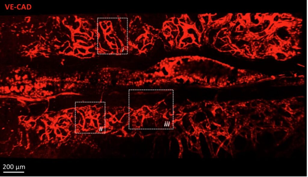
The calvarial bone marrow is an interesting model for studying hematopoiesis and vascular dynamics due to its unique anatomical features. Its thin structure allows for high-resolution imaging, facilitating the observation of cellular interactions and vascular networks. Additionally, it is easily accessible, making it ideal for experimental manipulations and real-time monitoring. This model provides valuable insights into the complex processes of blood cell formation and vascular development.
z-projection of tile scan view of the calvaria vasculature labeled by cdh5-DSRED – 2-photon microscope
Your team has developed a custom-made titanium cranial implant. What advantages does it offer compared to existing methods?
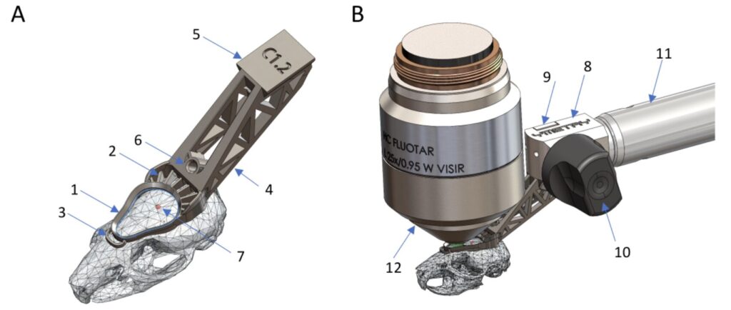
The stability of the imaging area has always been a major challenge in intravital microscopy. Indeed, the animal’s breathing and temperature variations complicate long-term acquisitions. Moreover, precise repositioning of the acquisition area over several days is essential for observing the evolution of the cellular environment. The development of titanium implants, as opposed to traditional resin 3D printing, allows for more robust fixation of the system to the microscope stage and, most importantly, limits the deformation of the implant.
(A) Parts of the implant in situ: 1 observation ring, 2 cementing feature, 3 stabilizing anchor, 4 tail, 5 dovetail, 6 threaded hole, 7 Bregma. (B) Connection of the head implant to the holder: 8 fixation body, 9 clamp, 10 eccentric lever, 11 structure, 12 microscope objective.
What perspectives does this method open for the understanding of hematological diseases, such as leukemia?
This method opens significant perspectives for understanding hematological diseases like leukemia by enabling detailed visualization of disease progression and cellular interactions. It allows researchers to study the impact of treatments in real-time, enhancing the development of targeted therapies. Additionally, it facilitates the exploration of the bone marrow microenvironment’s role in disease pathogenesis.
What are your upcoming projects?
Following the same principle, we are collaborating with the company Ymetry to develop similar appendages adapted for soft organs. Our goal remains to maintain the acquisition area for as long as possible without any drift.
We’re proud to announce the official integration of our two new cutting-edge imaging Nodes into the French Node of Euro-BioImaging: Normandie and Rhône-Alpes!
With this upgrade, the French Node now spans 10 geographical sites and provides access to 30 state-of-the-art imaging facilities, supporting both national and transnational users.
Why does this matter for the Euro-BioImaging community?
These new Nodes significantly expand the scope and excellence of the French Node:
Pioneering technologies in biomechanics and mechanobiology
Rare capacities in spatial transcriptomics, adaptive optics, and metabolic imaging
Deep expertise in large-volume 3D EM with integrated image analysis pipelines
These new capabilities fill critical technological and geographic gaps and will benefit users across Europe seeking access to next-generation imaging and expert support.
Users can expect powerful collaborations, robust training opportunities, and access to highly specialized platforms.
Researchers and imaging facility staff from low- and middle-income countries (LMIC) are invited to apply for access to cutting-edge biologicial and biomedical imaging technologies, training and hands-on experiences funded by the Wellcome Trust and coordinated by Global-BioImaging.
Why should you apply?
You should apply of you want to:
Explore new imaging techniques for your biological samples
Gain hands-on skills with advanced microscopy
Develop expertise in image analysis and imaging facility management
Imaging 4 All offers three grants and sub-tracks addressing different access needs and objectives:
i4A Access Grant: to access imaging facilities and laboratories of your choice to use imaging technologies and related services on offer, and to benefit from their imaging expertise.
i4A Pro Grant: to advance your skills in biological or biomedical imaging technologies and/or applying advanced imaging techniques to your research.
i4A Training Grant: to participate in a national or an international in-person training workshop or course of your choice
Who can apply?
Any researcher or imaging facility staff or management affiliated with a not-for-profit organisation (e.g. university, institute) located in a LMIC is welcome to apply. Please check if the country of your home organisation is classified as an LMIC by using this link.
The programme will include a seminar by Kate Miroshnikova (NIDDK/NIH, Bethesda USA and Max Planck Institute for Molecular Biomedicine, Münster, Germany), presentations by participants and practical mechanobiology workshops (a choice of 4 workshops from a dozen: optical tweezers, micro/nano-fabrication, microfluidics, AFM, micropipette aspiration, mechanical confinement, force measurements, etc…).
Preliminary program:
Day 1 – 26/03/2026, Paris 5e (IBPS / Curie / IPGG)
Morning : scientific presentations, small group discussion
Afternoon : 2 sessions of practical workshops on real set-ups in participating labs (Paris 5e)
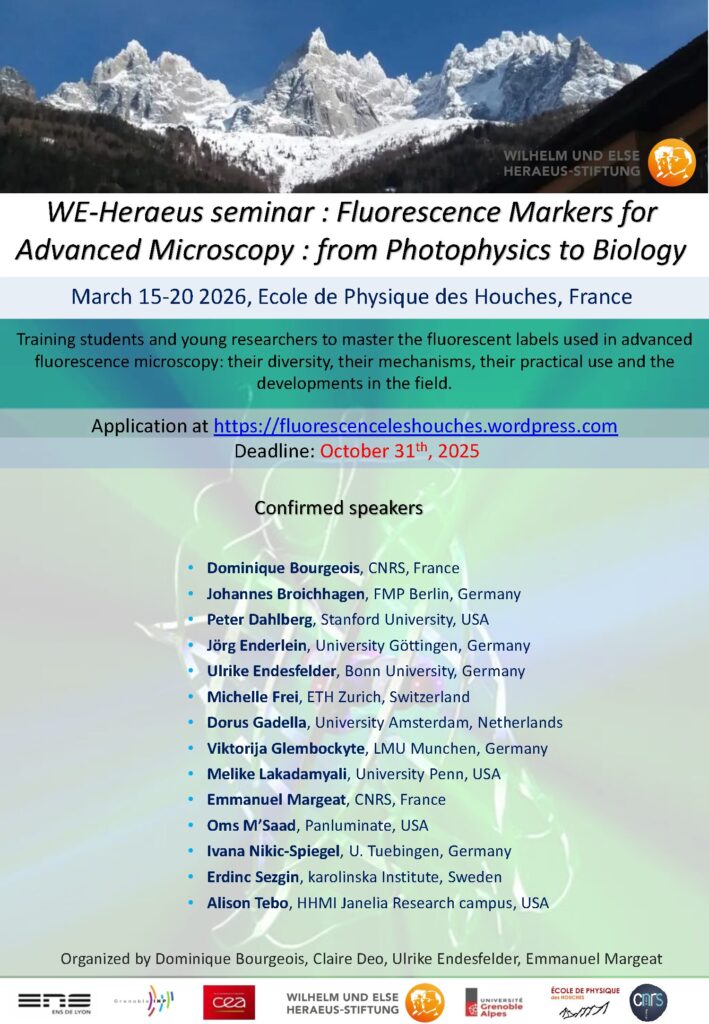
The “Ecole physique des Houches” is hosting the 4th edition of its winter seminar dedicated to biophysical research, from March 15th to 20th, 2026. This year will be focused on the development and application of fluorescent markers for advanced fluorescence microscopy.
What is the objective?
This interdisciplinary training event aims to provide in-depth theoretical and practical knowledge on selecting and using fluorescent biomarkers for advanced microscopy techniques, such as high-resolution fluorescence imaging and single-molecule studies in living cells.
Who can apply?
The seminar is open to:
PhD students
Early-career scientists
Research engineers
Experienced researchers looking to explore new research areas
What is on the program?
A rich and interactive program including:
2 introductory lectures
12 advanced lectures by internationally renowned experts
A round table
A flash talk session*
2 poster session evenings*
Find the complete program and all the experts below!
*All participants are required to bring a scientific poster showing their current scientific work and to participate in the flash talk session.
How to join?
When? March 15th-20th 2026 Where? Ecole de Physique des Houches Fees? Participation is free of charge!
Applications are open until October 31th, 2025!Click here to fill your application.
The French Institute of Bioinformatics is organizing its next conference, focused on the use of generative AIs to support programming and scripting for biology, on June 13, 2025.
This event, intended for bioinformaticians and biologists, aims to share experiences, tools, and perspectives on the use of generative AIs (ChatGPT, Copilot, Devin, etc.) for developing scripts and software in biology..
Program
9:00 AM – 12:30 PM: Presentations and roundtable featuring developers, researchers, educators, and trainers sharing concrete feedback and experiences
2:00 PM – 5:30 PM: Hands-on workshops and closing session
Workshops are optional and limited to 50 participants. If demand is high, selection will be based on responses provided in the registration form.
Join us on June 3rd at 14:00 for the next FBI.data webinar, where the France-BioImaging team will share the latest updates and improvements to their data management solution, and answer all your questions!
Agenda
Project Background and History: Overview of deployments across the France-BioImaging’s Nodes
Théo Barnouin: System Security Enhancements – Implementation strategies at the Nodes and collaboration with Information Security Officers (RSSI)
Guillaume Gay: New Project Developments – Data import scenarios and insights from user testing
The Executive Board of the Rhône-Alpin Node of France-BioImaging is pleased to invite you to the event “Imaging & Microscopy Day in Rhône-Alpes – Image Analysis” – pre-program attached.
It will be held on Tuesday, July 1st at the Faculté Rockefeller, 69008 Lyon
As the number of seats is limited, please register as soon as possible to best organize the final program!
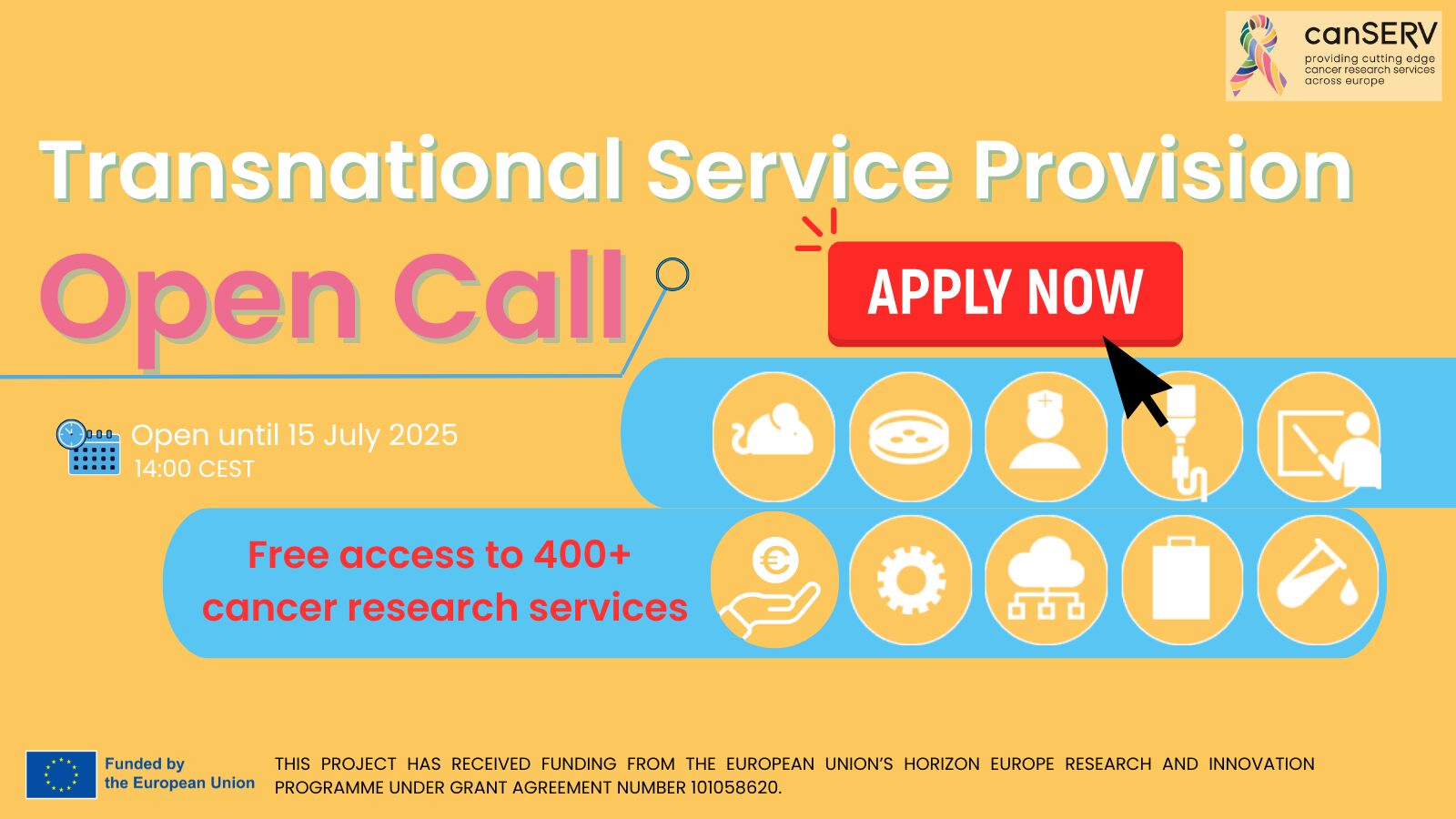
Last weeks to apply to the last canSERV Open Call, a unique opportunity designed specifically for early career cancer researchers worldwide. This initiative aims to provide these researchers with access to cutting-edge services and training, helping them advance their groundbreaking work in cancer research.
Who can apply?
This call is open to:
First-stage researchers: PhD students and junior researchers without a PhD.
Recognised researchers: Postdoctoral fellows, assistant professors or young investigators.
What’s on offer?
Selected applicants will gain free access to imaging services and expertise provided by 36 Euro-BioImaging Nodes, as well as state-of-the-art image data analysis resources. This support empowers researchers to elevate their projects through innovative technologies and expert guidance.
Research focus areas
Your project should address one or more of the following:
Cancer research topics, spanning discovery science, translational research, personalised oncology or clinical studies.
At least one of the four strategic goals of the EU Cancer Mission:
We use cookies on our website to give you the most relevant experience by remembering your preferences and repeat visits. By clicking “Accept All”, you consent to the use of ALL the cookies. However, you may visit "Cookie Settings" to provide a controlled consent.
This website uses cookies to improve your experience while you navigate through the website. Out of these, the cookies that are categorized as necessary are stored on your browser as they are essential for the working of basic functionalities of the website. We also use third-party cookies that help us analyze and understand how you use this website. These cookies will be stored in your browser only with your consent. You also have the option to opt-out of these cookies. But opting out of some of these cookies may affect your browsing experience.
Necessary cookies are absolutely essential for the website to function properly. These cookies ensure basic functionalities and security features of the website, anonymously.
Cookie
Duration
Description
cookielawinfo-checkbox-analytics
11 months
This cookie is set by GDPR Cookie Consent plugin. The cookie is used to store the user consent for the cookies in the category "Analytics".
cookielawinfo-checkbox-functional
11 months
The cookie is set by GDPR cookie consent to record the user consent for the cookies in the category "Functional".
cookielawinfo-checkbox-necessary
11 months
This cookie is set by GDPR Cookie Consent plugin. The cookies is used to store the user consent for the cookies in the category "Necessary".
cookielawinfo-checkbox-others
11 months
This cookie is set by GDPR Cookie Consent plugin. The cookie is used to store the user consent for the cookies in the category "Other.
cookielawinfo-checkbox-performance
11 months
This cookie is set by GDPR Cookie Consent plugin. The cookie is used to store the user consent for the cookies in the category "Performance".
viewed_cookie_policy
11 months
The cookie is set by the GDPR Cookie Consent plugin and is used to store whether or not user has consented to the use of cookies. It does not store any personal data.
Functional cookies help to perform certain functionalities like sharing the content of the website on social media platforms, collect feedbacks, and other third-party features.
Performance cookies are used to understand and analyze the key performance indexes of the website which helps in delivering a better user experience for the visitors.
Analytical cookies are used to understand how visitors interact with the website. These cookies help provide information on metrics the number of visitors, bounce rate, traffic source, etc.
Advertisement cookies are used to provide visitors with relevant ads and marketing campaigns. These cookies track visitors across websites and collect information to provide customized ads.