The Grenoble Institute of Neurosciences celebrates science!
Author: Marine Beraud
On the occasion of the “Fête de la Science”, the Grenoble Institute of Neurosciences is organizing a neuroscience photo exhibition from October 2 to 11, 2026.
This event offers the public the opportunity to explore the inside of the brain through images of cells, tissues and brain structures, revealing a universe invisible to the naked eye.
Four new invited speakers have been announced for SFBS 2026, further enriching the scientific programme dedicated to super-resolution microscopy for biological imaging.
Here’s few places left, you can still register for the event!
Rainer Heintzmann, studied physics and computer science in Osnabrück and finished his Dr. rer. nat. in Physics at Heidelberg University, Germany. As a postdoctoral fellow at the Max Planck Institute of biophysical Chemistry in Göttingen and as a group leader at the Randall Division, King’s College London he continued to work on developing fluorescence microscopy methods.
Currently he is a professor of Physical Chemistry at the Friedrich-Schiller UniversityJena, and heads the microscopy research unit at the Leibniz Institute of Photonic Technology in Jena, Germany.
His research focuses on methods for imaging cellular function at high resolution and developing techniques to measure multidimensional information in small biological objects such as cells, cellular organelles or other small structures of interest.
He developed structured illumination, pointillism, image inversion interferometry and optical photon reassignment and is highly interested in computer-based reconstruction and inverse modelling methods such as deconvolution.
Bei Liu
Dr. Bei Liu joined the College of Future Technology at Peking University as an Assistant Professor in 2022. Prior to this appointment, he conducted his postdoctoral research with Professor Klaus M. Hahn at the University of North Carolina at Chapel Hill from 2015, and was promoted to Research Assistant Professor in 2019.
His laboratory develops novel molecular tools and microscopy techniques to visualize and control cell behavior across scales, with a particular focus on biosensors and optogenetics.
The lab takes a multidisciplinary approach, integrating high-throughput, advanced imaging with AI-driven protein design, image analysis, and laboratory automation.
His work has been published in leading peer-reviewed journals, including Cell, Nature Chemical Biology, and Nature Communications.
Flavie Lavoie-Cardinal
Flavie Lavoie-Cardinal is an Associate Professor in the Department of Psychiatry and Neuroscience at Université Laval and Deputy Director of the Cellular and Molecular Research Axis at the CERVO Brain Research Centre in Québec City, Canada.
She is Scientific Director of the AI-for-Health Platform PREDIS, and of the Canadian Optical Nanoscopy Platform.
She holds the Canada Research Chair in Intelligent Nanoscopy of Cellular Plasticity and leads interdisciplinary research at the intersection of optical microscopy, neuroscience, and artificial intelligence.
Her laboratory develops AI-assisted super-resolution microscopyand computational approaches to uncover how nanoscale synaptic organization and heterogeneity shape neuronal function and plasticity across biological scales.
Fan Xu
Fan Xu is a Professor in the School of Optics and Photonics at Beijing Institute of Technology (Lab website: www.xulab.cc). Fan received the PhD degree in computer science from Institute of Computing Technology, Chinese Academy of Sciences in 2017 and then worked as a postdoctoral fellow and senior research associate in the Weldon School of Biomedical Engineering at Purdue University
His research focuses on the development and application of super-resolution microscopy techniques, especially in 3D super-resolution imaging and single molecule localization analysis.
He combines novel imaging instruments and physical model or data-driven approaches to investigate cellular/tissue structures and dynamics that are below the diffraction limit of light and therefore inaccessible to conventional microscopy.
His publications include Nature Methods, Cell Research, Nature Communications, Protein & cell, and Biomedical Optics Express.
Don’t miss this opportunity to meet these experts during SFBS 2026! Join us in Bordeaux from October 19 to 21, 2026!
From September 15 to 18, France-BioImaging joined the Euro-BioImaging All Hands Nodes Meeting 2026 in Turku, Finland, under the theme “Connecting the Communities”.
This meeting brought together imaging infrastructures, Nodes, researchers, technology experts and industry partners from across Europe to exchange experiences, showcase emerging technologies and discuss how to strengthen collaboration within the Euro-BioImaging community.
Several topics particularly resonated with the France-BioImaging community:
Multimodal & multiscale imaging From advanced microscopy and nanoscopy to X-ray imaging and other complementary approaches, the meeting highlighted the power of combining technologies to explore biological systems across scales.
AI & bioimage analysis AI is rapidly transforming imaging workflows. Discussions and presentations showcased new approaches for AI-assisted image analysis, scalable computational infrastructures and the development of more efficient and accessible imaging workflows.
Connecting infrastructures & communities A recurring message throughout the meeting: infrastructure is not only about technology. Training, expertise, coordination and collaboration are essential to make advanced imaging accessible and useful to a broad research community.
From research to innovation The meeting also explored opportunities to strengthen links between academic research, research infrastructures and industry – from collaborative projects and technology development to pathways towards innovation and industrial applications.
Building the future of bioimaging together The All Hands Meeting was a great opportunity for France-BioImaging to connect with European partners, exchange experiences and follow the exciting developments shaping the future of imaging research.
A big thank you to Euro-BioImaging ERIC and the Finnish hosts for bringing the community together in Turku!
Looking forward to the next opportunities to connect, collaborate and advance bioimaging across Europe!
PhysChemCell conference is back for another edition from November 16 to 18, 2026 in Université Paris-Saclay. During this event, participants will explore novel approaches at the crossroads of chemistry, physics and biology to study living systems, from single cells to entire organisms.
PhysChemCell is open to researchers, students and more widely scientists working at the interface of physics, chemistry and biology.
If you are interested, the registration is open until October 31, 2026. Participants can also apply to present an oral presentation or a poster, until October 1, 2026.
Scientific topics include
New chemical probes, labelling and activation strategies compatible with living systems
Biosensors and nanosensors: from design to imaging applications
Label-free chemical analysis and imaging
Multiscale observation of biological samples, from the nano/molecular scale to in vivo and real-time imaging
Plenary speakers
Stéphanie Descroix – Institut Curie, Paris
María García-Parajo – ICFO, Barcelona
YongKeun (Paul) Park – KAIST, Daejeon
Oliver Thorn-Seshold – Dresden University of Technology
A new study(1) published in Nature Communications shows that membrane enrichment with two polyunsaturated fatty acids, DHA and DPA, reduces agonist-induced endocytosis of the dopamine D2 receptor, a major target of antipsychotic drugs. By combining confocal microscopy and live-cell TIRF imaging, the researchers showed that D2R clustering at the plasma membrane is preserved, while β-arrestin2 recruitment to D2R endocytic sites and the formation of D2R-containing endocytic vesicles are reduced.
Linking membrane lipids and dopamine receptor trafficking
The brain is highly enriched in polyunsaturated fatty acids, or PUFAs, and altered PUFA levels have been associated with several neuropsychiatric disorders. Previous work had linked dietary PUFA deficiency in mice to behavioural changes, including altered motivation, involving neurons expressing the dopamine D2 receptor (D2R). In this new study, Silvia Sposiniet al. investigated whether PUFA levels in cellular membranes could directly affect the trafficking of the D2R, a G protein-coupled receptor involved in dopaminergic signalling and a major target of antipsychotic drugs.
By enriching cell membranes with two different PUFAs, DHA and DPA, the team showed that agonist-induced D2R endocytosis was strongly reduced. This effect was observed in HEK-293 cells and confirmed in cultured cortical neurons. Importantly, PUFA enrichment did not affect clathrin-mediated endocytosis in general, assessed using transferrin uptake, nor the internalization of several other GPCRs tested. These results highlight a specific sensitivity of D2R endocytosis to membrane PUFA levels.
Imaging the altered steps of D2R endocytosis
Microscopy played a key role in understanding this mechanism. Confocal imaging showed that fewer D2R-containing endosomes formed in PUFA-enriched cells after receptor activation. Live-cell TIRF microscopy then allowed the researchers to observe early endocytic events at the plasma membrane. While D2R clustering at endocytic sites remained unchanged, the recruitment of β-arrestin2, an essential protein for D2R endocytosis and signalling, was reduced. Using the pulsed-pH assay, the team further showed a lower frequency of D2R-containing endocytic vesicle formation.
Finally, the study identified two key residues in the second intracellular loop of D2R, serines 147 and 148, as essential for the sensitivity of D2R endocytosis to PUFA levels. When these residues were mutated, the receptor still internalized after activation, but PUFA enrichment no longer reduced its endocytosis.
Together, these results show that membrane lipids do not only influence general membrane properties such as fluidity or rigidity. They can also specifically regulate the trafficking of membrane proteins such as the dopamine D2 receptor. This study opens new perspectives on how membrane composition may influence D2R trafficking, with potential consequences for dopaminergic signalling in the brain.
(1)Sposini, S., Baccouch, R., Lescuyer, M. et al.Membrane lipid poly-unsaturation selectively affects dopamine D2 receptor endocytosis. Nat Commun17, 6661 (2026).https://doi.org/10.1038/s41467-026-73057-5
The Advanced Microscopies team at the Laboratory for Optics and Biosciences has shown that third harmonic generation (THG) microscopy, mainly used to visualize tissue structure, can also help distinguish certain biological pigments. By exploiting their response to different wavelengths, the researchers open new possibilities for label-free multiphoton imaging that can provide complementary information about sample composition.
Multiphoton microscopy is widely used to explore biological tissues in three dimensions. While many approaches rely on fluorescent labels to precisely identify specific structures or molecules, label-free techniques can also make use of the intrinsic properties of biological samples.
Among them, third harmonic generation (THG) microscopy can reveal interfaces and variations in optical properties within tissues. It is therefore particularly useful for providing information about tissue organization and morphology without requiring staining or fluorescent labeling. Until now, however, THG has mainly been used for structural imaging.
In this study1, Stella Dees et al. investigated another property of THG: its signal can be strongly enhanced in the presence of certain absorbing molecules when they are illuminated at specific wavelengths.
The researchers studied three representative biological pigments: hemoglobin in red blood cells, pteridines found in xanthophores (pigment cells in zebrafish) and melanin in human hair. For each of them, they measured how the THG signal changed depending on the excitation wavelength.
They observed signal enhancements ranging from 10- to 100-fold in these pigmented structures. Importantly, this enhancement did not occur in the same way for all pigments. Each showed a different response to the excitation wavelengths, providing information that can be used to distinguish them.
Distinguishing structures in a living sample
The team then applied this principle to the imaging of live zebrafish larvae. The researchers used an approach called third-order sum-frequency generation (TSFG), which provides simultaneous access to several spectral responses.
By taking advantage of the differences between the pigments, they were able to distinguish red blood cells containing hemoglobin from xanthophores and from surrounding structures.
This simultaneous readout is also particularly useful for imaging living and dynamic samples, as it provides spectroscopic contrast without requiring the excitation wavelength to be changed successively to acquire multiple images.
Color TSFG microscopy. (b) Tiled large-area TSFG image of a zebrafish larva (reanalysis of data used in Ref. 21). (c) Three-color image extracted from the same data, combining the following signals. RBC: red blood cells; xantho: xanthophores; noto: notochord interface.
FIG. S3. Color TSFG image of xanthophores and red blood cells in a zebrafish larva. Zoomed-in view of Fig. 5C showing the three individual channels (TSFG1, TSFG2, THG) in greyscale lookup table and the overlay in RGB.
Going beyond morphological information
These results show that THG could provide more than structural information alone. By exploiting the spectral properties of pigments naturally present in biological samples, THG and TSFG could add a degree of specificity to label-free multiphoton imaging.
This approach is not intended to replace fluorescence imaging, which remains particularly powerful for specifically targeting molecules or biological structures, but could provide complementary information without requiring additional labels.
The work therefore opens new perspectives for studying pigments and other naturally absorbing structures in biological samples, while also highlighting the need for further characterization of their spectral properties and for a better understanding of how light propagation through tissues influences the measured signals.
1Stella Dees, Júlia Ferrer Ortas, Pierre Mahou, Willy Supatto, Nicolas Olivier, Emmanuel Beaurepaire; Resonant third harmonic generation in biological pigments. APL Photonics 1 August 2026; 11 (8): 086116.https://doi.org/10.1063/5.0341777
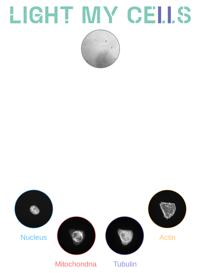
We are happy to announce that the new edition of our machine learning competition, Light My Cells 2, is now open! This Challenge completes the previous Light My Cells in 2024 with additional data and a new prediction task.
Light My Cells 2 is organized in two phases, the validation phase (September 21st – November 30th) and the evaluation phase (December 1st – December 13th).
The Challenge: a data machine learning competition
Since 2024, France-BioImaging organizes its data machine learning competition in the aim to respond to the microscopy community issues.
With Light My Cells 1, participants predicted fluorescence images of four cellular structures – nucleus, mitochondria, tubulin, and actin – from label-free transmitted-light microscopy images acquired using bright-field (BF), phase-contrast (PC), or differential interference contrast (DIC) modalities. This technique is also known as in-silico labelling.
This Challenge established a large-scale, multi-site benchmark for evaluating in-silico fluorescence prediction from transmitted-light unlabeled microscopy images under realistic imaging variability.
Light My Cells 2 introduces additional training data and a new prediction task to reconstruct the fluorescence maximum-intensity projection (MIP) from transmitted-light image stacks.
Light My Cells 2 tasks
To help the participants build their algorithm, they have access to a training dataset of 76 studies, which represents more than 150 000 images. From these acquisition-sets, participants will have to create an algorithm to:
Predict the best-focused output-images of four fluorescently labelled organelles from label-free 2D transmitted light input-images.
Predict the MIP output-images of four fluorescently labelled organelles from a stack of 2D input-images of label-free transmitted light.
Key informations
The Challenge is now open and you can register to access the training dataset and start building your algorithm!
From September 21st, 2026, the validation phase begins – you will be able to test your algorithm and the submission process, up to five test submissions.
From December 1st to 13th, 2026, the evaluation phase will start – participants will have one final submission.
The results will be announced on December 18th, 2026.
Four new invited speakers have been announced for SFBS 2026, further enriching the scientific programme dedicated to super-resolution microscopy for biological imaging.
Here’s few places left, you can still register for the event!
Laurent Cognet is a Research Director at the CNRS and Group Leader at the LP2N (Laboratory of Photonics, Numerical and Nanosciences), part of the Institute of Optics at the University of Bordeaux, France, where he also serves as Director of the research center. He earned his PhD in 1999 in quantum physics from Université Paris-Saclay under the supervision of Alain Aspect.
He then transitioned to nano- and biophotonics as a Marie Curie postdoctoral fellow at Leiden University (Netherlands) in the group of Thomas Schmidt. In 2006–2007, he was a Fulbright Visiting Scholar at Rice University.
Over the past 25+ years, Laurent Cognet has contributed to advances in single-molecule localization microscopy and carbon nanotube spectroscopy, applying these techniques to key questions in nanosciences and neuroscience. His current work focuses on developing innovative microscopy methods to study molecular diffusion in complex biological environments.
He has authored over 125 peer-reviewed publications and received multiple awards for his contributions.
Pingyong Xu
Dr. Pingyong Xu, a tenured professor at the Institute of Biophysics, Chinese Academy of Sciences, and faculty member at the College of Life Sciences, University of Chinese Academy of Sciences, earned his Ph.D. from Huazhong University of Science and Technology in 2004.
His research focuses on integrating spectroscopy, biophysical microscopy techniques, and protein design to develop innovative optical imaging tools, particularly photo-controllable fluorescent proteins (FPs).
Notably, Dr. Xu pioneered the development of the groundbreaking FP mEosEM for correlative light and transmission electron microscopy. He has also designed a range of high-performance FPs tailored for super-resolution imaging technologies such as PALM/STORM, SOFI, RESOLFT, and NL-SIM, significantly enhancing their temporal and spatial resolutions. Among these, mEos3.2 and Skylan-S stand out as premier FPs for PALM and SOFI imaging, respectively.
His most recent achievement is the development of mScarlet3-S2 for 3D STED microscope, and 3Snet CLID, a groundbreaking method capable of reconstructing SR images using just 1 frame of wide-field/spinning disk confocal raw images.
Anna Brachet
Anna Brachet is a CNRS researcher at the Interdisciplinary Institute for Neuroscience in Bordeaux, France. She was trained as a cell biologist in Marseille, where she studied the organization and dynamics of sodium channels at the axon initial segment.
During her postdoctoral work in Madrid, Spain, she investigated molecular mechanisms of synaptic plasticity, including AMPA receptor trafficking.
Her current work focuses on the neuronal membrane periodic skeleton and its role in maintaining cellular architecture while supporting plasticity. Combining advanced fluorescence microscopy, molecular perturbations, and functional assays, her research aims to understand how membrane-associated cytoskeletal structures organize and regulate the function of neurons and astrocytes.
Yongdeng Zhang
Dr. Yongdeng Zhang received his B.E. in Biomedical Engineering from Central South University in 2008 and his Ph.D. in Biophysics from Huazhong University of Science & Technology in 2013, through a joint program with the Institute of Biophysics, Chinese Academy of Sciences.
He then pursued postdoctoral research at Yale University School of Medicine from 2014 to 2020. In October 2020, he joined the School of Life Sciences at Westlake University as a Principal Investigator.
Dr. Zhang’s laboratory operates at the interface of technology and biology, advancing subcellular imaging through two complementary innovations. To map protein complexes with molecular precision, they developed 4Pi single-molecule localization microscopy (4Pi-SIMFLUX and me4Pi-SMLM), achieving isotropic 3D localization precision of 2–3 nm to resolve ultrastructural features below 10 nm in whole cells. To capture rapid subcellular events and inter-organelle interactions, they developed 4Pi structured illumination microscopy (4Pi-SIM) for two-color, time-lapse volumetric imaging, delivering ~100 nm isotropic 3D resolution at 1 Hz in living cells.
Together, these technologies constitute a scalable toolbox that bridges static molecular architecture and live cellular dynamics, enabling quantitative studies that were previously inaccessible to fluorescence microscopy.
Don’t miss this opportunity to meet these experts during SFBS 2026! Join us in Bordeaux from October 19 to 21, 2026!
This image contest is open to all within the imaging community: core facility staff and users, R&D labs teams and co-workers, students… Submit your best microscopy images for a chance to showcase your skills, research and creativity to the French bioimaging community and beyond, allowing people to see the visual appeal of the life sciences. Images from the contest will be featured on France-BioImaging communication tools, online and in print.
France-BioImaging and all the French community aims to develop and promote innovative imaging technologies and methods. But microscopy images can also take an artistic, creative look and make the invisible world beautiful.
We are all eager to see your work!
Prizes
First place: France-BioImaging will cover the registration fees for one 2027 microscopy related event of the winner’s choice (FOM, ELMI, EMC, COMULIS conference, etc.).
Top 3: Your images will be added to the CNRS and INSERM image librairies.
All participants: Your images will be used in our communication materials (digital calendar, flyers, social media posts, etc.), and some of you may also be invited for an interview about your research work.
Important: Only French or foreign participants affiliated to a French institution can enter the contest. Foreign participants non-affiliated to a French institution can submit images and will be featured in the gallery, but will not be evaluated as part of the contest.
Submission deadline: Sunday, November 22th, 2026, 23h59
For the 2026 edition of the CNRS “Visites Insolites”, four France-BioImaging member research institutes and imaging facilities will open their doors to the general public. The Institut Fresnel, IBDM, the IMAG’IC facility at Institut Cochin, and theImagoSeine facility at Institut Jacques Monod will welcome visitors in October for a series of hands-on activities and demonstrations exploring imaging, light and the hidden world of living systems. Find out more about each event below.
Please note: registration for all four events closes on 20 September 2026.
Institut Fresnel: Photonics is fantastic!
Hervé Rigneault’s MOSAIC R&D team, together with the Photonics facility, will introduce visitors to the fascinating possibilities of manipulating light to reveal what cannot be seen with the naked eye, through interactive experiments and demonstrations.
Institut Jacques Monod: Take a closer look at your plate with flow cytometry
The flow cytometry unit of the ImagoSeine facility will invite visitors to discover the hidden world of living organisms found in our food. Participants will also have the opportunity to visit the facility and discover some of its imaging equipment.
At IBDM, the visit will focus on developmental biology and the earliest stages of life. Through hands-on activities and observations using a stereomicroscope, visitors will explore this otherwise invisible world and discover how scientists study the first steps of development.
Institut Cochin: Biomedical research – a journey into the heart of cells
The IMAG’IC imaging facility will welcome visitors for an exploration deep inside cells using fluorescence imaging. Participants will also be able to build their own mini optical microscope, the Foldscope, which can be used with a smartphone.
And there’s more to come! The following weekend, several laboratories and imaging facilities will also open their doors for the Fête de la Science. Check what is happening near you and take the opportunity to discover even more behind-the-scenes science!
Ana Svetić is a PhD student in Nanotechnology at the University of Trieste (Italy). Her research focuses on developing a cell-derived plasma membrane model for correlative nanoscale imaging, with the aim of observing membrane nanodomains.
During a research stay abroad as part of her PhD, she visited theCBS AFM facility in Montpellier, where she used the PALM-AFM set-up to correlate AFM scanning with TIRF microscopy and investigate phase separation and nanodomains.
Through France-BioImaging User Access Support, Ana was able to acquire specific AFM tips that improved the quality of her scans and helped her obtain significant results. She also highlights the simplicity and efficiency of the access process and encourages other researchers to apply for support.
Could you introduce yourself?
Hi, my name is Ana Svetić, and I’m a biotechnologist turned biophysicist. I’m currently in the process of finishing my PhD in Nanotechnology at the University of Trieste and Elettra-Sincrotrone Trieste. My laboratory is called Nanoinnovation, and we do lots of Atomic Force Microscopy (AFM) as a supporting lab to the synchrotron.
What is your research project about?
My research project focuses on developing a cell-derived plasma membrane model, namely Giant Plasma Membrane Vesicles (GPMVs) and their patches (GPMV patches) for correlative nanoscale imaging – imaging with AFM and correlative techniques such as epifluorescence and TIRF microscopy, to ultimately observe the membrane nanodomains.
These nanodomains are, as the name suggests, nano-sized. In cells, they are just a few nanometers, very small and transient, and there hasn’t been a technique so far that allows the visualization and formation of said domains. But these ordered domains have been observed in synthetic models, namely Supported Lipid Bilayers (SLBs) and Giant Unilamellar Vesicles (GUVs).
So, my goal is to pass from more common simple synthetic membrane models (SLB and/or GUV) to a more complex one (GPMVs), in hopes to have a better, more biologically relevant model for observation of phase separation, which is responsible for the creation of previously mentioned nanodomains.
Which France-BioImaging’s facility did you visit, and which imaging technology did you use?
I was a visiting PhD student at the Centre de Biologie Structurale (CBS) – CNRS facility in Montpellier, where I’ve used the PALM-AFM set-up as a part of my PhD period abroad.
The PALM-AFM allowed me to correlate AFM scanning with TIRF (Total Internal Reflection Fluorescence), or more specifically, have the ability to measure the fluorescence intensity of the undermost layer of the GUV patches and GPMV patchesat the same time as performing an AFM scan of the same region of interest.
This ultimately allowed me to observe the phase separation and nanodomains in two ways – by seeing (TIRF) and by scanning (AFM) the surface of the membrane models.
Could you walk us through your experience accessing France-BioImaging?
I was made aware of the France-BioImaging opportunity by my mentor, Luca Costa PhD. He kindly told me about the platform, how to apply, and so I did. It was quite easy to fill out the form, and it took little-to-no-time to receive a positive reply!
I was astounded at the efficiency of the FBI and how smooth it all went, to be honest.
How did this access contribute to advancing your project?
I was already using the set-up before applying for the funding, but the funding helped me to buy and obtain AFM tips that I needed for my measurements. AFM tips that I needed were hard to get by, so I’m quite grateful for the funding of France-BioImaging. This in turn helped me get AFM scans of better quality, and at the end of the day obtain significant results for my research.
Thanks to TIRF, we were able to confirm different types of phase separations, which in turn shows up in nanodomain patterns.
AFM-TIRF image of a 20 mol% cholesterol GUV patch, same ROI. The correlative imaging demonstrates the existence of 2 phases. TIRF (left), AFM (right).
What are the next steps for your research project?
As I’ve already mentioned, I’m currently wrapping up my PhD project. Afterwards, I’m not sure, but I would like to explore more of what I’ve developed so far, and eventually localize meaningful proteins inside GPMVs and GPMV patches. This would let us know where exactly in nanodomains are proteins such as, for example, HER-2! It would be really, really cool.
What advice would you give to researchers who are considering applying for the FBI User Access Support?
Do it! Any support is meaningful and sometimes just a little bit can change the course of your research.
The CNRS technical networks RTmfm and FEMTO are organizing two thematic days dedicated to “Nonlinear microscopy: the latest advances in multiphoton microscopy”, on December 10–11, 2026, at the Bordeaux Imaging Center in Bordeaux.
The event will highlight the latest technological developments in nonlinear microscopy, including advances in laser technologies, aimed at simplifying these approaches, improving their performance and making them more accessible to users.
This event has received support from the Euro-BioImaging Sponsorship Programme!