24-27 May, 2016
Debrecen, Hungary

Download the programme here
Website and Registration

The annual Meeting of ELMI is an internationally recognized meeting on Light Microscopy combining lectures and hands-on industry workshops on cutting edge microscopy techniques and innovative applications in life sciences. In 2016 it will take place in Debrecen, Hungary, from the 24th to the 27th of May. In addition to plenary scientific lectures, ELMI 2016 offers a fantastic opportunity for users to enjoy the hands-on workshops in the latest developing instrumentation, and the chance to gain insight into the next generation of technologies. It also provides an excellent opportunity for networking with the industry and other devoted microscopists.

Auditorium of the François Jacob building
Institut Pasteur, 28 rue du Dr Roux, 75015 Paris, France
Tuesday March 8, 2016
Nota Bene
 
Registration (by e-mail):
chantal.martinez@pasteur.fr
Free of charge but mandatory

More information

The Education Department of the Institut Pasteur will be running a symposium on the same topic on Tuesday March 8, 2016, with the “Principles and Applications of Fluorescence Microscopy” course directors : David DiGregorio, Gael Moneron and Jost Enninga. This event will include a series of seminars to explore the latest technological developments in fluorescence microscopy. The symposium, which will take place in the auditorium of the François Jacob building, represents a unique opportunity to hear from the leading experts in fluorescence microscopy about their latest research findings. It is open to all scientists on campus. The event is free but prior registration is required.

bandeau_logos

From: October 18, 2015
To: October 23, 2015
Les Houches, FR

Download the programme here
Website

The school aims at providing the researchers the numerical tools enabling the acquisition, quantification and the modelling of multimodal and multi-scale biophysical data. The school will include introductory courses on spectroscopies and image acquisition methods together with conventional processing methods. They will be followed by advanced courses on recent data processing methods, such as compressed sensing, and the illustration of their applications in different fields such as mass-spectrometry, NMR and different imaging methods. Theoretical courses will be completed by extensive practical labs on personal computer.

Softwares: Ipython Nump/Scipy and ImageJ
Registration deadline: October 12th 2015
Registration cost: 450 Euros including food and accomodation.
Poster presentation opportunity.
Audience: PhD, post-doc, researchers and technical staff.

December 15, 2015
Paris, Jussieu

Download the programme here
Website and Registration

La Mission pour l’Interdisciplinarité du CNRS et les réseaux CALCUL, DEVLOG, RBDD, RCCM, REMISOL, RTMFM organisent le 15/12/2015 à Paris une journée d’exposés et de rencontres autour du traitement d’images en microscopie.

Cette journée traitera de l’ensemble du cycle de vie des images, au vu des évolutions techniques majeures récentes. En effet, le travail aux interfaces entre différentes disciplines est une des clefs pour lever les verrous technologiques.
 
Le programme s’articulera autour des 4 thèmes suivants :

    T1 : Workflow de traitements sur big data : extraction d’information, gestion de flux de tâches, fouille de données, codes de calcul
    T2 – Génie logiciel: Quel logiciel (développement sur mesure ou utilisation de solutions existantes) ? Comment développer, valider, diffuser ?
    T3 – Infrastructures de calcul et de stockage adaptées aux données images de microscopie
    T4 – Outils et méthodes d’enrichissement sémantique des images (quels standards de métadonnées et/ou ontologies pour le partage, l’indexation/le référencement de l’information ?)

Microscopy

November 30, December 1, 2015
London, UK

Download the programme here

 Website and Registration

Symposium on Super Resolution Microscopy, Functional Imaging, Correlative Light Electron Microscopy (CLEM), High Throughput & High Content Screening, BioImage informatics – Image Processing and Data Management, Training, Dissemination & Technological Transfer


The strength of this meeting lies in the mixture of scientific lectures on state-of-the-art, high-end microscopy combined with “hands-on” workshops and exhibition of the latest technology, organized by the leading companies in the field. This year we will maintain the format and focus on leading edge developments and its implementation to life science.

The goal of this workshop is to help mobilise and liven up the interest of theoreticians and modellers and present them with the most recent experimental results and new functional questions brought about by correlation (FCS, FCCS,…) or supra-resolution microscopies (STORM, PALM…), related to how biomolecules or vesicles move in the cell and its membranes. Our objective is to promote interactions between experimentalists and theoreticians/modellers working on the spatial or spatiotemporal dynamics of: + the transcriptional machinery + synaptic receptors + viral assembly + intracellular transports

Invited Talks:
R. Voituriez, UPMC Paris
A. Triller, ENS Paris
O. Thoumine, IINS, Bordeaux
H. Soula, INSA Lyon
J. Salamero, Curie Paris
J.-B. Masson, Pasteur Paris
C. Kervrann, INRIA Rennes
L. Héliot, Phlam Lille
L. Foret, ENS Paris
C. Favard, CPBS Montpellier
N. Destainville, UPS
Toulouse
A. Chauvière, UJF Grenoble
M. Coppey, Curie Paris
H. Berry, INRIA Lyon

Organizing committee:
L. Héliot, Phlam Lille
C. Favard, CPBS Montpellier
H. Berry, INRIA Lyon

Main Topics include:
Spatial dynamics of:
– transcription
– synaptic receptors
– viral assembly
– intracellular transports

Molecule Trajectories in Cellular Spaces:
Promoting interactions between
theoreticians and experimentalists

November 16-17, 2015
Ecole Normale Supérieure, Lyon
1 place de l’Ecole, 69007 Lyon, France
 
Links:

Traece Inria Website
Registration
Affiche (pdf)


AfficheTraece AfficheTraece

1396-advertisement

June 27-29, 2016
New Orleans, USA

OMICS International, (conference series) the World Class Open Access Publisher and Scientific Event Organizer is hosting “International Conference on physics” which is going to be the biggest conference dedicated to Physics. The theme “Highlighting innovations and challenges in the field of Physics” and it features a three day conference addressing the major breakthroughs, challenges and the solutions adopted. The conference will be held during June 27-29, 2016 at New Orleans, USA.

Programme:

  • Astrophysics and Cosmology
  • Atomic, Molecular and Optical Physics
  • Plasma Physics
  • Chemical physics
  • Particle physics & Phenomenology
  • Nuclear physics
  • Applied Physics
  • Computational Physics and Numerical Simulation
  • Mathematical Physics
  • Condensed Matter Physics

physics2016-89744

New microscope in the Imagopole:
Live-cell super-resolution with BioAxial CODIM100
Open day in Pasteur the 24/09/2015

pseudo logo Imagopole

Dear friends of imaging,

The BioAxial company and the Imagopole (the imaging platform of the Institut Pasteur) has collaborated for several years around the development of an entirely new approach to super-resolution imaging based upon conical diffraction. It is now a microscopy system, available commercially (http://www.bioaxial.com/products/codim100/product-description), the CODIM100.

PastedGraphic-1 We host such a system, and it will be made available at the Imagopole to all scientists, following the Imagopole access policy (as any regular system) by the end of September 2015. The CODIM100 is very simple to use, and can be operated by autonomous users, following a short training session.

We organize an open day for the system Thursday the 24th of September. You are cordially invited to bring your sample and try the CODIM100. We will be there all day to help you setting up the microscope to harvest the best images.

If you are interested in coming to this open day, please write to Jean-Yves Tinevez (tinevez@pasteur.fr) and state your needs. We will allocate a slot depending on the time you need. We have the facilities to host and prepare live samples.

If you are interested in super-resolution microscopy, keep reading on.
While there already exist several super-resolution modalities, this system is distinguished by the unique combination of several performance features. In brief,:

    • it is ideal for live-cell super-resolution imaging (even long term time-lapse imaging);
    • it works with any fluorescent sample without any special preparation;
    • it has a very good sensitivity even to weakly labelled samples.

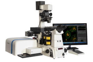
It is attached to a normal laser scanning confocal microscope. The typical usage is to image a sample with normal LSCM mode, then draw a ROI over the acquired image to rescan it in super-resolution. Here are some quick specifications of the super-resolution module:

    • Excitation lines: 488, 561 and 640nm
    • Objectives: 40x 0.95 air / 60x 1.2 water / 60x 1.49 oil.
    • Acquisition speed: a few seconds for a 25µm2 ROI.
    • Labels: any that can be excited with the wavelength mentioned above
    • Sample prep: any confocal or live sample with 0.17mm glass bottom coverslip. No restrictions on mounting media for fixed samples.
    • XY resolution: 110 nm with 488nm laser/0.95 NA, 90 nm with 488 nm laser/1.20 NA
    • Very low excitation power: less than 1 µW/µm2.
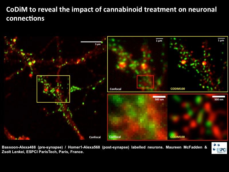
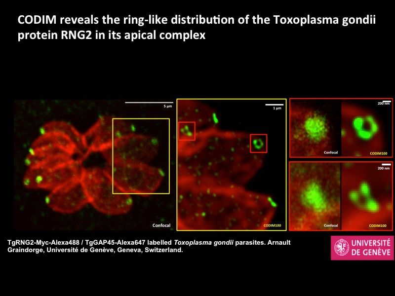
Some examples:
CODIM100_example1
CODIM100_example2

affiche 237 copie
New trends in super-resolution optical microscopy: unraveling the ultra-structure and dynamics of molecular assemblies in cells and tissues

PHASE I
September 30 to October 2, 2015
Bordeaux

WebsiteAfficheInscriptions

 
This Workshop will present the theoretical basis, the latest developments and the acquisition and data analysis procedures on super-resolution microscopy techniques used to investigate the ultrastructure of molecular assemblies and their dynamics in living cells and tissues.

Registration Deadline: March 27, 2015
Organizers:
Maxim DAHAN (Institut Curie, Paris), Orestis FAKLARIS (Institut Jacques Monod, Paris) and Marcelo NOLLMANN (Centre de Biochimie Structurale, Montpellier)
 

Phase I : critical assessment
  • Introduction to super-resolution techniques
  • Application and new developments in single-molecule super-resolution microscopy
  • Labeling, Theoretical and computational methods
  • In-depth super-resolution microscopy, STED, SIM microscopies
Phase II : technical workshop

18 trainees will be selected among phase I participants.
 

inserm.fr (2)Renseignements et inscriptions
Pôle des Ateliers – Inserm
ateliers@inserm.fr
Tél : 33(0)1 44 23 62 03 ou 04

November 17, 2014, November 18, 2014 in Institut Curie Research Center

Workshop on Cell Biology, Biochemistry, Neurobiology, Physiology, Chemistry or Chemical Biology & Image Processing and Data Management


 Website, program and registration

Affiche-2nd-high-throughput-cell-biology-2014

The strength of this meeting lies in the mixture of scientific lectures on state-of-the-art, high-end microscopy combined with “hands-on” workshops and exhibition of the latest technology, organized by the leading companies in the field. This year we will maintain the format and focus on leading edge developments and its implementation to life science.

June 30, 2014, July 2, 2014 in IMT Toulouse

 
flyer_journee_thematique_2014
 

Download the programme here


 Website and registration


The strength of this meeting lies in the mixture of scientific lectures on state-of-the-art, high-end microscopy combined with “hands-on” workshops and exhibition of the latest technology, organized by the leading companies in the field. This year we will maintain the format and focus on leading edge developments and its implementation to life science.

14th international ELMI Meeting

20-23 May 2014 – Oslo, Norway


slide1

The 2014 ELMI meeting will be following the proven successful format of research talks, company workshops and exhibition.


Download the programme here


Website


ELMI was first set up in 2001 in order to promote the quickly developing field of light microscopy as a fundamental research tool for the life sciences and to strengthen the channels of communication between researchers and industry. The annual ELMI meeting, which has been running for the last 12 years at various venues across Europe, has an excellent reputation within the microscopy community, making this meeting a key event in the calendar of hundreds of scientists and developers.
 

The strength of this meeting lies in the mixture of scientific lectures on state-of-the-art, high-end microscopy combined with “hands-on” workshops and exhibition of the latest technology, organized by the leading companies in the field. This year we will maintain the format and focus on leading edge developments and its implementation to life science.