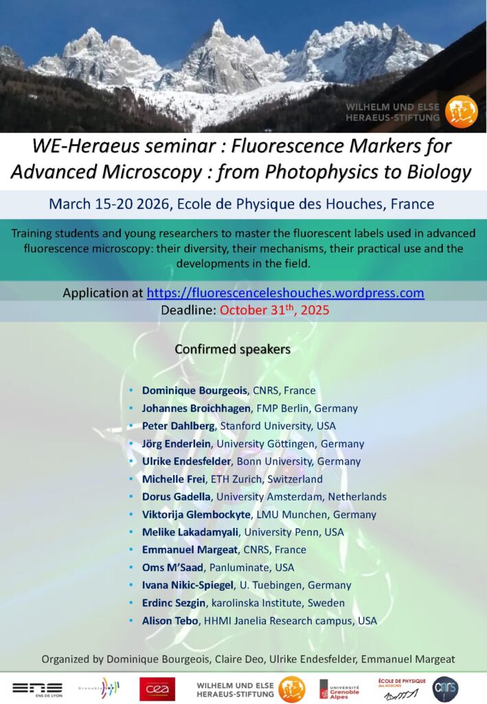
The use of fluorescence microscopy (wide field, confocal, multiphoton, and now superresolution) in combination with genetically encoded fluorescence probes comprise a powerful set of scientific tools to study live cells. However, surprising little practical and theoretical training in such methods exists within standard curricula, particular at the early stage of training (Masters or Doctorate level). This course offers to cover basic optics principles necessary to understand the origin of microscope resolution and design. Participants will get hands-on experience implementing simple optical configurations to illustrate these fundamental principles. Subsequently, participants will perform experiments on state-of-the-art imaging equipment provided by microscope vendors.

Lectures will cover, in depth, the principle behind traditional high resolution imaging methods such as confocal, multi-photon, and the recently developed super-resolutions methods. Students will also learn about fundamental properties of synthetic and genetically encoded fluorescence indicators used for cellular morphology imaging and signaling recording. Finally guest lectures will demonstrate applications of some of the discussed methodologies.

This course is intended for Master’s students and international PhD students who expect to be using advanced live cell fluorescence microscopy for future studies.

Master 2 students from partner Universities (Sorbonne University and University of Paris Cité) MUST NOT register online and must submit their registration request DIRECTLY to contacts in charge of their university course. These students have free access to the course.

All other students must register online and pay their registration fees.

Biogenouest has the pleasure to invite you for Gen2Bio 2024 at the Palais du Grand Large in Saint-Malo!

Gen2Bio, the annual biotech congress organized by Biogenouest, is aimed at all biotechnology stakeholders: researchers, engineers and doctoral students in life and environmental sciences, innovative biotech companies, laboratories or research centers, competitiveness clusters, technology parks and technical centers, players in valorization, etc.

In particular, you can discover the service offerings of the network’s 38 technological platforms and can thus follow innovations in life sciences in the West side of France.

In 2024, the 2-day formula will offer 5 plenary conferences, 24 technological workshops divided into 6 sessions, an exhibition space on a stand for our partners and a BtoB space.

Le RIC (Réseau d’Imagerie Cellulaire Paris-Saclay) a le plaisir de vous convier prochainement à une visioconférence d’une heure.

Le D. Slavka Kascakova développera ses thématiques de recherche dans le domaine biomédicale ainsi que ses approches technologiques faisant appel à l’utilisation de différentes lignes du Synchrotron SOLEIL (Source Optimisée de Lumière d’Énergie Intermédiaire du LURE) à St Aubin.

Actuellement Maître de Conférences à la faculté de médecine (Université Paris-Saclay) et représentante académique de l’UPSaclay au sein de l’alliance universitaire européenne EUGLOHSlavka Kascakova travaille en tant que chercheuse au Centre Hépato-Biliaire de l’Hôpital Paul Brousse à Villejuif. Ses principaux axes de recherche ont pour but de diagnostiquer des maladies hépatiques, de caractériser les greffons de foies destinés aux patients et de mettre au point des stratégies thérapeutiques contre l’hépatite fulminante et les cancers primitifs du foie grâce notamment à la mise en œuvre d’approches spectroscopiques multimodales au sein du Synchrotron.


The next Euro-BioImaging User Forum will take place online on Thursday, October 12, 2023, from 2 pm-5 pm CEST. The topic is “Understanding plant biology.” This event will highlight how cutting-edge imaging technologies can support research into the structure and function of plants, shed light on plant health, resilience and adaptability, and help answer agroecology research questions. Applications of diverse imaging technologies and plant biology research contexts will be highlighted by two keynote speakers and presentations from users of the Euro-BioImaging services, showcasing the specific expertise available at the Euro-BioImaging Nodes. We will also provide information on funding opportunities to access Euro-BioImaging services for agroecology-related research projects.

Full program & registration:
https://www.eurobioimaging.eu/news/euro-bioimaging-user-forum-understanding-plant-biology/

The Advanced Light Microscopy initiative is organizing a technical symposium involving several microscopy techniques either emerging or identified as lacking awareness from a wider audience. This symposium will take place at the Institut Pasteur in Paris on the 26th of October 2023. This day will orbit around stimulating exchanges between system building scientists and Life Science Researchers in need of new insight to their current available possibilities.  

The confirmed speakers as well as the final program will be available soon.

More information on: research.pasteur.fr/fr/event/advanced-light-microscopy-symposium-26th-of-october-2023

Registration (free but mandatory) right here

France-BioImaging was at the Euro-BioImaging‘s All Hands Nodes Meeting at EMBL in Heidelberg. It was a pleasure to share this unique moment with all the Euro-BioImaging nodes! We had great discussions from passionate people around building the future of the infrastructure and providing open access to high-end technologies and expertise.

This was the perfect time to hear about the latest news and opportunities from every European nodes. Several FBI members were there:

  • Caroline Thiriet, our External affairs manager, was a panelist at a discussion about “Funding for national imaging communities”, highlighting the history of France-BioImaging and how the french infrastructure works.
  • Perrine Paul-Gilloteaux, our Image data mission officer, gave a fantastic talk about “Linking and analyzing correlative image datasets”.
  • Fabrice Cordelières, our Training mission officer, and Alban Belloir, Communication officer, presented two posters on training and about our infrastructure’s structuring activities.
  • Melina Petrel, as an Electron #microscopy specialist, represented the FBI core facility staff.

Thanks to all Euro-BioImaging team for organizing and hosting this wonderful event! We are glad to be part of this amazing international community working together as a European Research Infrastructure!

Find further information on Euro-BioImaging’s article: www.eurobioimaging.eu/news/all-hands-nodes-meeting-2023–more-

The PFIC facility organizes a technological seminar on “Quantitave Phase Imaging – exploring the unseenApril 20th at 10:00 am in Espace Maurice Tubiana at Gustave Roussy, Villejuif.

Quantitative Phase Imaging (QPI) is a novel label-free microscopy technique bringing a completely new contrast into the live-cell imaging field. It allows extracting of information-rich quantitative data from unlabeled cells and monitoring their dry mass in non-invasive experiments.

This seminar will explain the principle of technology and present broad range of applications focusing on the label-free analysis of cell growth, cell death, cell migration etc. Apart from cell biology research, studies of biomolecular condensates and biomaterials will be discussed.

The seminar will be held in English by Zuzana Nováková (Telight application specialist).

Hope to see you all you interested by this technology there!

For any supplementary information you can contact me at tudor.manoliu@gustaveroussy.fr

This is the first edition of our Summer School outside of France, going to South America in synchrony with the IEEE SPS-EMBS ISBI Conference in Colombia.

The spirit of our Summer School was established in French Brittany in 1994 (by Christian ROUX and Jean-Louis COATRIEUX). This Summer School has become a worldwide reference with international lecturers from 20 countries and accessible to young scientists from all around the world. Our Summer School is an open yet privileged place for exchanges and discussions on major on-going research and technologies. Informal and warm, we always select a location and design a program where ample time is dedicated to interactions between lecturers and students.

The Summer School is open to graduate students (MSc., PhD), post doctoral scientists, radiologists, biologists, researchers and engineers in industry.

The next Euro-BioImaging User Forum will be taking place on 21.03.2023 from 2-5 pm CEST, focusing on the topic of “Cardiovascular Research”. 

Register here

Euro-BioImaging is looking forward to featuring some of the excellent science supported by the work of EuBI nodes via presentations from your users. The presentations will be 15 min long and will include the opportunity to briefly introduce your Node. In addition the event will feature two keynote presentations.

Abstracts can be submitted here – https://forms.gle/XriAc5HTMiLAhACG6 
The deadline for abstract submission is on February 6th. 

All users who are working in the area of cardiovascular research are welcome ! The topic is broad as it includes vascular and cardiac development and/or regeneration, development of cardiovascular disease, inflammation in response to cardiovascular injury, etc. The users also do not have to be Euro-BioImaging users.

Euro-Bioimaging is looking forward to receiving your abstracts!

The Thematic Institute of molecular and structural biology of the French Alliance for life science (Aviesan) is programming a 2 days symposium dedicated to RNA biology and applications, that is a very active field with emerging topics.

Coding and non-coding RNAs are today central players in biology and medicine. RNA binding proteins act as modulators of RNAs functions, dysregulated RNA-protein interactions being linked to human pathologies, leading for example to misfolding or aggregations in membraneless organelles. In addition to human RNAs, bacterial RNAs and RNA viruses, are regarded as drug targets.

Epitranscriptomics, with modifications found in all RNAs and associated proteins have also come to the focus of intense research revealing their roles in many aspects of RNA biology and diseases. Finally, RNA medicines are actively developed for different therapeutic applications. RNA-based drugs are coming of age as a consequence of the improved knowledge of RNA biology combined with the development of sophisticated technologies.

This symposium will take place in Paris, on February 2-3 2023 end in Amphi Buffon, 15 Rue Hélène Brion, 75013 Paris. The meeting is built around different scientific sessions, namely RNA therapeutics, RNA/RNP biogenesis and structure, RNA modifications and new technological developmentsThe meeting is for structural, molecular and cellular biologists to have an overview of the latest results and concepts that are currently under development in the field.

A poster session will be organized and short talks will be selected.

Deadline for abstracts submission: January 12th, 2023.

Dowload the preliminary program

Registration

The organizing committee:

Edouard Bertrand, Christiane Branlant, Carine Giovannangeli, Chantal Pichon, Carine Tisne.

Imagerie-Gif core facility, from our Ile-de-France Sud node, is pleased to announce the acquisition of a Scanning Ion Beam Electron Microscope (FIB-SEM) and a Lattice Structured Illumination Microscope (SIM) Elyra 7. For the occasion, the core facility is organizing “3D Res/volution“, a scientific event on high-resolution 3D imaging on December 15, 2022 from 2:00 pm to 5:00 pm at B21 amphitheatre. This event will be a great opportunity to introduce to you the possibilities of these 2 new systems available at Imagerie-Gif.

Free but mandatory registration: https://evento.renater.fr/survey/3d-res-volution-day-8pdzetbi

Ranging in size from 20 microns to 1 milimetre, Meiofauna is a crucial link between micro- and macro- marine ecosystems. Valentin Foulon, a research engineer in marine biology, is part of the Blue Revolution program that aims to develop a taxonomic identification protocol for these tiny creatures. He approached the Bretagne-Loire node of France-BioImaging, in Nantes, where a Single Plane Illumination Microscopy (SPIM) light-sheet available in open access helped him to further reach his goal.

Valentin Foulon loves imaging and microscopy – and he loves meiofauna. He works on the Blue Revolution program, whose goal is to accelerate the taxonomic identification of meiofauna using artificial intelligence algorithms. But while the confocal-microscopy-based protocol worked well for small plankton, the researchers quickly realized that it had to be adapted to work with meiofauna. 

Choosing the right microscopy approach

Identification of Meiofauna is usually operated manually on a classical microscope, which is “incredibly challenging” says Valentin. “And we only have one focal plane. To see important details, we need a 3D view of organisms.” explains Valentin. “That’s why we got curious about light-sheet imaging, because we would be able to see the organisms in 3D.” As France-BioImaging provides this type of microscopy in open-access in Nantes, “we clearly saw an opportunity there,” explains Valentin. His project proposal, which was submitted to the Euro-BioImaging pilot User Access fund, was fully funded by France-BioImaging, in a generous initiative introduced by France-BioImaging to fund all user projects to its facility received by Euro-BioImaging for the pilot User Access fund. 

“I had never tried light sheet microscopy before,” says Valentin, “And in Nantes, the lab didn’t have experience with marine organisms. Together, we found a compromise between different imaging parameters such as the resolution or the acquisition time.” Once the set-up was fine-tuned, he sent his samples, and the engineer at the lab did the imaging work. “It was a very nice experience,” expresses Valentin.  “I discovered a new technique, which is always enriching. I also think the lab was happy to work with a new sample type. And finally, we validated our proof-of-concept, proving that it is possible to image these organisms in 3D.

Image data management

But the project doesn’t end there. “The next step is data management. We imaged between 200-300 different organisms. It’s not enough for automatic classification, but nevertheless, we have 6-7 TB of data. Now we must process the data for this classification with machine learning. It’s an essential part of the project, to make the link between sample imaging, and data processing. We are working to close that gap.” 

“The outcome of this project is to validate a proof-of-concept, from the sample collection to the image classification. With this new taxonomic identification protocol, we will be one step forward in the comprehension of meiofauna, these mysterious marine organisms, who may hold the key to understanding the impact of human activity on marine ecosystems,” concludes Valentin. 

Thank you Marianna Childress, communication officer of Euro-BioImaging, for the original article.