The RIC (Réseau d’Imagerie Cellulaire Paris-Saclay) is pleased to invite you to a one-hour
visioconference.
D. Slavka Kascakova will discuss her research themes in the biomedical field and her
technological approaches using different lines of the SOLEIL Synchrotron (Source Optimisée
de Lumière d’Énergie Intermédiaire du LURE ) in St Aubin.
Currently Senior Lecturer at the Faculty of Medicine (Université Paris-Saclay) and UPSaclay’s
academic representative on the European university alliance EUGLOH, Slavka Kascakova
works as a researcher at the Centre Hépato-Biliaire of the Hôpital Paul Brousse in Villejuif.
Her main lines of research are aimed at diagnosing liver diseases, characterizing liver
transplants for patients, and developing therapeutic strategies for fulminant hepatitis and
primary liver cancers, thanks in particular to the use of multimodal spectroscopic approaches
at the Synchrotron.
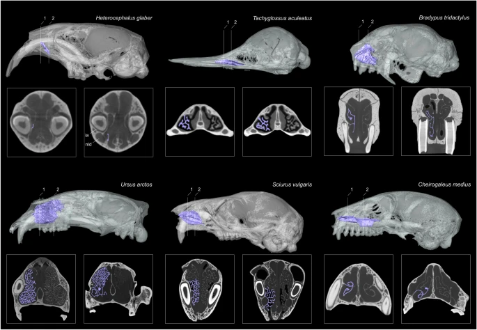
Most mammals can maintain a relatively constant and high body temperature. This is considered to be a key adaptation for theses species, enabling them to successfully colonize new habitats and survive harsher environments. Scientists from the Institut des Sciences de l’Evolution de Montpellier (ISEM) investigate the possible correlation between the maxilloturbinal in the anterior nasal cavity and the body temperature maintenance by using Micro Tomography (or MicroCT) at the MRI core facility (FBI Montpellier node). This technique was essential in this study as it rebuilt the hypothesis around body temperature maintenance. Here is what they found.
MicroCT: Image in a non-destructive way
First of all, what is Micro Tomography? Micro Tomography, or Micro-CT, is a 3D imaging technique using X-rays to see inside biological material, at a small animal or body part level. Slice by slice, this technology scans the object in a series of 2D images that are reconstructed in a 3D model. Micro CT is, thus, non-destructive. This means that it can be used to image a sample without having to cut it! Not only your material is still in one piece but you can use it for further experiments.
Phylogenetic studies as an example of application
In this study examining the correlation between skull structure and the stabilization of body temperature, MicroCT was the key. The presence and the relative size of the maxilloturbinal has been proposed as a hypothesis that reflects the endothermic conditions and basal metabolic rate in extinct vertebrates. Among bony structures, respiratory turbinals (e.g., maxilloturbinal) are interesting anatomical structures that may offer important insights to the origins of endothermy, in other words to the origin of warm-blooded animals. Indeed, respiratory turbinals are highly vascularized, which amplifies the surface area and offers an effective mechanism to avoid loss of internally-produced and costly heat.
You probably figured it out: scientists needed to compare the structure of the maxilloturbinal in order to take conclusion. This is when Micro Tomography was very useful. They scanned 424 individuals from 310 mammal species using high-resolution X-ray micro-computed tomography, with approximatively half of the samples imaged at MRI, part of our Montpellier node. Using the obtained comparative 3D µCT dataset, they explored the anatomical diversity of the maxilloturbinal based on relative surface area, morphology and complexity. They specifically tested the relationship between multiple parameters such as the size-corrected basal metabolic rate (cBMR), the relative surface area of the maxilloturbinal (Maxillo RSA) or body temperature.
And the results surprisingly showed that…
…there is no evidence to relate the origin of endothermy and the development of some turbinal bones! Even though scientists used a comprehensive dataset of Micro CT-derived maxilloturbinals spanning most mammalian orders, they demonstrate that neither corrected basal metabolic rate nor body temperature significantly correlate with the relative surface area of the maxilloturbinal. These results challenge the hypothesis of thermal regulation being linked to respiratory bone structure.
So, what could be linked with the thermoregulation of mammals? Researchers proposed 3 more hypothesis. First of all, environmental conditions could have a bigger role: “the maxilloturbinal function could have a more prominent heat/moisture exchange role in species that face harsh environmental conditions, thus helping to limit spurious heat and moisture loss”. Another major role of the maxilloturbinal is water conservation. As an example, the naked mole-rat avoid breathing through the mouth when performing energy intensive digging because the lips close behind the digging incisors and this species has the lowest value of predicted Maxillo RSA of the entire sample. But most of all, the factor could be a multifactorial physiological question. What is the relation of the maxilloturbinal with the overall nasal cavity? Do other functions play a role in the evolution of this body part, such as its protective role against toxic elements? Is it linked with brain cooling?
Well, imaging will certainly give them an answer in the future!
Get access to one of our services!
You need Micro-CT or another imaging technology or expertise that France-BioImaging provides? To get open access, please login via Euro-BioImaging website! You just have to choose the technology you want to use, then submit your proposal. All applications will be processed by the Euro-BioImaging Hub in close relation with France-BioImaging. And of course, all scientists regardless of their affiliation, area of expertise or field of activity can benefit from open access services! Users whose projects will be validated by Euro-BioImaging will benefit from a waiver for the access cost on France-BioImaging core facilities (https://france-bioimaging.org/access/).
Martinez, Q., Okrouhlík, J., Šumbera, R. et al. Mammalian maxilloturbinal evolution does not reflect thermal biology. Nat Commun 14, 4425 (2023). https://doi.org/10.1038/s41467-023-39994-1
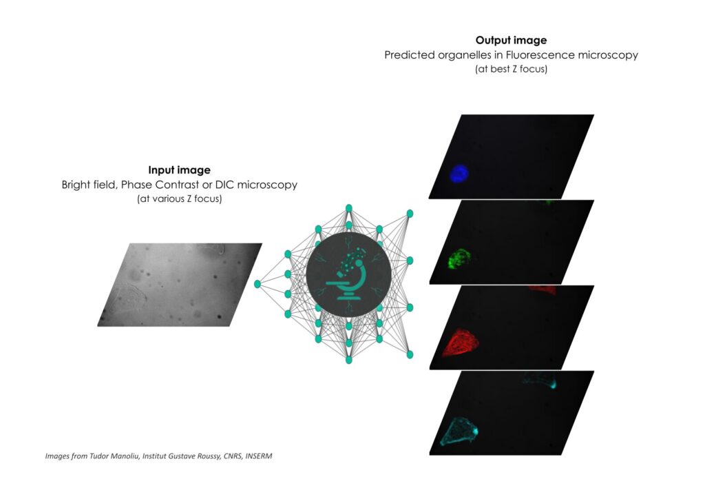
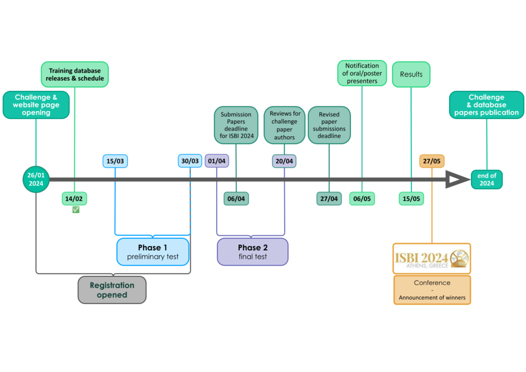
In order to answer image data analysis demands, France-BioImaging is launching its first data machine learning competition: welcome to the Light My Cells challenge!
The challenge
The Light My Cells France-Bioimaging challenge aims to contribute to the development of new image-to-image ‘deep-label’ methods in the fields of biology and microscopy. Basically, the goal is to predict the best-focused output-images of several fluorescently labelled organelles from label-free transmitted light input-images. And we need you for that!
More information about the challenge: lightmycells.grand-challenge.org/lightmycells
How to participate
We have defined the challenge as a single task with two phases:
- A preliminary test phase (on 30 images) to familiarize with the algorithm submission procedure, with the possibility to have five submissions (with a maximum of one by week)
- The final test phase (on 300 images) with only one submission accessible will not give the possibility to evaluate their algorithms before submitting.
So, you have until the end of the first phase, on March 30, 2024, to register and participate at this Light My Cells challenge. Nonetheless, you can start working on your preliminary algorithm and tests on February 14st, 2024 (with the release of the training database)!
Register now! lightmycells.grand-challenge.org/lightmycells
Prizes
For top 3 winners:
- Award certificate
- A challenge paper will be written with the organizing team’s members for submission to journals
- Invitation to publish their methods in the proceedings of the IEEE International Symposium on Biomedical Imaging 2024s
- Support and integration of open source code into open science image processing and analysis software (e.g. BioImage Model Zoo, Napari)
For the 1st:
- Invitation to 2024 France-Bioimaging annual meeting
- Graphic card
- Android tablet
For the 2nd:
- Graphic card
- Android tablet
For the 3rd:
- Android tablet
Why launching a challenge?
To develop powerful methods that will then end up in creating public databases, standards & benchmarks in the field of bioimaging! The FBI challenge is hinged on a double contribution: from core facilities engineers and from data scientists. The first group acquired a large number of images to build a dataset, that will later be used by the algorithms. These images were produced by microscopy engineers & technicians from FBI’s platforms. As for the second contribution, this is where the challenge starts! The challenge is then published to have a maximum of data scientists to work on the algorithms that best fulfill the analysis task.
The first project is also based on four pillars:
- Open source + FAIR (Findable, Accessible, Interoperable, Reusable)
- Supervised learning, it involves annotated datasets to maintain control over performances.
- In silico annotations, a computer labeling method to avoid manual annotation and its drawbacks.
- Image-to-image analysis tasks, an image analysis tasks which aim to predict an output image from the input one.
For any questions, please contact Dorian Kauffmann: dorian.kauffmann@france-bioimaging.org.
France-BioImaging and all the French community aims to develop and promote innovative imaging technologies and methods. But microscopy images can also take an artistic, creative look and make the invisible world beautiful, allowing people to see the visual appeal of the life sciences.
We enjoyed the diversity of the images submitted with many different microscopy techniques, models and applications represented. A big thank you to all the participants!
The National Coordination Team and the Executive Board are proud to announce the winners of the FBI Image Contest 2023:
- 1st Place: Laurent LE, Lévêque-Fort Team, Institut des Sciences Moléculaires d’Orsay
“In the blink of an eye”
COS7 fixed cell. Alpha-tubulin labeled with DNA-PAINT and imaged with Atto 647N. Axial information is obtained by virtual-SAF measurement known as DONALD.
SMLM Fluorescence Microscopy with DNA-PAINT with DONALD detection
- 2nd Place: Gonzalo QUIROGA-ARTIGAS, Team Contrôle cytoplasmique de la stabilité du génome, Centre de recherche en Biologie Cellulaire de Montpellier
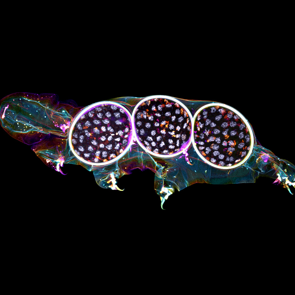
“Tardigrade embryos protected by mother’s molt”
Tardigrades commonly align the time of molting with egg laying. In this image we observe a tardigrade molt covering three developing embryos (DNA in white). The microscopy technology applied was confocal microscopy, and the research aimed to investigate the synchronization of embryo development in tardigrades.
Confocal microscopy
- 3rd Place: Hugues LELOUARD, Gorvel team, Centre d’Immunologie de Marseille Luminy
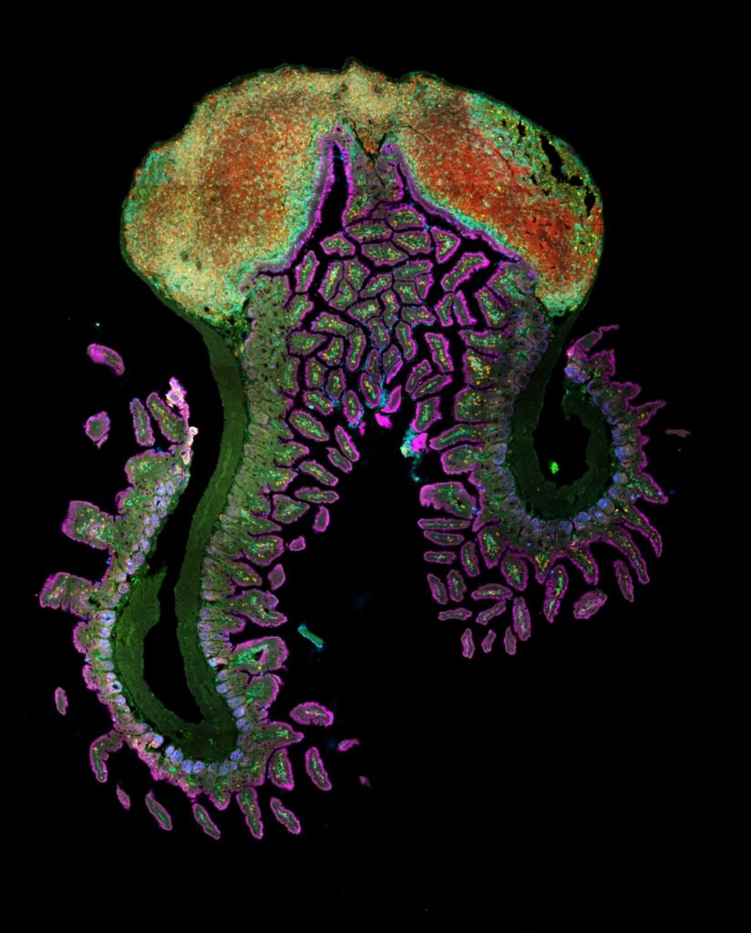
“Intestinal octopus”
Small intestine section from a LyzM-eGFP mouse containing one Peyer’s patch and stained for proliferative cells (Ki-67, yellow), Paneth cells (UEA-I, blue), epithelial cells (EpCAM, magenta), naive B cells (IgD, red), T cells (CD3, orange), helper T cells/macrophages (CD4, cyan), phagocytes (CD11c, turquoise), monocyte-derived phagocytes (GFP, green).
10-color spectral confocal microscopy
Congratulations to the winners!
Explore all the images submitted here:


As stated in the Terms & Conditions of the contest, foreign participants non-affiliated to a French institution are featured in the gallery, but were not evaluated as part of the contest.
The FBI Working Group Correlative Light-Electron Microscopy is organizing a workshop!
This workshop will take place at the Bordeaux Imaging Center (FBI Bordeaux node) from January 31st to February 2nd, 2024.
Correlative Light and Electron microscopy (CLEM) increases our capacity of biological investigation. By combining light microscopy and electron microscopy, this complementary approach takes advantages of both techniques. Light imaging provides valuable functional information thanks to its labeling power, whereas Electron microscopy excels at high resolution.
The Bordeaux Imaging Center has developed several workflows such as In-resin fluorescence and Array tomography. Both helps to determine 3D ultrastructure of a targeted area or a whole sample at different resolutions. In the framework of the FBI CLEM workshop, participants can choose one of these 2 different practicals, In-resin fluorescence and Array Tomography, in which a local specialist will show them the workflows.
Workshop 1: “In-Resin Fluorescence” – From HPF to tomography, by way of freeze-substitution and on-section fluorescence observation.
Workshop 2: “Array tomography” – From serial section to acquisition with confocal and SEM.
- It is firstly intended to scientists with expertise in electron microscopy, who are expected to use the chosen technique.
- 2 workshops of 4 people each (you can apply only to one workshop)
- A practical manual will be provided, covering every step.
You want to attend this workshop? Please fill the following form before December 22nd, 2023:
The candidates whose research projects suit the workshop the best will be selected. We will get in touch with you after the Christmas break to confirm your participation at this workshop.
This form is currently closed for submissions.
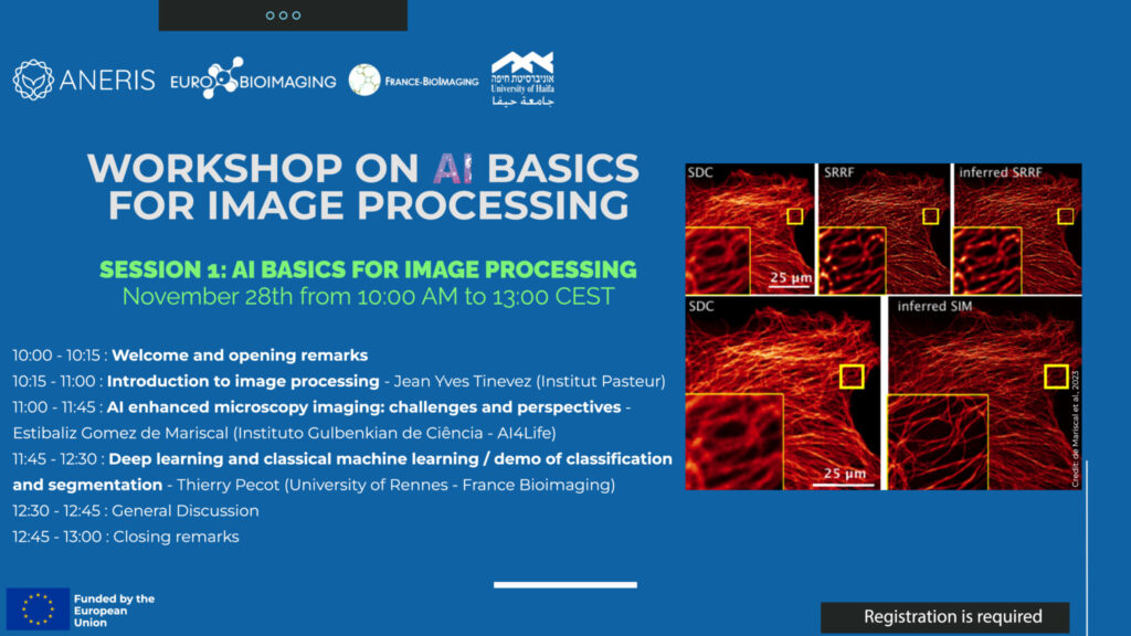
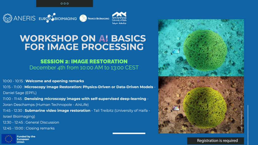
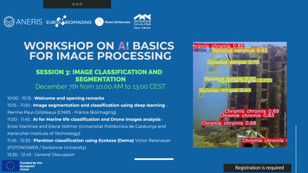
ANERIS is thrilled to announce a series of three workshops centered around the use of AI technology in image processing.
Discover the potential of Artificial Intelligence in the field of image processing during the dynamic workshop series organised within the ANERIS project. These three informative sessions (AI basics for image processing, Image restoration, and Image classification and segmentation) provide a thorough exploration of AI and how it can be utilised to improve and interpret visual data.
Together with our colleagues from Euro-Bioimaging ERIC and University of Haifa, we will take participants through expert-guided sessions on:
Session 1: AI basics for image processing November 28th from 10:00 to 13:00:
Session 2: Image restoration December 4th from 10:00 to 13:00:
Session 3: Image classification and segmentation December 7th from 10:00 to 13:00:
Launched earlier this year in coordination with the African BioImaging Consortium and Imaging Africa and within the framework of the Horizon Europe Programme, the Africa-France Joint Initiative for Biological Imaging aims at extending its partnership with colleagues in Africa that have interest in using advanced microscopy approaches for their own research programs and projects. With this in mind, we have previously designed two calls for funding: one for access to FBI’s bioimaging core facilities, the other as a twinning program.
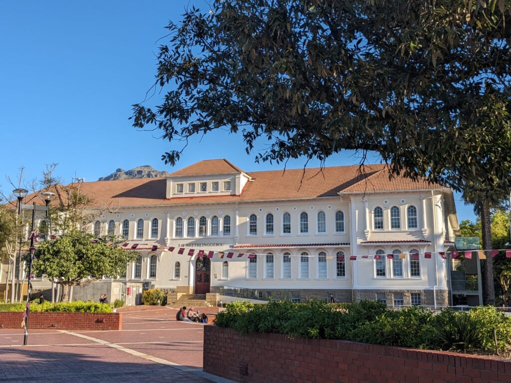
Good news! Our first project has started! Granted by our second call, the Twinning program has begun between Stellenbosch University and FBI-Paris Node. A fantastic experience based on sharing practices, knowledge transfer and many fruitful discussions on image analysis and correlative approaches between light sheet and serial block face microscopy techniques. For the South African partner, Madelaine Frazenburg (Stellenbosch University), it is the opportunity to see how other microscopy laboratories in France works but also to learn more about cryo-SEM and to study new kind of sample preparation methods. From the French side, Ludovic Leconte (Institut Curie, FBI Paris-Centre node) is indeed very interested in gaining new experience in electron microscopy mainly in Serial Block Face, another tissue section imaging that is not available on his site and for which the Stellenbosch imaging platform has the mastery.
Our warmest thanks to Lize Engelbrecht, Professor Ben Loos and Janica Conradie for making this event possible and for the warm welcome they extended. The second stage of this “Twinning” project will take place at Institut Curie next spring. We look forward to welcoming Madelaine!
This event co-organized by the ALPHA-RLH competitiveness cluster, Bordeaux Imaging Center (from FBI Bordeaux node) and the Interdisciplinary Institute of Neurosciences will be dedicated to advances and to the promotion of innovation in the bioimaging sector.
The main experts in bioimaging – researchers and industrialists – will be reunited around three main themes: microscopy, cell biology and image analysis, from the perspective of the main fields of application: neuroscience, oncology, bioengineering…
Designed for everyone interested in bioimaging, specialists or not, this event is open to researchers, clinicians, engineers, industrialists and students. The day will be interspersed by conferences, a poster session and moments for discussions and networking.
– Research and development organisations:
Bordeaux Imaging Center, Institut Interdisciplinaire de Neurosciences, Institut Bergonié, Institut d’Optique Graduate School, Laboratoire de Biogenèse Membranaire, Institut des Sciences Moléculaires d’Orsay, IPREM, XLIM, SATT.
– Industrials:
TreeFrog Therapeutics, Imagine Optic, Spark Lasers, Argolight, Abbelight.
Program here
Free but mandatory registration right here
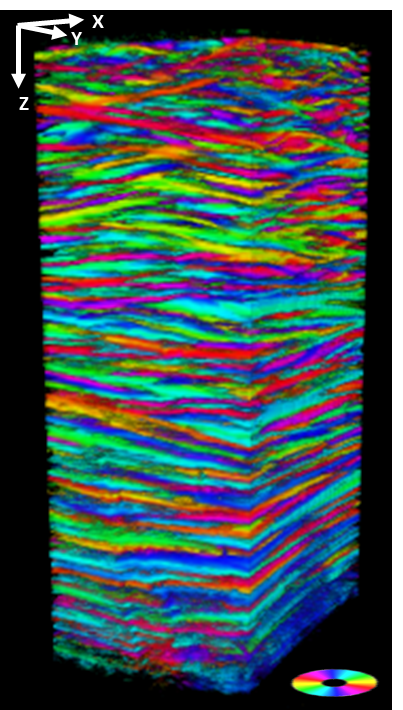
A key property of the human cornea is to maintain its curvature and consequently its refraction capability. Although we know that it is related to its stacked collagen lamellae structure, the distribution, size, and orientation of these lamellae along the depth of the cornea are poorly characterized up to now. A team from the Laboratory for Optics and Biosciences (LOB) has optimized a recent technology which combines Second Harmonic Generation microscopy and polarimetry (P-SHG) to image the lamellar microstructure of human corneas and more!
Acquire the structure in depth
Imaging the cornea is essential to understand how visual acuity works. This part of the eye is characterized by its transparency and refractive power but also by its unique mechanical properties. To answer the many questions of the cornea structure, the Second Harmonic Generation microscopy is the perfect technique. This technology is based on the sample capacity to generate second harmonic light, which has half the wavelength of the light entering the material. However, the SHG is working on well-aligned assemblies of non-centrosymmetric molecules which fits perfectly with the collagen!
Apart from being specific to this kind of macromolecules, the SHG microscopy offers multiple advantages. First of all, as it does not involve the excitation of molecules, molecules do not suffer from phototoxicity or photobleaching effects. Moreover, no markers are necessary which makes this type of microscopy noninvasive. Finally, Second Harmonic Generation microscopy allows the visualization of in-depth structure of thick samples. As a matter of fact, it is, nowadays, the gold standard technique for in situ visualization of collagen 3D organization in unstained biological tissues.
Add polarimetry and get orientation information
P-SHG first offers all the advantages of usual SHG microscopy: 3D optical imaging in depth and high specificity and sensitivity to collagen without any labeling. In this study, scientists took advantage of the light polarization to reveal the direction of the collagen fibrils that make up the lamellae of the cornea in their SHG microscopy acquisition. This recent technology is called: polarization-resolved SHG microscopy (P-SHG). The main novelty of this study was to implement P-SHG in depth to analyze intact human corneas along their full thickness (up to 600 µm). 3D reconstructions of P-SHG data show in a unique way the stacking of collagen lamellae with different orientations all along the thickness of the cornea. Here, imaging helped confirm that these lamellae are roughly organized parallel to the cornea surface, with different collagen orientations in sequential lamellae, and provided new information about the variation of these orientations along the depth of the cornea.
Aside from being the first quantitative characterization of the lamellar structure of the human cornea continuously along its entire thickness with micrometric resolution, this imaging technique could be a huge step forward in vivo diagnosis as it uses the detection of the reflected signal. Furthermore, this study opens the way to promising new characterizations of the cornea, such as mapping the size and distribution of lamellae as a function of depth, but also as a function of position (center or periphery of the tissue). This information will feed into mechanical modelling of corneal behavior during variations in intraocular pressure or healing processes. Finally, the study of pathological tissues will clarify the role of the corneal defective structure in certain diseases.
These results show the unique potential of P-SHG microscopy for imaging of collagen distribution in thick dense tissues. And of course, this approach is readily applicable to more than just cornea! It may be used for instance to decipher the structure of collagen in fibrotic pathologies or in other proteins that exhibit SHG, namely myosin and tubulin, or in starch and cellulose in plants. This shows the unique potential of P-SHG microscopy for imaging thick collagen-rich tissues.
Get access to one of our services!
Polarization SHG microscopy is not available in open access but we are open to collaborations!
You need classic SHG microscopy or another imaging technology or expertise that France-BioImaging provides? To get open access, please login via Euro-BioImaging website! You just have to choose the technology you want to use, then submit your proposal. All applications will be processed by the Euro-BioImaging Hub in close relation with France-BioImaging. And of course, all scientists regardless of their affiliation, area of expertise or field of activity can benefit from open access services! Users whose projects will be validated by Euro-BioImaging will benefit from a waiver for the access cost on France-BioImaging core facilities (https://france-bioimaging.org/access/).
Sources: https://www.inp.cnrs.fr/fr/cnrsinfo/cartographier-la-structure-de-la-cornee-humaine
https://www.ip-paris.fr/en/news/unveiling-structure-human-cornea
Raoux, C., Chessel, A., Mahou, P. et al. Unveiling the lamellar structure of the human cornea over its full thickness using polarization-resolved SHG microscopy. Light Sci Appl 12, 190 (2023). https://doi.org/10.1038/s41377-023-01224-0
Nous vous invitons au premier webinaire sur l’Initiative Commune Afrique-France pour l’Imagerie Biologique qui se déroulera le 6 décembre 2023 à 11h00 CET!
En coordination avec l’African BioImaging Consortium et Africa Microscopy Initiative et dans le cadre du programme Horizon Europe, l’Initiative Commune Afrique-France pour l’Imagerie Biologique vise à étendre et renforcer les collaborations entre collègues africains et français intéressés par l’utilisation de microscopie avancées pour leurs propres projets de recherche. C’est dans cette optique que nous avons lancé deux appels à projet : l’un favorisant l’accès aux plateformes de bioimagerie de France-BioImaging, l’autre consistant à un programme de jumelage.
Ce webinaire sera notamment l’opportunité d’en apprendre plus sur les projets sélectionnés et sur les bénéfices de cette initiative pour les scientifiques africains et français.
Webinaire
Starts: November 10, 2023 • Ends: December 31, 2023
FBI opens a call for the recruitment of its next Scientific Director (2024-2028)
(Deadline is 31st of December 2023)
France BioImaging-FBI (laureate of the INBS program of the PIA in 2011) is the National Research Infrastructure in Biological Imaging. FBI is built on 8 geographical Nodes identified on the basis of strong relationships between R&D labs and imaging Core Facilities. Each Node shows a specialization of a local expertise in methods and biological topics. This crossover between imaging technologies and expertise in scientific topics is a characteristic of the complementarity between FBI Nodes. A 9th Transversal Node gathers FBI strengths and resources in Image Analysis and DATA management. The Operating Coordination is done under the umbrella of the UAR 3426.
Our motto is “Innovation-Training-Access”
(i) Invent and disseminate new imaging technologies
(ii) Training users and facility staff on existing and new technologies
(iii) Make them accessible to as many people as possible.
Role of the Scientific Director of FBI
-He/She is the Strategic Manager of the Infrastructure
–He/She leads the National Coordination (NC), composed of an adjunct director for international affairs, a manager of internal affairs and a manager of external affairs. The Infrastructure also benefits from several support functions: a communication officer, a business developer and an accounting officer. The NC leads the Executive Committee (EC) to manage the Infrastructure. With the Help of an international Scientific Advisory Board (SAB), he/she reports the overall Infrastructure policy and strategy to the Institutional Committee (Steering Committee, SC) which is the “decision maker”.
–He/She is responsible for the arbitration of recruitment proposals (in interaction with the other governance bodies), equipment investments (PIA, TGIR, other National and International common actions…) and new service opening, in relation to the development objectives (R&D, service offers…) and the overall infrastructure strategy at national and international levels.
–He/She is responsible, with the staff concerned, for the inventory of the different activities/tasks of the infrastructure: links with Europe and International, work with the different committees, web site/communication, training, animation of the “FBI-community”, scientific and financial reports…
– He/She is responsible, in interaction with the EC, for managing interactions/collaborations (R&D, service providing, partnerships, technology watch…) with other PIA Research Infrastructures.
Entry into function is planned for the 1st of July 2024.
If you are interested, please send a short CV and a letter of intent (2 pages) indicating your motivation, vision and strategy, at direction@france-bioimaging.org
BEFORE the 31st of December 2023
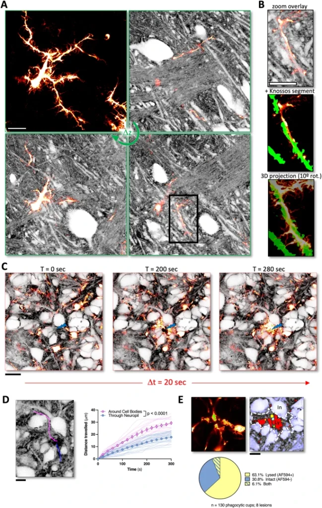
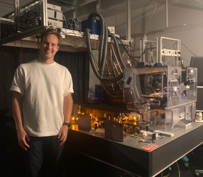
In 2022, in a project funded by the Euro-BioImaging pilot User Access Fund, Andrew Boyce, postdoctoral fellow at the University of Calgary, used state-of-the-art microscopy techniques at the Bordeaux Imaging Center, part of the Bordeaux node of France-BioImaging, for panoptical visualization of brain tissue in vivo. His work just got published in a Nature Communications article!
The advantages that shadow imaging has in visualizing brain tissue
Progress in neuroscience research hinges on technical advances in visualizing living brain tissue with high fidelity and facility. Unfortunately, current neuroanatomical imaging approaches either require tissue fixation (electron microscopy), do not have cellular resolution (magnetic resonance imaging) or only give a fragmented view (fluorescence microscopy).
In this study, scientists have shown how regular light microscopy together with fluorescence labeling of the interstitial fluid in the extracellular space provide the information lacking in the previous techniques. Basically, it’s like looking at the negative of a photo! Called Shadow Imaging, they have demonstrated the power of this approach on revealing neurons, microglia, tumor cells and blood capillaries together with their complete anatomical tissue contexts.
The perfect combination between the right technology and the right expertise
How did Andrew Boyce contribute to this study? Interested in blebbing neuron dendrites during cell death caused by a stroke, he only had, at that time, results from how cells in culture reacted during this kind of events but no information about the neighboring cells.
Thanks to Euro-BioImaging pilot User Access Fund, he came to Bordeaux to use a combination of live cell 3D stimulated emission depletion (3D-STED) microscopy and super-resolution shadow imaging (SUSHI), and adapt shadow imaging approaches to conventional confocal microscopy (COSHI). Shadow imaging allows you to visualize fine details of cell-cell interactions and the extracellular space in an unbiased manner. Brain tissue is complex and shadow imaging is a technique that allows researchers to visualize all of the cells in this very complex architecture in an unbiased manner so it was the perfect match for Andrew’s research project!
A career-boosting experience
“This has really been an incredible experience. It’s amazing to be trained by the person and team who pioneered this technique. I’m very thankful for this opportunity and humbled to be over here,”reflects Andrew from Bordeaux.
When asked how his visit to Bordeaux might impact his career, Andrew explains that he can certainly imagine doing shadow imaging in the future. He would eventually like to run his own lab at an academic institution in Canada. Being able to bring an exciting and novel technique like this back to Canada would be really impactful for his career, and for science in general.
“Shadow imaging is a very exciting technique for studying brain tissue, but getting started on sample preparation and adapting this technique to your research question can be daunting. Coming to the Bordeaux Imaging Center to work with the person who developed this approach and get expert training is a dream come true and will make a big difference to my research. This is an amazing opportunity made possible by the Euro-BioImaging Pilot User Access fund with the goal of making shadow imaging more accessible across diverse platforms,” concludes Andrew.
at the Bordeaux Imaging Center (www.eurobioimaging.eu/news/using-super-resolution-live-cell-imaging-to-understand-cell-death-during-stroke)
Get access to one of our services!
You want to be the next user to get access to state-of-the-art technologies that France-BioImaging provides? Please login via Euro-BioImaging website! You just have to choose the technology you want to use, then submit your proposal. All applications will be processed by the Euro-BioImaging Hub in close relation with France-BioImaging. And of course, all scientists regardless of their affiliation, area of expertise or field of activity can benefit from open access services! Users whose projects will be validated by Euro-BioImaging will benefit from a waiver for the access cost on France-BioImaging core facilities (france-bioimaging.org/access)
Thanks to Marianna Childress and Andrew Boyce for the original article!
Dembitskaya, Y., Boyce, A.K.J., Idziak, A. et al. Shadow imaging for panoptical visualization of brain tissue in vivo. Nat Commun 14, 6411 (2023). https://doi.org/10.1038/s41467-023-42055-2