1st edition of the International Symposium of Institut Curie: From Basic Science to Cancer Research
▽ Scroll down
Author: Alban Belloir
The 1st edition of the International Symposium of Institut Curie, From basic science to cancer research will be held on May 15, 16 and 17, 2024, at the Maison de la Chimie, in Paris. This Symposium aims to bring together 700 participants, biologists, chemists, physicists, clinicians, experts in basic science or in cancerology, from the United States, Europe or Asia. All will meet in a historic and prestigious venue: the Maison de la Chimie, in the heart of Paris to attend internationally renowned speakers’ conferences. You will have the opportunity to meet and exchange with Charles Swanton from the Francis Crick Institute (UK), Edith Heard from the EMBL (DE) or Yasmine Belkaid from Institut Pasteur (FR), and many others to be found here: https://www.curiesymposium.fr/speakers/.
For its first edition, the symposium will include four sessions dedicated respectively to evolution, genetics, immunity and systems biology, as well as a final session entitled “Science has great beauty” in homage to Marie Curie.
Cellular junctions are essential to the integrity of epithelia, which cover most of our organs. In an article published in the journal PNAS, scientists, with among them members of our FBI Marseille node, reveal the existence of a new category of cell junctions. Using Stimulated emission depletion (STED) microscopy, they have put forward the need to reconsider the organization of intestinal cell junction described as such for more than 40 years.
Imaging intestine with STED
Stimulated emission depletion (STED) microscopy is a super-resolution technique that bypasses the diffraction limit of light microscopy to increase resolution. In our case, scientists were able to resolve the organization of complexes located at cell junctions with a resolution of a few tens of nanometers thanks to STED. Moreover, STED tripled the spatial resolution in the junctional plane and, using cryosections, they achieved imaging with a seven times greater spatial resolution compared to approaches that would use confocal microscopy and thus, without physical sectioning.
Although the resolution of STED is at least an order of magnitude lower than that of electron microscopy, the combination of STED with immunostaining reveals organization up to then unknown as multiple proteins can be efficiently labeled at the same time.
Three types of intestinal cell junctions
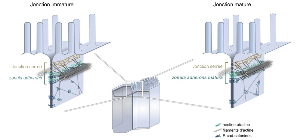
The intestine is covered with cells, most of which absorb the nutrients we ingest. These cells are joined together by three types of junctions which coexist and provide different functions, ranging from the selective filtration of certain ions to the mechanical maintenance of the epithelial layer. These junctions, the tight junction, the adherens junction, also called zonula adherens, and the desmosomes, were discovered in the 1960s and their constituent elements as well as their organization were proposed during the 1980s and 1990s.
The adherens junction in particular is established as being organized into a belt of adhesion proteins anchored to the membrane, the cadherins, and supported by filaments, the actin filaments. This junction has an important mechanical role in the cell, for example by impacting the shape of the cell. The zonula adherens (ZA), a fundamental module of epithelial cell–cell adhesion initially observed in intestinal cells, is believed to comprise a single contractile actin belt linked via E-cadherin-catenin to the ones of neighboring cells.
How did microscopy help reevaluate our current knowledge?
By observing the adherens junction of epithelial cells obtained from human intestinal biopsies, or from human cells in culture using STED super-resolution microscopy, scientists have made a very surprising discovery. They show that the ZA consists of two distinct belts of adhesive complexes, a basal one with E-cad-catenin and an apical one with nectin–afadin. Contrary to the prevailing view, the major actin belt aligns with nectin and afadin, not E-cad-catenin.
The authors further demonstrate that this organization depends on the cell maturation state and that the classical ZA found in textbooks corresponds to a less mature state of the intestinal junction. Therefore, they decided to call the junction found in mature cells the zonula adherens matura. Genetic and physical perturbations show that afadin is essential for force transmission across cell junctions. This work redefines the intestinal ZA architecture and prompts a reevaluation of how forces propagate within an epithelial sheet.
Not only, these results are important to better understand the adhesion and mechanics of epithelial cells, but these two essential characteristics of the epithelia are particularly affected in cancers of epithelial origin, which represent 80% to 90% of current cancers. This discovery is, thus, a step forward to the comprehension of cancers and to their treatment.
Get access to one of our services!
You need FRAP, two photon FLIM-FRET, PALM/dSTORM at France-BioImaging? To get open access, please login via Euro-BioImaging website! You just have to choose the technology you want to use, then submit your proposal. All applications will be processed by the Euro-BioImaging Hub in close relation with France-BioImaging. And of course, all scientists regardless of their affiliation, area of expertise or field of activity can benefit from open access services! Users whose projects will be validated by Euro-BioImaging will benefit from a waiver for the access cost on France-BioImaging core facilities (https://france-bioimaging.org/access/).
Mangeol, P., Massey-Harroche, D., Sebbagh, M., Richard, F., Le Bivic, A., & Lenne, P. F. (2024). The zonula adherens matura redefines the apical junction of intestinal epithelia. Proceedings of the National Academy of Sciences, 121(9), e2316722121. https://doi.org/10.1073/pnas.2316722121
My name is Samy Al-Bourgol and I recently joined the France-BioImaging team as a Business Engineer as part of the CNRS “Transfer Engineer” program. In a few words, my mission consists of forging solid links between the academic and the industrial world by facilitating partnerships and finding a common ground.
What’s your scholar and professional background?
I originally have a Master’s degree in Genetics and Cell Biology with a specialization in BioImaging at the University of Lyon 1. Subsequently, I had the opportunity to carry out a thesis in Saint-Étienne, on the conservation of corneal grafts integrated into bioreactors. The objective was to improve the storage conditions of the grafts, in order to provide the patient with a better quality and with a significantly increased lifespan. My experiences in the biomedical field, and in the field of lasers, led me, following my thesis, to join a core facility specialized in femtosecond lasers, named Alphanov in nearby area of Bordeaux.
For almost two years, I worked as a research engineer developing a proof of concept to integrate femtosecond lasers into operating theaters for maxillofacial bone surgeries, with the aim of replacing saws and surgical hammers, very traumatic tools for patients during operations. After my position as a research engineer, I undertook an MBA (Master of Business Administration) in marketing and business development at the ESG Bordeaux business school with the aim of developing my functions.
Why did you move from research to business?
I have always been interested in the business side of science. Originally, even before the obtention of my PhD, I wanted to enroll in a Technical-Commercial Engineering Master’s degree, with the aim of developing a dual skill in science and commerce. Ultimately, fate decided otherwise, but in reality, this ambition never really left my mind. If anything, the many interactions I’ve had with biomedical and pharmaceutical companies have only reinforced this idea over time.
The moment when I really decided to train in commerce and the valorization of science was during a discussion with surgeons at the Bordeaux University Hospital, an experience which, in my humble opinion, illustrates the purpose of research: to be able to reach and serve society.
For humanity to truly benefit from research projects, it is necessary to involve a plurality of actors and skills. Only in this way can we hope to exploit the work of scientists to their full potential.
“I firmly believe that these collaborations can bring tangible benefits to all parties involved, a notion that I consider not only feasible but also extremely valuable in the current national and international research environment.”
What kind of missions do you have? What’s your job’s objectives?
Researchers and research engineers carry out excellent work every day, demonstrating unrivaled expertise in their respective fields. However, science is a demanding art that requires total involvement. It becomes complicated to find the time to respond to funding or to participate in the co-construction of collaboration with manufacturers who have sometimes restrictive specifications. My role is to relieve facilities and academic laboratories that wish to collaborate with industrials. In a nutshell, my overall objective is to create win-win collaborations, bringing tangible benefits, both scientifically and financially, to the platforms and laboratories integrated into France-BioImaging.
What’s your vision about the future of core facilities and research infrastructures?
Research infrastructures represent essential tools in a scientific and technological environment that is constantly becoming more complex, especially in an increasingly demanding economic context. They offer their members valuable support by promoting the pooling of technologies and knowledge and by providing undeniable financial advantages. However, in my opinion, each research infrastructure is still missing an essential characteristic: a common culture, a true feeling of belonging, while respecting the specific identity of each entity that composes it.
If we can unify these cultures and encourage mutual understanding and effective cooperation, then research infrastructures can realize their full potential. Indeed, the best technologies and knowledge, when used individually, cannot match the potential and results that sharing and cooperation between platforms and research laboratories can offer.
What do you expect from this new professional adventure?
This work is located at the border between the worlds of science, commerce, communication and valorization/innovation. Professionally speaking, it is a real opportunity to be at the interface of these very different but complementary fields of work. My main ambition in this professional adventure is to be able to serve as a bridge between actors and expertise coming from various professional backgrounds, and to succeed in mobilizing collaborations that are beneficial for all parties involved.
Persistent prejudices about the academic and industrial sectors can sometimes generate resistance to possible collaborations, even though they could lead to exceptional results. Having the opportunity to consolidate or establish contacts between these two worlds, with all the benefits that will ensue, represents both my greatest expectation and one of the greatest challenges of this adventure.
We would like to introduce to you one of our France-BioImaging external users: Sonhita Chakraborty! Post doctoral researcher at the Umeå Plant Science Center, in Sweden, she is interested in studying how plant can adapt to temperature stresses in a context of climate change.
As she needed high-end imaging technology and expertise to move her research to the next step, Sonhita has asked for access to one of France-BioImaging core facilities, the Bordeaux Imaging Center.
Meet her in this short video and see how it was a great experience both scientifically and humanly!
You can now apply for a new call to get free open access to instruments and services at one of our nodes’ core facility!
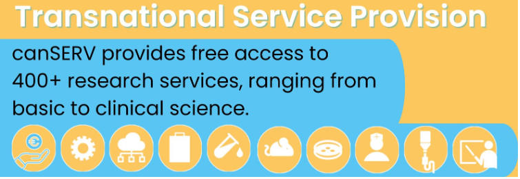
We are happy to announce the 2nd Open Call from the Horizon Europe-funded CanSERV project! Cancer Researchers are invited to apply for FREE state-of-the-art services and training at several European Research Infrastructures, including Euro-BioImaging ERIC. Within this project, 28 Euro-Bioimaging Nodes (- Yes, France-BioImaging is part of it too! -) offer access to their expertise. It’s an amazing opportunity for the cancer research community to access a wide-ranging portfolio of services.
All user projects – ranging from basic discovery science to translational science and into personalised oncology on any type of cancer – are eligible. The total indicative funding volume of this call is 1 Million Euro across the entire canSERV consortium.
The full call text, including user guidelines and other useful information, and the canSERV catalog of services are linked here and can be accessed via canserv.eu and via eurobioimaging.eu/content/canserv.
Submission deadline is May 21st, 2024, 14:00 CEST.
Qu’est-ce que la science ouverte? Un processus FAIR? Un PGD? Le RGPD? Un entrepôt de données?
Où? Comment? stocker, gérer mes données, mes programmes, mes publications?
Quels sont les usages? Les règles? Les outils?
Rendez-vous à la Journée Cycle de Vie des Données en Biologie le 4 Avril 2024 pour tenter de répondre à ces questions.
avec le soutien d’UniCA, du Labex SignaLife, de 4DOMICs, France-BioImaging, EMBRC-FR et du RTmfm
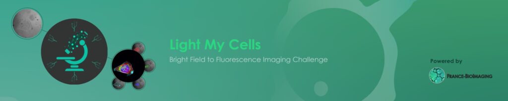
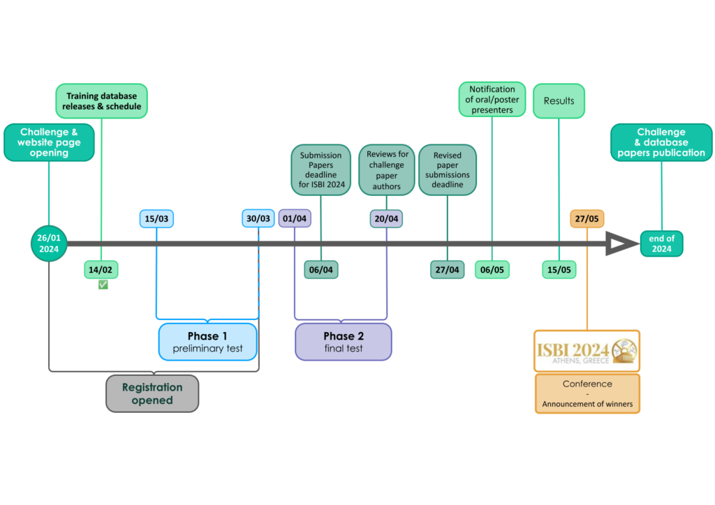
Phase 1 submissions for the Light My Cells challenge are now open to participants! This is a preliminary test phase to prepare for the final phase and familiarize participants with the algorithm submission procedure, with the possibility of making five submissions up to March 30.
Would you like to take part? Registration is open until March 30!
What is the challenge?
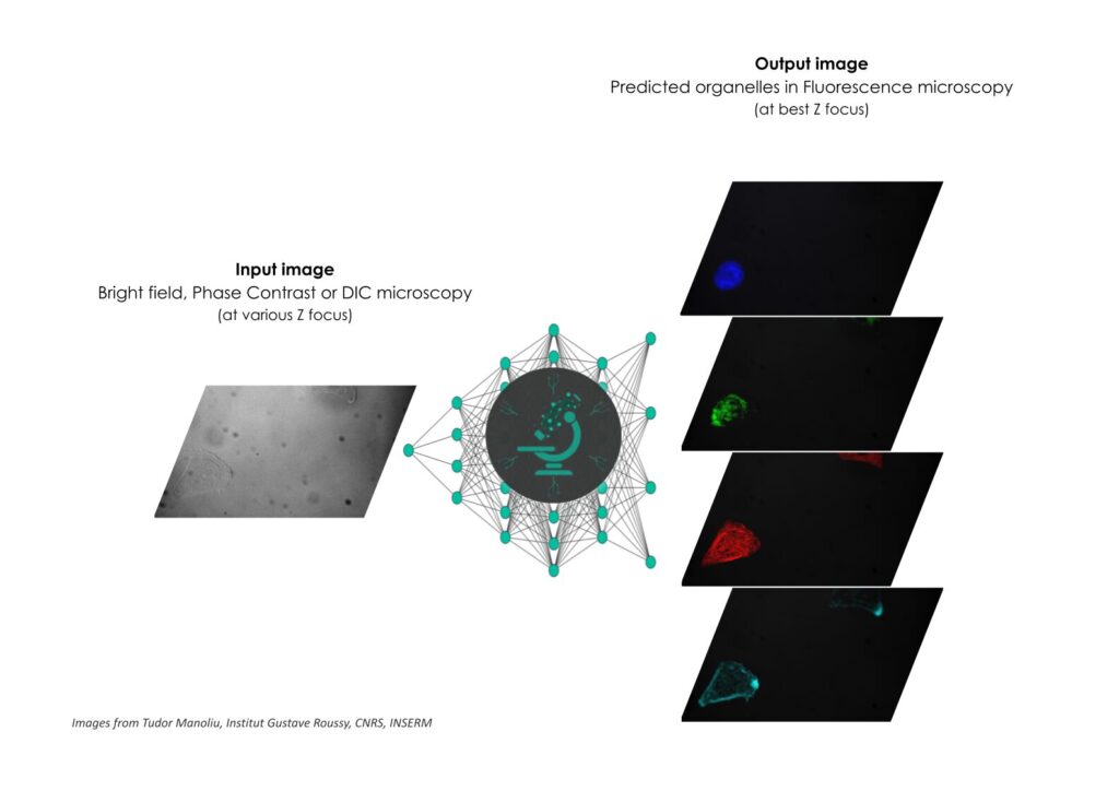
The Light My Cells France-Bioimaging challenge aims to contribute to the development of new image-to-image ‘deep-label’ methods in the fields of biology and microscopy. Basically, the goal is to predict the best-focused output-images of several fluorescently labelled organelles from label-free transmitted light input-images. And we need you for that!
We have defined the challenge as a single task with two phases:
A preliminary test phase (on 30 images) to familiarize with the algorithm submission procedure, with the possibility to have five submissions (with a maximum of one by week)
The final test phase (on 300 images) with only one submission accessible will not give the possibility to evaluate their algorithms before submitting.
So, you have until the end of the first phase, on March 21, 2024, to register and participate at this Light My Cells challenge.
A challenge paper will be written with the organizing team’s members for submission to journals
Invitation to publish their methods in the proceedings of the IEEE International Symposium on Biomedical Imaging 2024s
Support and integration of open source code into open science image processing and analysis software (e.g. BioImage Model Zoo, Napari)
For the 1st:
Invitation to 2024 France-Bioimaging annual meeting
Graphic card
Android tablet
For the 2nd:
Graphic card
Android tablet
For the 3rd:
Android tablet
Why launching a challenge?
To develop powerful methods that will then end up in creating public databases, standards & benchmarks in the field of bioimaging! The FBI challenge is hinged on a double contribution: from core facilities engineers and from data scientists. The first group acquired a large number of images to build a dataset, that will later be used by the algorithms. These images were produced by microscopy engineers & technicians from FBI’s platforms. As for the second contribution, this is where the challenge starts! The challenge is then published to have a maximum of data scientists to work on the algorithms that best fulfill the analysis task.
The first project is also based on four pillars:
Open source + FAIR (Findable, Accessible, Interoperable, Reusable)
Supervised learning, it involves annotated datasets to maintain control over performances.
In silico annotations, a computer labeling method to avoid manual annotation and its drawbacks.
Image-to-image analysis tasks, an image analysis tasks which aim to predict an output image from the input one.
Coorganised by NeurATRIS, France Life Imaging (FLI) and France BioImaging (FBI), the 5th edition of the Transnational Neuroscience Days will be held on the 24th and 25th of May, 2024, in Pornichet, Domaine Ker Juliette.
For this new edition, the event will focus on multiscale imaging applied to transnational research in neuroscience. These days are hinged on interactive moments between public and private science stakeholders in neuroscience, especially for young researchers.
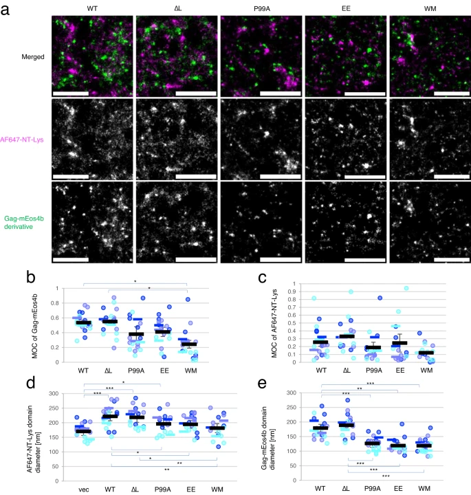
HIV type 1 virus has a lipid envelope enriched with host cell sphingomyelin and cholesterol. In order to understand the mechanism of this enrichment, the FBI Alsace node (Laboratoire de Bioimagerie et Pathologies from Université de Strasbourg and the Imaging Center PIQ-QuESt) has participated in a study recently published in Nature Communications about HIV-1 virus assembly. Indeed, they have investigated the interplay between the HIV-1 Gag protein and the host cell lipids at the plasma membrane. This work has greatly benefited from the use of a great combination of different quantitative (FLIM-FRET and FRAP) and super-resolution (PALM/STORM) custom-made microscopes with specific probes.
Using FRAP to characterize mobile and immobile molecules
The Fluorescence recovery after photobleaching (FRAP) quantifies the two-dimensional lateral diffusion of fluorescently labeled molecules of interest. This technique is very useful in biological studies of cell membrane diffusion and protein binding as it not only reports on the diffusion rates of mobile fractions of molecules but also provides information about the proportion of immobile molecules.
In our case, FRAP experiments indicated that the expression of Gag significantly decreased the mobile fraction of sphingomyelin (SM)-rich domains. Besides, the technique showed that cholesterol (Chol)-rich domains were intrinsically immobile, even in the absence of Gag. It is speculated that the association of Gag with SM-rich domains restricts the lateral diffusion of the lipid domains, resulting in an increase of the immobile fraction in FRAP measurements.
Using PALM/dSTORM to localize molecules at high resolution
The Photo-activated localization microscopy (PALM) is a widefield fluorescence microscopy imaging method that provides images with a resolution beyond the diffraction limit. By collecting a large number of images each containing just a few active isolated fluorophores, the collection of these images allows to stochastically activate each fluorophore and thus to obtain a global image of the sample with high resolution.
The Stochastic optical reconstruction microscopy (STORM) works on the activated state of a photo-switchable molecule that leads to the consecutive emission of sufficient photons to enable precise localization before it enters a dark state or becomes deactivated by photobleaching.
Coupling these two techniques, scientists next investigated at high resolution the localization of Gag and SM-rich or Chol-rich domains, both labeled with specific fluorescently labeled lipid binding proteins.
PALM/dSTORM visualized domains of different sizes labeled with the two lipid binding proteins, showing that the expression of Gag induced the formation of larger SM-rich domains but not the formation of larger Chol-rich domains. The main hypothesis is that the formation of large lipid domains may be due to the coalescence of smaller lipid domains.
Using FLIM-FRET to identify molecule proximity and interaction
And last but not least, the Fluorescence-lifetime imaging microscopy (FLIM) is an imaging technique for producing an image based on differences in the fluorescence-lifetime rather than its intensity. By quantifying variations in the exponential decay rate of the fluorescence from a fluorescent sample (fluorescence-lifetime) it is possible to report on molecule proximity. Since the fluorescence-lifetime is insensitive to changes in fluorophore intensity or concentration, it is the most quantitatively precise technique to report on fluorescence resonance energy transfer (FRET).
FRET is a mechanism describing energy transfer between two light-sensitive molecules (chromophores). A donor chromophore, initially in its electronic excited state, may transfer energy to an acceptor chromophore through non-radiative dipole-dipole coupling. FRET is extremely sensitive to small changes in distance and therefore an excellent reporter on molecule proximity and interaction.
In this third and final part, to better understand the possible effect of Gag on the lipid distribution in the plasma membrane, scientists investigated by two-photon FLIM-FRET the interaction of Chol-rich lipid domains with SM-rich lipid domains and its dependence on Gag multimerization. These last results showed that Gag multimerization induces SM-rich and Chol-rich domains to be in close proximity and that membrane curvature affects the apposition of SM-rich and Chol-rich domains.
A great example of application on how a combination of high-end technologies in microscopy can help you understand multiple aspects of a biological mechanism. So, what are you waiting for? Dive into the true potential of microscopy!
You need FRAP, two photon FLIM-FRET, PALM/dSTORM at France-BioImaging? To get open access, please login via Euro-BioImaging website! You just have to choose the technology you want to use, then submit your proposal. All applications will be processed by the Euro-BioImaging Hub in close relation with France-BioImaging. And of course, all scientists regardless of their affiliation, area of expertise or field of activity can benefit from open access services! Users whose projects will be validated by Euro-BioImaging will benefit from a waiver for the access cost on France-BioImaging core facilities (https://france-bioimaging.org/access/).
Tomishige, N., Bin Nasim, M., Murate, M. et al. HIV-1 Gag targeting to the plasma membrane reorganizes sphingomyelin-rich and cholesterol-rich lipid domains. Nat Commun14, 7353 (2023). https://doi.org/10.1038/s41467-023-42994-w
As the FBI Correlative Light-Electron Microscopy workshop is currently happening at the Bordeaux Imaging Center (FBI Bordeaux node), what a better occasion to highlight a correlative microscopy technique: The Array Tomography.
Correlative Light and Electron microscopy (CLEM) increases our capacity of biological investigation. By combining light microscopy and electron microscopy, this complementary approach takes advantages of both techniques. In fact, light imaging provides valuable functional information thanks to its labeling power, whereas Electron microscopy excels at high resolution.
Where array tomography (AT) is special is that this technique is based on serial ultramicrotomy (cutting many sections less than a micrometer in thickness) of the sample, section collection onto support, and serial scanning EM (SEM) imaging.
An array of microscopy modes
Array tomography is a versatile microscopy method that offers opportunities to explore cell and tissues in three dimensions. This technique is well suited to image large tissue volumes of your sample with fine structural and molecular details.
Different modes are available, each having its own specificity and benefits:
The fluorescence microscopy AT mode (FM-AT) delivers volumetric resolution and molecular marker multiplexing highly superior to traditional fluorescence microscopies.
The electron microscopy AT mode (EM-AT) captures three-dimensional ultrastructure at size scales that would require prohibitive effort using traditional serial-section EM methods.
And of course, you can combine both modes in a unique one FM/EM-AT with three-dimensional light and electron images acquired in perfect volumetric data.
Why you should consider this technique next time?
The use of FM-AT should be considered for volumetric fluorescence imaging of fixed tissue specimens whenever there is need for very high resolution, high-order molecular multiplexing and/or rigorously depth-independent quantification of fluorescence signal intensities. Use of EM-AT offers perhaps the most convenient approach to volumetric electron microscopy available. Moreover, even though fields of applications are numerous, these attributes establish AT as an ideal choice for the most demanding analyses of diverse cellular architectures within mature and developing tissues such as brain tissue (neuroscientists, this technique is for you!).
Finally, while originally developed for EM, physical cutting of ultrathin sections was found to improve the axial resolution and provide accessibility to the sample for molecular labeling, beneficial for both, EM and light microscopy. Need another argument? The same or adjacent sections can be imaged with different modalities in correlative or even conjugate microscopy!
Get access to one of our services!
You need Array Tomography or another imaging technology or expertise that France-BioImaging provides? To get open access, please login via Euro-BioImaging website! You just have to choose the technology you want to use, then submit your proposal. All applications will be processed by the Euro-BioImaging Hub in close relation with France-BioImaging. And of course, all scientists regardless of their affiliation, area of expertise or field of activity can benefit from open access services! Users whose projects will be validated by Euro-BioImaging will benefit from a waiver for the access cost on France-BioImaging core facilities (https://france-bioimaging.org/access/).
The LCI core facility at Karolinska Institutet has the pleasure to invite you to follow the microscopy course called Microscopy: improve your imaging skills – from sample preparation to image analysis.
The course starts next Monday (29 Jan) and runs until the 16th of February.
As usual, all the course lectures are broadcasted live on Zoom. It is free of charge and you do not need to register.
The aim for this course is to improve the microscopy skills of students and researchers who have already used a microscope to acquire digital images of fluorescent samples but feel that more knowledge could help them.
Here is a selection of what we will talk about:
Optics and image formation,
Anatomy of a microscope
Objectives and refraction index
Cameras and detectors, Noise and background, Bit depth and saturation
Sample preparation, Immunostaining, Clearing and expansion
Resolution and contrast, Nyquist sampling, Microscope settings
Data handling, OMERO.figure, Requirements for image analysis, Colocalization
As part of the scientific activities that it wishes to carry out and in continuation of its 1st seminar (Dec. 2022), the FBI “Preclinical Microscopy” Working Group is organizing its 1st webinar on February 1 from 2:00 pm to 4:00 pm. The objective of these scientific meetings is to present research projects, technologies and regulatory frameworks related to light microscopy from the organ/organoid to the living animal.
Program
2:00 pm – Welcoming & Introduction
2:15 pm – “Visualization of the ticking of circadian clock cells in freely moving mice” by Xavier Bonnefont (Institute of Functional Genomics, Montpellier).
3:00 pm – “Regulatory aspects of the use of animals for scientific purposes” by Isabelle Bardou (Cyceron, Biomedical Imaging Platform, Caen).
We use cookies on our website to give you the most relevant experience by remembering your preferences and repeat visits. By clicking “Accept All”, you consent to the use of ALL the cookies. However, you may visit "Cookie Settings" to provide a controlled consent.
This website uses cookies to improve your experience while you navigate through the website. Out of these, the cookies that are categorized as necessary are stored on your browser as they are essential for the working of basic functionalities of the website. We also use third-party cookies that help us analyze and understand how you use this website. These cookies will be stored in your browser only with your consent. You also have the option to opt-out of these cookies. But opting out of some of these cookies may affect your browsing experience.
Necessary cookies are absolutely essential for the website to function properly. These cookies ensure basic functionalities and security features of the website, anonymously.
Cookie
Duration
Description
cookielawinfo-checkbox-analytics
11 months
This cookie is set by GDPR Cookie Consent plugin. The cookie is used to store the user consent for the cookies in the category "Analytics".
cookielawinfo-checkbox-functional
11 months
The cookie is set by GDPR cookie consent to record the user consent for the cookies in the category "Functional".
cookielawinfo-checkbox-necessary
11 months
This cookie is set by GDPR Cookie Consent plugin. The cookies is used to store the user consent for the cookies in the category "Necessary".
cookielawinfo-checkbox-others
11 months
This cookie is set by GDPR Cookie Consent plugin. The cookie is used to store the user consent for the cookies in the category "Other.
cookielawinfo-checkbox-performance
11 months
This cookie is set by GDPR Cookie Consent plugin. The cookie is used to store the user consent for the cookies in the category "Performance".
viewed_cookie_policy
11 months
The cookie is set by the GDPR Cookie Consent plugin and is used to store whether or not user has consented to the use of cookies. It does not store any personal data.
Functional cookies help to perform certain functionalities like sharing the content of the website on social media platforms, collect feedbacks, and other third-party features.
Performance cookies are used to understand and analyze the key performance indexes of the website which helps in delivering a better user experience for the visitors.
Analytical cookies are used to understand how visitors interact with the website. These cookies help provide information on metrics the number of visitors, bounce rate, traffic source, etc.
Advertisement cookies are used to provide visitors with relevant ads and marketing campaigns. These cookies track visitors across websites and collect information to provide customized ads.