Funding for Access
France-BioImaging is the French Node of the European research infrastructure Euro-BioImaging.
As such, France-BioImaging invites all its external users* to register through Euro-BioImaging web portal to access any equipment available on France-BioImaging facilities and labs.
By registering through the EuBI web portal, France-BioImaging external users will:
- have access to 30 biological imaging facilities and laboratories specializing in R&D for imaging in 10 regional nodes
- [Temporarily suspended] receive a waiver for the costs of instrument access (1) (up to 750€ per week for a maximum of 3 weeks) on FBI facilities or labs (2).
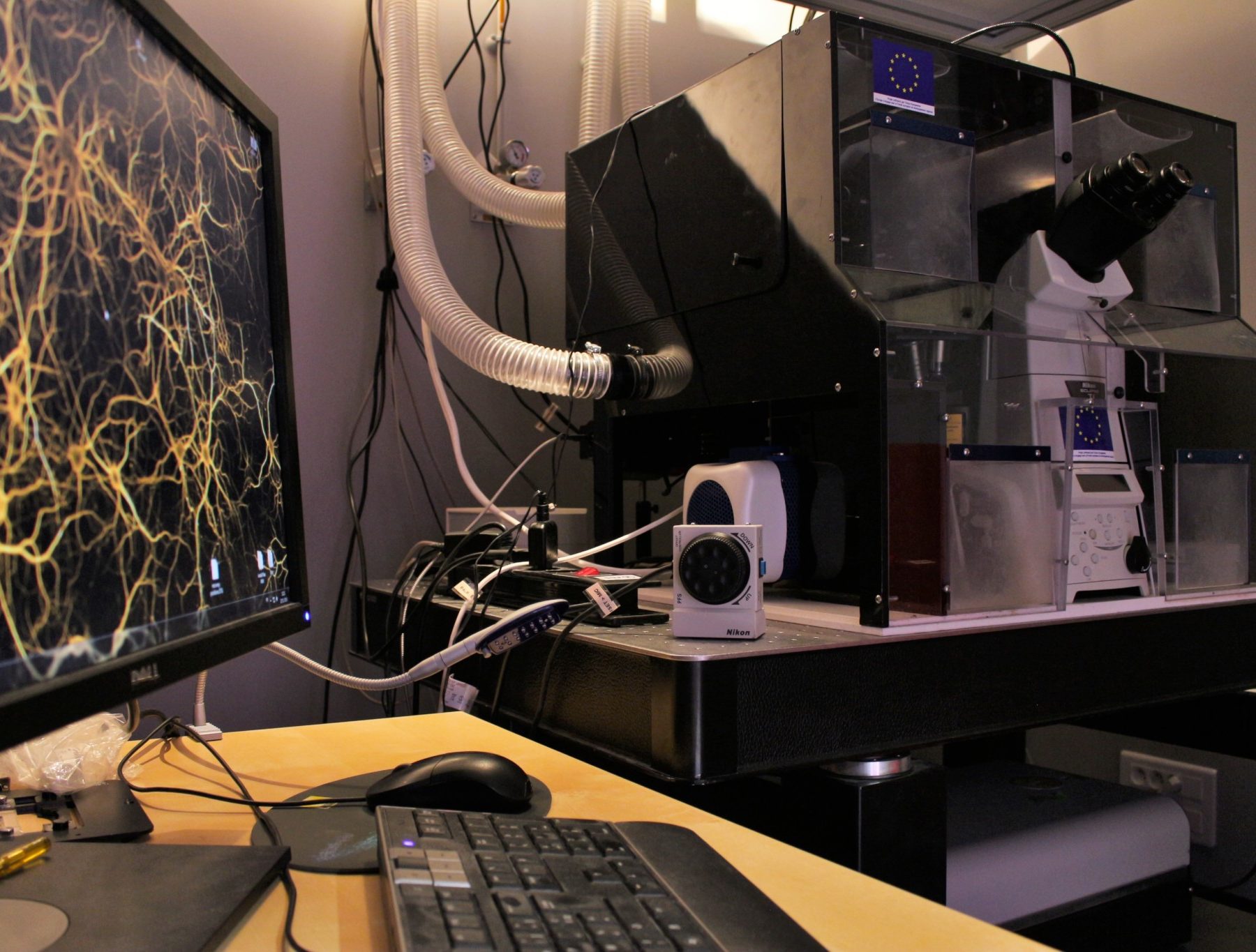
- have access to a wide range of technical and methodological expertise: Single Particle Tracking & Super Resolution / Multimodal & Quantitative fluorescence Microscopies / Multiscale & Correlative Microscopy / New Contrast & In-Depth Imaging / High Throughput & High Content Screening / Probe development, Optomanipulation & Optogenetics / BioImage Informatics, Image Processing & Data Management
- receive a strong support in computational analysis, provide quantitative measures and integrative understanding of a wide range of cell and tissue activities in biological models, from the simplest, to small animals in normal and pathological situations
Submit a proposal
To Request Access to France-BioImaging services, external users have to fill out a simple online form on the Euro-BioImaging Web portal to create a research proposal.
Timeframe: The application and review process is efficient, transparent and quick. The whole process is estimated to take 2-3 weeks.
When to apply: Applications for access can be submitted at any time. Periodically, special calls for access will be published with a defined deadline. These special calls carry specific criteria, so please read the calls carefully.
Proposal application steps:
- Go to the Euro BioImaging website: https://www.eurobioimaging.eu/service, click on “biological Imaging”, select the technology that will be used and the host node (French BioImaging Node). Then you will have to click on “select” (available service section).
- “Name_of_the_Technology @ France – French BioImaging Node” will appear and you will have to click on “make a proposal”. See the list of technologies available below.
- You will then be redirected to the Life Science Login page to create an account and register your project. You should find your home institution in the institution list, otherwise there are alternatives (Google, ORCID, etc.)
- Submit your proposal: Once you are logged in through Life Science Login, fill in and submit the simple proposal form (see https://www.eurobioimaging-access.eu/upload/Euro-BioImaging%20Access%20form%20questions%202024-06-05.pdf for an overview of the form). NB: It is important to highlight the imaging aspects of the project and their importance, as well as to mention that contact has already been established with the France BioImaging facility/lab and the name of your contact on site. Also, if the project has already undergone an external scientific evaluation (i.e. for a grant) it is important to mention it, since it will greatly speed up the acceptance by Euro BioImaging.
*This registration process is open to:
- any users from outside the institutional perimeter of France-BioImaging nodes (i.e. from outside the partner university of the Node) who would like to use imaging technologies in one of FBI nodes: Alsace, Toulouse, Paris Centre, Paris Ile-de-France-Sud, Marseille, Montpellier, Bordeaux, Bretagne-Loire, Normandie, Rhône-Alpes. They can be French or international users – EU and non-EU
- or users from one France-BioImaging regional Node who want to access an equipment available in another FBI regional node.
Questions? Contact us: France BioImaging coordination team
Offered technologies @France – French BioImaging Node
Fluorescence Microscopy
- Two-photon microscopy (2P)
- Deconvolution widefield microscopy (DWM)
- Image Scanning microscopy (ISM)
- Lattice light-sheet (LLS)
- Laser scanning confocal microscopy (LSCM/CLSM)
- Objective-coupled planar illumination (OCPI)
- Random Illumination Microscopy (RIM)*
- Spinning disk confocal microscopy (SDCM)
- Structured illumination microscopy* (SIM)
- Total internal reflection fluorescence microscopy (TIRF)
Functional Imaging and specialised methodologies
- Anisotropy/Polarization Microscopy (AM)
- Imaging at Biosafety Level >1 (BSL>1)
- Expansion Microscopy (ExM)*
- Fluorescence (cross)-correlation spectroscopy (FCS/FCCS)
- Feedback microscopy (FDBKM)*
- Fluorescence Lifetime Imaging (FLIM)
- Fluorescence Recovery after Photobleaching (FRAP)
- Fluorescence Resonance Energy Transfer (FRET)
- High-speed Imaging (HSI)*
- High throughput microscopy/high content screening (HTM/HCS)
- Imaging Flow Cytometry (IFC)*
- Intravital Microscopy (IVM)
- Microdissection (µDis)*
- Multiplexing imaging (MXI)*
- Photomanipulation (Pmanip)
- Single molecule FRET (smFRET)*
- Tissue Clearing (TC)*
- Voltage/pH/Ion Imaging *
Fluorescence Nanoscopy
- Reversible optical fluorescence transitions (RESOLFT)
- Single Molecule localization microscopy (SMLM)
- Light-sheet mesoscopic imaging (SPIM/dSLSM)
- Stimulated emission depletion microscopy (STED)
Label-free Imaging
- Polarization microscopy (PM)
- Quantitative Phase Imaging (QPI)*
- Raman Spectroscopy (RS)
- Second/Third Harmonic Generation (SHG/THG)
Mesoscopic Imaging
- Optical projection tomography (OPT)
- Macro Serial Blockface Fluorescence imaging* (S-BFI)
Scanning Electron Microscopy (SEM)
- Scanning Electron Microscopy (SEM)
Ultrastructural analysis in 2D
- Large scale EM (lsTEM)
- TEM of chemical fixed samples (TEM)
Ultrastructural analysis in 3D (volume EM)
- Array tomography (AT)
- EM tomography (ET)
- Focused Ion beam SEM (FIB-SEM)
- Serial Blockface SEM (SBF-SEM)
- serial section TEM (ssTEM)
- STEM tomography (STEM)
Cryo-EM
- Cryo Electron Tomography (Cryo-ET)*
- Cryo Transmission Electron Microscopy* (Cryo-TEM)
Correlative Light Microscopy and Electron Microscopy
- 3D Correlative Light and Electron Microscopy (3D-CLEM)*
- post-embedding CLEM (post-emb CLEM)
- pre-embedding CLEM (pre-emb CLEM)
Live-cell imaging and EM
- live-cell Correlative Light and Electron Microscopy (live-cell CLEM)
X-ray and EM
- Correlative X-ray and EM (CXEM)*
Ultrastructural localization of molecules
- Genetic encoded EM probes (GE probes)
- post-embed Correlative Light and Electron Microscopy (on-section CLEM)
- pre-embed Correlative Light and Electron Microscopy (pre-embed CLEM)
- pre-embedding immunolabelling (pre-embed IL)
- Immuno-gold EM on resin sections (resin-EM)
- Immuno-gold EM on thawed cryo-sections (Tokuyasu-EM)
Sample characterisation
- Atomic Force Microscopy (AFM)*
- Traction Force Microscopy (TFM)*
Biological Image Data Services
- Image Analysis -bio (IA bio)*
Small animal and plant Imaging
- in vivo optical imaging (OI)
- micro-CT
* Technologies under Proof of concept studies in Euro-BioImaging portfolio.
Questions about the available technologies and technical feasibility should be addressed directly to the heads of the France BioImaging facilities.
General conditions
Waiver of costs
(1)The following costs are eligible for financial support:
- Contribution to the verifiable incurred costs for measurement – data acquisition
- Imaging related sample preparation (special chemicals, labels, etc.)
- Data processing and analysis
France BioImaging financial support will be transferred directly to the France-BioImaging core facility/lab in which the project will be performed. The support does not include travel costs, accommodation and daily allowances. This has to be covered by the user.
(2)Restrictions: This waiver does not apply for users from Industry.
Acknowledgements
Publications resulting from work undertaken at France-Bioimaging facilities/labs have to contain the following acknowledgement:
“The National Infrastructure France-BioImaging (https://ror.org/01y7vt929) supported by the French National Research Agency (ANR-24-INBS-0005 FBI BIOGEN)”
Depending on the context, we suggest three model sentences:
- For the publications of scientists using one of the France-BioImaging Imaging Facilities: “We acknowledge the X (name of imaging facility), member of the national infrastructure France-BioImaging (https://ror.org/01y7vt929) supported by the French National Research Agency (ANR-24-INBS-0005 FBI BIOGEN)”
- For the publications by FBI members (technological development or scientific projects and collaborations) the sentence must appear in tributes as follows: “We acknowledge France-BioImaging infrastructure (https://ror.org/01y7vt929) supported by the French National Research Agency (ANR-24-INBS-0005 FBI BIOGEN)
- For the FBI external users (EuBI users) whose access to FBI facilities/lab was subsidized by France-BioImaging: “The study was supported by the France-BioImaging (https://ror.org/01y7vt929) User Access Fund”