Service
The Bordeaux node is centered on the Bordeaux Imaging Center core facility that provides service training and R&D in photonic and electronic imaging, surrounded by 9 R&D teams. The strength of the Bordeaux node lie in the strong integration of physics at the LP2N (IOGS), imaging, chemistry and neurobiology at IINS and plant science at LBM. Bordeaux is a flagship center for super resolution imaging, both using photonics and electronic microscopies, applied to Neuroscience and Plant biology.
The BIC (Bordeaux Imaging Center) offers resources in photonic and electronic imaging, mainly in life, health and plant sciences. It is a core facility identified at the national level as IBISA that gathers 12 highly skilled engineers. It has the ISO9001 label. The different components of the BIC are: PHOTONIC imaging, ELECTRONIC imaging, PLANT imaging. The Bordeaux Imaging Center offers access to the most advanced bio-imaging techniques for fixed and live cell imaging such as video-microscopy, confocal microscopy, multiphoton microscopy, transmission electron microscopy and scanning electron microscopy. The BIC provides a unique set of high-end equipment for super- resolution microscopy such as STED confocal microscopy, FRAP video-microscopy, lifetime imaging FLIM for the measurement of molecular interactions. We also provide access to equipment for sample preparation such as ultra-microtoms, high pressure freeze (HPF) and we can host live samples.
The “Nanophotonics” group research activities aim at understanding, mastering and using light-matter interactions at the nanometer scale. For this purpose, the group develops ultra-sensitive optical nanoscopy techniques for the detection of individual nanoscale objects, perform the spectroscopy of their electronic excitations, and explore their applications in physics and biophotonics.
The general ambition of the team is at the crossroad of nanoscience, imaging and biology through the design and study of innovative optically active synthetic nanostructures as well as the investigation of complex and dynamic biological systems at the nanoscale. One of our focus is to work with living biological tissues.
The majority of our samples of interest are engineered, and more specifically, generated using the Cellular Capsule Technology (CCT), which was invented and patented in the lab and led to the creation of a startup company (TreeFrog Therapeutics). The CCT relies on the use of 3D-printed microfabricated or microfluidic and allows to produce organoids encapsulated in hollow alginate spherical or tubular micro compartments. These multicellular aggregates are highly relevant to model disease and in regenerative medicine, but they are challenging to image in live conditions, without labelling or in a high throughput format. We thus develop imaging and environmental set-ups dedicated to their observation, in conditions that are dictated by the biological or medical question.
The team develops several research topics, combining neuroscience, imaging, chemistry and protein
engineering in order to unravel the dynamics and nanoscale organization of multimolecular receptor
complexes and their functional role in glutamatergic synaptic transmission. We develop 3 main
research axes to understand the interplay between AMPA type glutamate receptor nanoscale
dynamics, synaptic plasticity and memory formation in the healthy and diseased brain: 1) dynamics
and physical-chemistry of the macro-molecular complexes of the synapse, 2) nano-scale organization
and dynamics of synaptic proteins and membrane trafficking, 3) impact of the dynamic of synapse
organization on synaptic physiology.
We specifically investigated the trafficking and function of the NMDA receptor (NMDAR), as well as dopamine receptors, since their physiological activity is central for the maturation of networks, synaptic plasticity, learning and memory, and is currently one of the most studied targets for neuropsychiatric disorders. Indeed, a deficit in NMDAR signaling in the brain is currently proposed as the core molecular dysfunction of brain circuits in psychotic disorders. The team contribution to the field has been to demonstrate that i) these receptors are highly dynamic at the surface of neurons, ii) the dynamics is regulated by intracellular, transmembrane, and extracellular interactors, iii) their dynamics is essential for activity- and hormone-dependent synaptic plasticity and associative memory, and iv) this surface dynamics is strongly dysregulated in psychotic disorders (e.g. autoantibodies). Our discoveries were possible thanks to interdisciplinary efforts, in which for instance we developed single nanoparticle tracking in live brain tissue to define receptor behavior and the extracellular space ex vivo.
The team develops and applies advanced super-resolution microscopy approaches to study the
molecular and cellular mechanisms of cell-to-cell communication and its dynamics in the mammalian
brain, using the mouse as a model system. Notably, we invented super-resolution shadow imaging
(SUSHI) to visualize the extracellular space of the brain in the living state.
It is an interdisciplinary team composed of physicists, computer scientists and bioengineers having a strong interest in biology. The team is developing cutting edge quantitative imaging and analysis techniques to decipher protein organization and dynamics with high spatial and temporal resolution and high content screening context. Recent developments include analysis software for single molecule-based super-resolution microscopy (SMLM), innovative single objective light-sheet microscopy, SMLM-based high content screening platform and correlative STED-SMLM super-resolution platform. The Quantitative Imaging of the Cell has an important academic and industrial transfer technology activity.
Our goal is to decipher at the molecular level the spatiotemporal and mechanical mechanisms controlling the architecture and dynamics of motile structures. Exploration of these new dimensions requires an innovative and multidisciplinary approach combining cell biology, biophysics, biomechanics, super-resolution microscopy, single protein tracking and quantitative image analysis.
This team is a joint research lab between IINS and the company Alvéole (www.alveolelab.com). By joining their research efforts in a joint research laboratory (JRL), Alvéole and IINS expect to become key players in the development of human in vitro models for fundamental research, disease modeling and drug testing. The team develops instruments and methods to prototype and image advanced in vitro cellular models.
The team studies the nanostructure of exocytosis and endocytosis sites at synapses, the cellular and molecular mechanisms of endocytosis. We extend the topic to diverse synapse types (glutamate, dopamine…) and how these processes are affected in early stages of neurodegenerative diseases (synucleinopathies). We develop new probes (pH sensors) to detect exo- and endocytosis events at the highest resolution possible. We also develop fluorescence-based strategies to purify synaptosomes for cellular and molecular omics analyses.
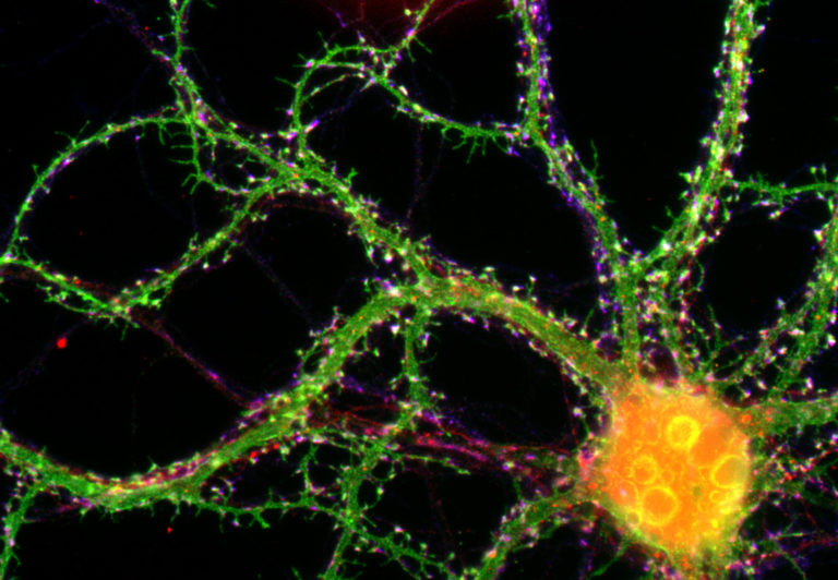
The team investigates then mechanisms of synapse assembly in neuronal culture systems with a focus on the role of adhesion molecules, using a combination of experimental approaches ranging from single molecule imaging to optogenetics and electrophysiology. Recent developments include the design of small fluorescent probes for super- resolution imaging of synaptic adhesion molecules (Chamma et al., Nat Comms 2016), the release of a simulator of single molecule dynamics for fluorescence live-cell imaging (Lagardère et al., Sci Reports, in press), and the optogenetic stimulation of phosphotyrosine pathways for the control of synapse differentiation (Letellier et al., eLife 2020).
The team studies the function of organelle tethering in plant cell to cell communication via plasmodesmata pores, and develops tools and methods to 1) image these structures at a nanometer resolution 2) measure cell-cell trafficking in a non-invasive way in planta. Plant samples present cytological particularities (auto-fluorescent components, presence of cell wall, large cells with an important vacuole compartment, air flats) that require developments for high-resolution microscopy approaches. Recent developments include the adaptation transmission electron tomography to reveal plasmodesmata ultrastructure at unprecedented resolution.
The ‘autophagy’ team was recently created within the Laboratoire de Biogenèse Membranaire. The team studies the function of lipids in the formation of the autophagy vesicles in plant cells, the autophagosomes. The team combines biochemistry and imagery to decipher the nature, dynamics and lateral heterogeneity of lipids within autophagosome membranes.
|
|
|
|
|