Advancing multimodal microscopy of thick samples for preclinical studies

The new France-BioImaging preclinical microscopy Working Group, whose aim is to advance multimodal microscopy of thick samples for preclinical studies, is organizing its first meeting! The event will take place on Monday December 12th 2022 in Nantes.
The event will present three aspects of preclinical microscopy:

  1. Imaging technologies (correlative microscopy, thick tissue or organoid microscopy, light sheet microscopy, intravital imaging and label free imaging)
  2. Image analysis (whole slide, correlative and 3D analysis)
  3. Regulatory frameworks (animal testing ethics and best practices in preclinical development)

Preliminary program

Registration Form

Deadline to register 04/12/2022 at 23:59

This form is currently closed for submissions.

The meeting will cover three aspects of preclinical microscopy with presentations of 30mn and a final round table:

  1. Imaging technologies
    • Correlative microscopy
    • Thick tissue imaging
    • 3D whole organ macroscopy
    • Intravital imaging (imaging chambers, exposed organs in living animal..)
    • Label free imaging
  2. Image analysis
    • Whole slide imaging analysis
    • Correlative image analysis
    • 3D reconstruction (distortion, alignment problems…)
  3. Regulatory frameworks
    • Animal testing ethics (3R, Ethics Committee…)
    • Preclinical developments (GMP, 21 CFR Part 11 Compliance…)
  4. Closing
    • Round table
    • Synthesis

Poster

France-BioImaging PreClinical Working Group

The PreClinical topic is usually connected to animal studies, but one needs to take into account the following aspects :

  1. Preclinical development, also termed preclinical studies or nonclinical studies, is a stage of research that begins before clinical trials (testing in humans) and during which important feasibility, iterative testing and drug safety data are collected, typically in laboratory animals.
  2. Preclinical imaging usually refers to whole animal imaging techniques allowing longitudinal studies such as high-frequency ultrasound, magnetic resonance imaging (MRI) and computed tomography (CT) which are usually used for anatomical imaging, while optical imaging (fluorescence and bioluminescence), positron emission tomography (PET), and single photon emission computed tomography (SPECT) which are usually used for molecular visualizations. This macro imaging scale is proposed within the infrastructure France Life Imaging.
  3. Multimodal imaging including the microscopy scale is largely developing. Technologies developed within FBI could benefit to the preclinical community
  4. The communities of cell, tissue and intravital microscopies need to know each other better.

Our Goal

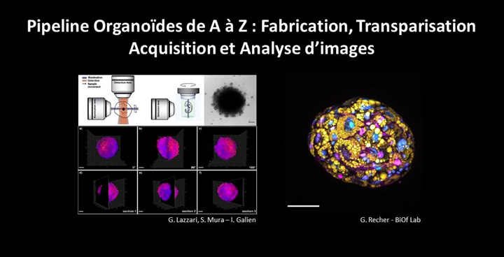
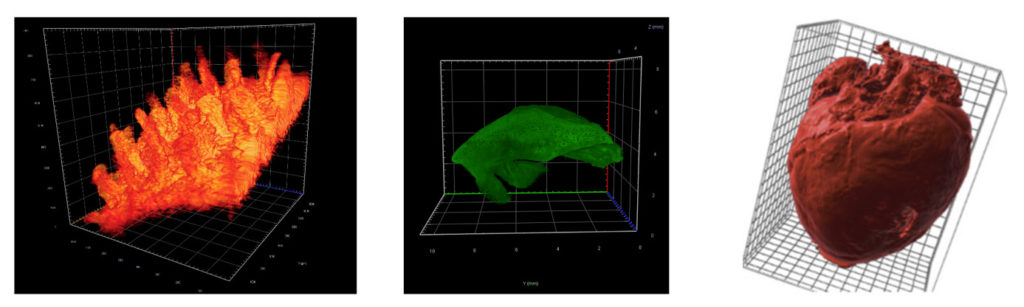
The new France-BioImaging Working Group wants to focus on the microscopic aspect of the “preclinical imaging”. It aims to develop microscopy technologies for preclinical studies used to explore highly resolved information at the organ level. It includes, for example, whole slide quantitative tissue histology, correlative microscopy, 3D from sections as well as Light sheet fluorescence microscopy for 3D whole organ imaging, intravital organ imaging or multi photon microscopy. The goal is to bridge the gaps between communities dealing with either fixed sections or living organs.

Call for speakers

The Working Group would like to call on speakers interested in presenting one of the several aspects that will be highlighted during the meeting and contributing to the Working Group (open to everyone). If you are interested, please get in touch:

This form is currently closed for submissions.

Save the date! The Electron Microscopy facility of Imagerie-Gif (I2BC, France-BioImaging), the Cryo-Electron Microscopy facility (I2BC, FRISBI) and the Cimex facility of Ecole Polytechnique are organizing a 5-day workshop from October 3rd to October 7th, 2022 on Transmission Electron Microscopy to explore the architecture of a virus in all its forms.

The aim of this workshop is to propagate knowledge about transmission electron microscopy applications and to outline the advantages of transversal studies combining structural biology and cell biology. Indeed, structural biology and cell biology approaches both use TEM but are rarely merged in the same studies.

The workshop will focus on the advantages of combining both approaches, which can be easily performed with the same equipment. The workshop will focus on a biological object whose study requires such multiscale approaches: a virus. The virus will be studied in vitro to resolve its high-resolution 3D structure, and will be observed in infected cells to determine the infection and replication mechanisms in situ.

The workshop targets students and young researchers. The training will focus on a given biological object, a virus, which will be studied by two complementary approaches:

  • Single particle analysis by cryo-electron microscopy, allowing high-resolution 3D reconstruction of particles purified in vitro. This part will be performed on a 200kV TEM on the Cimex facility.
  • Cellular tomography of infected cells with observation of the virus replication sites in situ and analysis of its interaction with cellular membranes. This workshop will cover the workflow from sample preparation and resin sections realisation, to acquisition and analysis of tomograms with a 120kV TEM.

Attendees will have a theoretical and practical overview of these two complementary techniques. The practical training will be particularly emphasised, to ensure that attendees will be able to apply the knowledge acquired in the workshop for their further research projects.


Susbscription here: https://www.azur-colloque.fr/DR04/inscription/preinscription/245

Preliminary programme

The first edition of “Digital Spaces for Research and Medicine” will take place at Institut Curie on July 7th, 2022. This event will be dedicated to providing scientists and clinicians insight into Virtual Reality, Augmented Reality and Mixed Reality based tools that offer novel and powerful ways for visualizing and analysing data in research and medicine. This day will include presentations of the tools followed by hands-on demonstrations. The day will close with an open forum for questions and discussing future perspective. The following tools will be presented:

  • MorphoNet-VR (Serpico-STED, Inria-UMR144 Institut Curie and LIRMM): Visualizing live microscopy data (3D+T) in VR
  • Genuage (Team LOCCO, UMR168, Institut Curie): visualization and analysis of multidimensional point cloud data, such as single molecule data, in virtual reality
  • DIVA (Institut Pasteur, Institut Curie): 3D reconstructions of raw experimental image stacks that are integrated in virtual reality for volumetric analysis
  • Tamed Cloud (ENSADLab): immersive VR experience to interact with cloud of images
  • HoloTracks (TEAM INRAE-Unité MaIAGE / AVIZ Inria, Paris Saclay): Immersive (AR) visualization of dynamic compounds in living cells
  • UnityMol A&VR (UPR 9080, IBPC) : deep inside molecules – digital twins at the nanoscale

Presentations of the tools will be at amphitheater Burg and online (click here to join)

Registration is mandatory for hands-on demonstrations : register here to book your slot!

Program

9 – 9.15 am Welcome coffee

9.15 – 11.30 am Presentations of the tools

11.30 – 1 pm Demonstrations part 1

2 – 5 pm Demonstrations part 2

5 – 6 pm Discussion “Future and perspectives”

6pm Cocktail

GRenoble OliGomérisation GROG will cover the latest innovations
in imaging techniques and analysis methods to quantify oligomerization and
clusterization in live cells.
The meeting will mostly deal about Single molecule localization microscopy and Fluorescence fluctuation methods and will be concluded with a round table.

Les inscriptions pour la seconde édition du TED ImaBio lundi 2 mai à Montpellier sont ouvertes.

Le GDR ImaBio vous propose de venir decouvrir les dernières innovations des partenaires industriels du GDR , et pour les etudiants un focus sur les métiers du domaine.

Programme

The 7th edition of OPTOGEN will bring developers and end-users in the field of Optogenetics and Neurophotonics together. It will be a great occasion to meet in person for stimulating discussions and to reignite collaborations.

Registration and abstract submission are open.

Early registration will close on April 15th.

Thanks to the support of the IHU Foresight and of the sponsors, inscription is free for students registered before April 15th and some travel grants will be available.

Euro-BioImaging is organizing a third online User Forum on Tuesday, April 5, 2022 from 14:00-17:00 CEST. The topic is “At the Forefront of Neuroscience.”

This event will highlight the importance of cutting-edge imaging technologies in support of brain research and showcase the specific expertise available at our Nodes across Europe through case studies presented in tandem with the research community. 

In addition, keynote presentations from Hervé Boutin, University of Manchester, and Selma Ding, Max Planck Institute for Animal Behavior, will further reveal the potential of biological and biomedical imaging technologies to boost brain research. 


Register here: https://us02web.zoom.us/meeting/register/tZ0ucuytpj8iE92A-JjvPbvpNnmTF4Z7iRaC

More information: https://www.eurobioimaging.eu/news/join-us-for-the-third-euro-bioimaging-user-forum-at-the-forefront-of-neuroscience/

Programme

Deadline: April 15th, 2022

The three national infrastructures ProFi, France-BioImaging and FRISBI along with the GIS IBiSA are pleased to announce a call for a funded access to IBiSA-labelled facilities.

Our aim is to promote IBiSA facilities networking through interdisciplinary research projects.

Applications should request access to at least two different IBiSA facilities from two disciplines (structural biology, Biological imaging and proteomics). The call is open to any academic laboratory.

The amount of the financial support will be up to 5000 € per application to cover facility costs.

Applications should be submitted to: Call-IBISA-FBI-FRISBI-PROFI@i2bc.paris-saclay.fr
using the template document https://mycore.core-cloud.net/index.php/s/VNLU2jgfcP4o0NN


The deadline for this inter-infrastructure access call is 15 April 2022.

All submitted proposals will be peer-reviewed by independent experts and the final funding will be approved by a committee comprising 2 representatives from each infrastructure as well as representatives
from GIS IBISA. We advise the project PI to contact the chosen facilities in order to set-up the optimal experimental design.

Call description

The France-BioImaging LSFM workgroup is pleased to announce an INSERM workshop on Light-Sheet Fluorescence Microscopy imaging technics. 

This workshop will be divided in two parts:

  • A first theoretical part in Bordeaux from the 16th to the 18th of May 2022 which will cover basic principles, applications and challenges of LSFM imaging through seminars,
  • A second practical part along June 2022 where you will have the possibility to choose the set-up on which you want to be trained amongst many systems available within the France-BioImaging community.

If you want to discover, learn and/or deepen your knowledge about this vast and powerful family of 3D imaging technics, this training is for you!


You will find all the information (program, registration details, venue and accommodations, …) about this event on the following poster and website (Atelier 268): https://evenium-site.com/site/atelier-de-l-inserm-268

REGISTRATION DEADLINE: March 4, 2022

Programme

Poster

Understanding how development is coordinated in multiple tissues and gives rise to fully functional organs or whole organisms necessitates microscopy tools. Over the last decade numerous advances have been made in live-imaging, enabling high resolution imaging of whole organisms at cellular resolution. Yet, these advances mainly rely on mounting the specimen in agarose or aqueous solutions, precluding imaging of organisms whose oxygen uptake depends on ventilation.

Engineers from the Institut Curie and Institut Jacques Monod implemented a multi-view multi-scale microscopy strategy based on confocal spinning disk microscopy, called Multi-View confocal microScopy (MuViScopy).

MuViScopy enables live-imaging of multiple organs with cellular resolution using sample rotation and confocal imaging without the need of sample embedding. They illustrated the capacity of MuViScopy by live-imaging Drosophila melanogaster pupal development throughout metamorphosis, highlighting how internal organs are formed and multiple organ development is coordinated. They foresee that MuViScopy will open the path to better understand developmental processes at the whole organism scale in living systems that require gas exchange by ventilation.

3D fusion reconstruction of eight different angles at 10x magnification. Animation of a 3D reconstruction after fusion of images acquired from eight angles by the MuViScope of an Ecad:3xGFP Drosophila pupa at 28 hAPF with a 10x objective. The animation starts with a 180° rotation along the A-P axis and then sequentially shows the eight different angles from a top view: 0° (red), 45° (orange), 90° (yellow), 135° (green), 180° (light blue), 225° (dark blue), 270° (purple) and 315° (pink). Fusion was performed using Huygens Fuser (SVI) and 3D visualization with Imaris software. The acquisition parameters are detailed in Table S1. Scale bar: 200 μm.

Olivier Leroy, Eric van Leen, Philippe Girard, Aurélien Villedieu, Christian Hubert, Floris Bosveld, Yohanns Bellaïche, Olivier Renaud; Multi-view confocal microscopy enables multiple organ and whole organism live-imagingDevelopment 15 February 2022; 149 (4): dev199760. doi: https://doi-org.insb.bib.cnrs.fr/10.1242/dev.199760

The MuViScope was co-funded by France-BioImaging.

Le Réseau d’Imagerie Cellulaire Paris-Saclay organise en avril prochain une visioconférence de 2 heures portant sur le thème de l’imagerie des organoïdes.

Titre : Pipeline Organoïdes de A à Z : Fabrication, Transparisation, Acquisition et Analyse d’images.

Thématique : Le RIC Paris-Saclay vous propose une sorte de pipeline sur l’imagerie des modèles organoïdes qui vous permettra de mieux appréhender l’ensemble du processus de leurs fabrications, leurs modes de transparisation, l’analyse des images 3D associées à ce type d’échantillon épais, et pour finir, l’importance de l’étude de ces modèles chez l’homme pour développer de nouveaux outils thérapeutiques.

Date : 14 avril 2022
Horaire : 14h à 16h

Programme : Auto-organisation cellulaire en systèmes 3D par imageries optiques — Tissue clearing : Adapted protocols for small samples — Analyses d’images et reconstruction 3D — Applications en Recherche translationnelle (Organoïdes hépatiques humains).

Inscription gratuite et obligatoire sur www.ric-paris-saclay.fr

Programme

Affiche