As the 2024 edition of the France-BioImaging Image Contest is still running, we wanted to highlight our previous winners and their projects. Here is a quick throwback to our 2023 winners.
Before getting to the heart of the matter, we want to remind you that you still have time (before November 8th) to submit your best images and try to win your registration fees for one 2025 microscopy-related event! Please make sure you upload your images on the following link:
Last year, we enjoyed the winning images submitted for their artistic take and their quality. Thanks to Laurent LE, Gonzalo QUIROGA-ARTIGAS and Hugues LELOUARD for their beautiful images!
- 1st Place: Laurent LE, Lévêque-Fort Team, Institut des Sciences Moléculaires d’Orsay
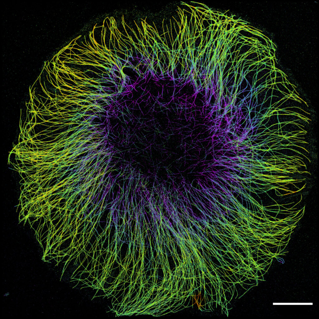
“In the blink of an eye”
COS7 fixed cell. Alpha-tubulin labeled with DNA-PAINT and imaged with Atto 647N. Axial information is obtained by virtual-SAF measurement known as DONALD.
SMLM Fluorescence Microscopy with DNA-PAINT with DONALD detection
Laurent LE is a Post-Doc researcher in the Sandrine Lévêque-Fort Team at Institut des Sciences Moléculaires d’Orsay. His goal is to develop new techniques for time-resolved microscopy and single molecule localization microscopy. During his PhD he especially learned to perform 3D SMLM with SAF light measurement named DONALD. This technique enables to have an isotropic three-dimensional localization precision of 20 nm within an axial range of around 150 nm above the coverslip.
During one of his trainings on the DONALD setup, he acquired this image with a nice round shape that looks like an eye. This image represents the alpha-tubulin of a fixed COS7 cell, labelled with DNA-PAINT and imaged with Atto 647N. The axial information is obtained by DONALD.
Thanks to the France-Bioimaging Image Contest, Laurent participated to the international conference FOM (Focus On Microscopy) in Genoa (Italy). This event is the most important one for his research community.
- 2nd Place: Gonzalo QUIROGA-ARTIGAS, Team Contrôle cytoplasmique de la stabilité du génome, Centre de recherche en Biologie Cellulaire de Montpellier
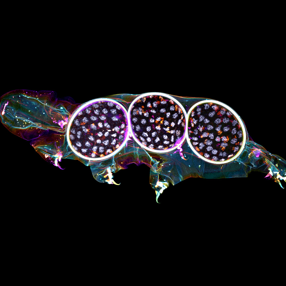
“Tardigrade embryos protected by mother’s molt”
Tardigrades commonly align the time of molting with egg laying. In this image we observe a tardigrade molt covering three developing embryos (DNA in white). The microscopy technology applied was confocal microscopy, and the research aimed to investigate the synchronization of embryo development in tardigrades.
Confocal microscopy
Gonzalo QUIROGA-ARTIGAS is a Post-Doc scientist in the Team “Cytoplasmic Control of Genome Instability” of the Centre de Recherche en Biologie cellulaire de Montpellier (CRBM-UMR5237). His research background is centered on Evolution and Developmental Biology (EvoDevo), a broad field that integrates knowledge from phylogenetics, cell and molecular biology, genetics and genetic engineering, as well as developmental biology and evolution. He is currently undertaking his second postdoctoral position, focusing on the study of the tardigrade Hypsibius exemplaris. Tardigrades are microscopic animals known for their extreme resistance to various stressors, and his research aims to uncover the mechanisms that enable them to endure such conditions. He is conducting this research in Dr. María Moriel-Carretero’s lab at the CRBM in Montpellier. His goal is to become a CNRS researcher in the years to come.
His image depicts the cuticle (or exoskeleton) of a tardigrade protecting three developing embryos. Tardigrades typically couple the moment of cuticle shedding with that of egg laying, a strategy designed to provide an extra layer of protection, ensuring the eggs survive and successfully develop into the next generation. The study aimed to assess the synchrony of embryonic development in eggs laid by the same mother within the same cuticle. This image was captured after staining the animal with a live dye (MemBrite Cell Surface Staining Kit), which highlights the cuticular structures, and a DNA stain (DAPI). A confocal Z-stack of each channel was processed using different depth LUTs in ImageJ, then merged to create the final image.
France-Bioimaging sponsored his participation to the Euro Evo Devo congress (EED) in Helsinki, Finland, in June 2024. This event brought together nearly 700 researchers from around the world, all sharing a passion for this research focus, to gather here for several days and share their ongoing projects. He attended the Arthropod satellite meeting, where he presented a current project using tardigrades as experimental models and received plenty of feedback. He also presented a poster at the main meeting and unlocked numerous potential collaborations, as many researchers showed interest in this animal model.
“Altogether, this meeting has been a fantastic opportunity for me. I was able to share my ongoing projects with the EvoDevo community, which has greatly stimulated my research progress. Additionally, the excellent atmosphere throughout the congress, the impeccable organization, and the collaborations I unlocked have made this experience wonderful both personally and professionally, opening new networking possibilities for my present and future endeavors” says Gustavo Quiroga-Artiguas.
- 3rd Place: Hugues LELOUARD, Gorvel team, Centre d’Immunologie de Marseille Luminy
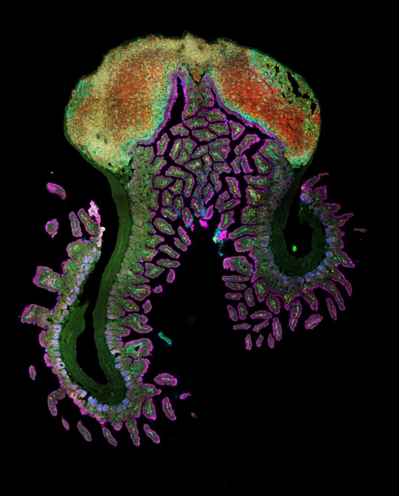
“Intestinal octopus“
Small intestine section from a LyzM-eGFP mouse containing one Peyer’s patch and stained for proliferative cells (Ki-67, yellow), Paneth cells (UEA-I, blue), epithelial cells (EpCAM, magenta), naive B cells (IgD, red), T cells (CD3, orange), helper T cells/macrophages (CD4, cyan), phagocytes (CD11c, turquoise), monocyte-derived phagocytes (GFP, green).
10-color spectral confocal microscopy
Confocal microscopy
Hugues LELOUARD is a CNRS research director working in the field of mucosal immunology at the Centre d’Immunologie de Marseille Luminy (CIML). His research focuses on understanding the role of Peyer’s patch phagocytes in initiating the intestinal immune response.
His image depicts a section of the small intestine from a LyzM-eGFP mouse, featuring a Peyer’s patch, captured using a Zeiss LSM 980 spectral confocal microscope with a mosaic setting of 20 tiles. Spectral confocal microscopy enables the simultaneous imaging of multiple parameters without requiring iterative acquisition steps. Here, staining highlights proliferative cells (Ki-67, yellow), Paneth cells/goblet cells/mucus (UEA-I, blue), epithelial cells (EpCAM, magenta), naïve B cells (IgD, red), T cells (CD3, orange), helper T cells/macrophages (CD4, cyan), phagocytes (CD11c, turquoise) and monocyte-derived phagocytes (GFP, green). This image illustrates the spatial distribution of phagocytes (green and turquoise) within the complex microenvironment of Peyer’s patches. Understanding their role in pathogen uptake, degradation and antigen presentation to effector immune cells is crucial for unravelling the mechanisms that initiate the intestinal adaptive immune response.
Want to be the next winner of our FBI Image Contest? Apply through the following link before November 8th, 2024: france-bioimaging.org/fbi-special-events/france-bioimaging-image-contest-2024