Learn more about our 2025 France-BioImaging Image Contest winners!
As the 2026 edition of the France-BioImaging Image Contest is still running, we wanted to highlight our previous winners and their projects. Here is a quick throwback to our 2025 winners.
Before getting to the heart of the matter, we want to remind you that you still have time (before November 22th) to submit your best images and try to win your registration fees for one 2027 microscopy-related event!

Last year, we enjoyed the winning images submitted for their artistic take and their quality. Thanks to Nicolas BAROIS, Vishwadeep MANE and Simli DEY for their beautiful images!
1st Place: Nicolas BAROIS

Gut Flower-Flora
Scanning Electron Microscopy
Nicolas Barois, Inserme research engineer, is in charge of the electron microscopy facility at the Pasteur Institute in Lille, France, since 2010. The facility belongs to the BioImaging Center of Lille (BICeL), which is part of the Lille Life Sciences and Health Research Facilities (PLBS, UAR 2014 – US 41). He is preparing samples for and observing them by transmission or scanning electron microscopy (TEM and SEM) and correlative light and electron microscopy (CLEM). Nicolas is working mostly on infection, immunity and diseases by virus (hepatic C virus, norovirus), bacteria (Yersinia pestis, Neisseria meningitidis), parasites (toxoplasma gondii, Schistosoma mansoni), as well as on cancers, neurodegenerative and cardiovascular diseases for examples.
Gut Flower-Flora represents a cross-sectional view of a thin slice of mouse gut and its associated flora. Cutting the gut into a very thin section resulted in an inversion of the usual topology, with the inner surface exposed outward. Typically, the gut is processed as small tubular segments that are longitudinally opened prior to SEM observation. When preparing the tubular segments, this particular piece, normally discarded, was kept and imaged out of scientific curiosity.
This was a project from a scientist working on intestinal flora in different diet conditions. The project was to observe presence or absence as well as the type of bacteria (cocci, rod-shaped or filamentous bacteria) for each diet condition. SEM was quite obvious to make these observations. He made first the serious work by observing all the small tubular segments from the different conditions, taking several images at different magnifications for each segment. Then he looked at this particular piece and he immediately saw he had a cool image. He spent the rest of the afternoon to obtain a high quality and resolution image. The scan took about 30 minutes. They see the overview of the intestine looking like a flower and by zooming in they see the filamentous bacteria anchored into the epithelium.
2nd Place: Vishwadeep MANE
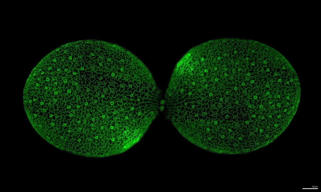
The Puzzled Awakening
Confocal Laser Scanning Microscopy
Vishwadeep Mane is a PhD student at the Indian Institute of Science (IISc), Bengaluru, in the lab of Prof. Utpal Nath, Department of Microbiology and Cell Biology. He also hold a visiting affiliation with the MechanoDevo group at the Plant Reproduction and Development Laboratory (RDP), ENS de Lyon, under Prof. Olivier Hamant.
His thesis examines how JAW-TCP transcription factors coordinate growth across tissue layers and organ surfaces to produce reproducible, flat leaf organs in Arabidopsis thaliana. I combine genetic approaches with mechanical and imaging tools to understand how cotyledons and leaves develop into robust, flat shapes despite genetic or mechanical perturbation.
The Puzzled Awakening shows the surface of a cotyledon shortly after germination, as the pair of embryonic leaves breaks free of the seed coat and unfolds into a flat, nearly circular lamina for photosynthesis. At the cellular scale, the image reveals the characteristic interlocking, puzzle-piece-shaped pavement cells of the epidermis, interspersed with stomata. These convoluted cell shapes are thought to help the tissue balance internal turgor pressure and relieve mechanical stress while growth is coordinated into a robust, harmonious organ shape which is directly the phenomenon his thesis investigates: how growth is coordinated at the cell and tissue-layer level to produce a reliably flat leaf. Between the two cotyledons, the first true leaf can be seen emerging, foreshadowing the next stage of the plant’s development.
Vishwadeep used Confocal Laser Scanning Microscopy. His team used the plants which have fluorescently labeled membrane that allows us to visualise boundaries of cells. This technique lets him resolve individual epidermal cell outlines and stomata in 3D with high spatial resolution on a living, intact seedling, non-destructively essential for capturing the puzzle-cell geometry and surface topology that this image. The image is taken in sectors and then stitched using a software called MorphoGraphX.
3rd Place: Simli DEY
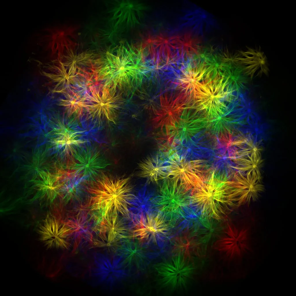
Kaleidoscope
Total Internal Reflection Fluorescent Microscopy
Simli Dey is a post-doctoral researcher, currently working at Physics of Cells and Cancer (UMR 168), Institut Curie (Paris) in Patricia Bassereau’s team. Her research uses in vitro reconstitution to investigate the mechanism by which actin generates force on a saddle-shaped curved membrane during clathrin-mediated endocytosis.
Actin assembly generates forces to facilitate membrane reshaping processes in cells, such as endocytosis, where vesicle formation involves the emergence of a vesicular bud connected to the plasma membrane via a saddle-shaped neck. However, mechanisms governing its spatial organization and force generation remain poorly understood. To understand this, they developed a reconstituted system that recapitulate saddle-shaped membrane geometry required for actin nucleation.
Kaleidoscope shows Atto 488-labelled branched actin, pseudocoloured for visualization, emanating from saddle-like membrane necks at in vitro-assembled Ω- shaped clathrin sites. Each membrane neck appears as a small central toroidal structure within the corresponding actin patch.
The imaging was performed using a Total Internal Reflection Fluorescence Microscope (TIRF). The process under investigation i.e. actin polymerization from curved membrane surfaces—is, by experimental design, confined to immediate vicinity of the coverslip. And TIRF microscopy is known to illuminate a thin region adjacent to the coverslip (100-200 nm), providing high-contrast, high-temporal-resolution imaging of events.
 Marine Beraud • October 15, 2026