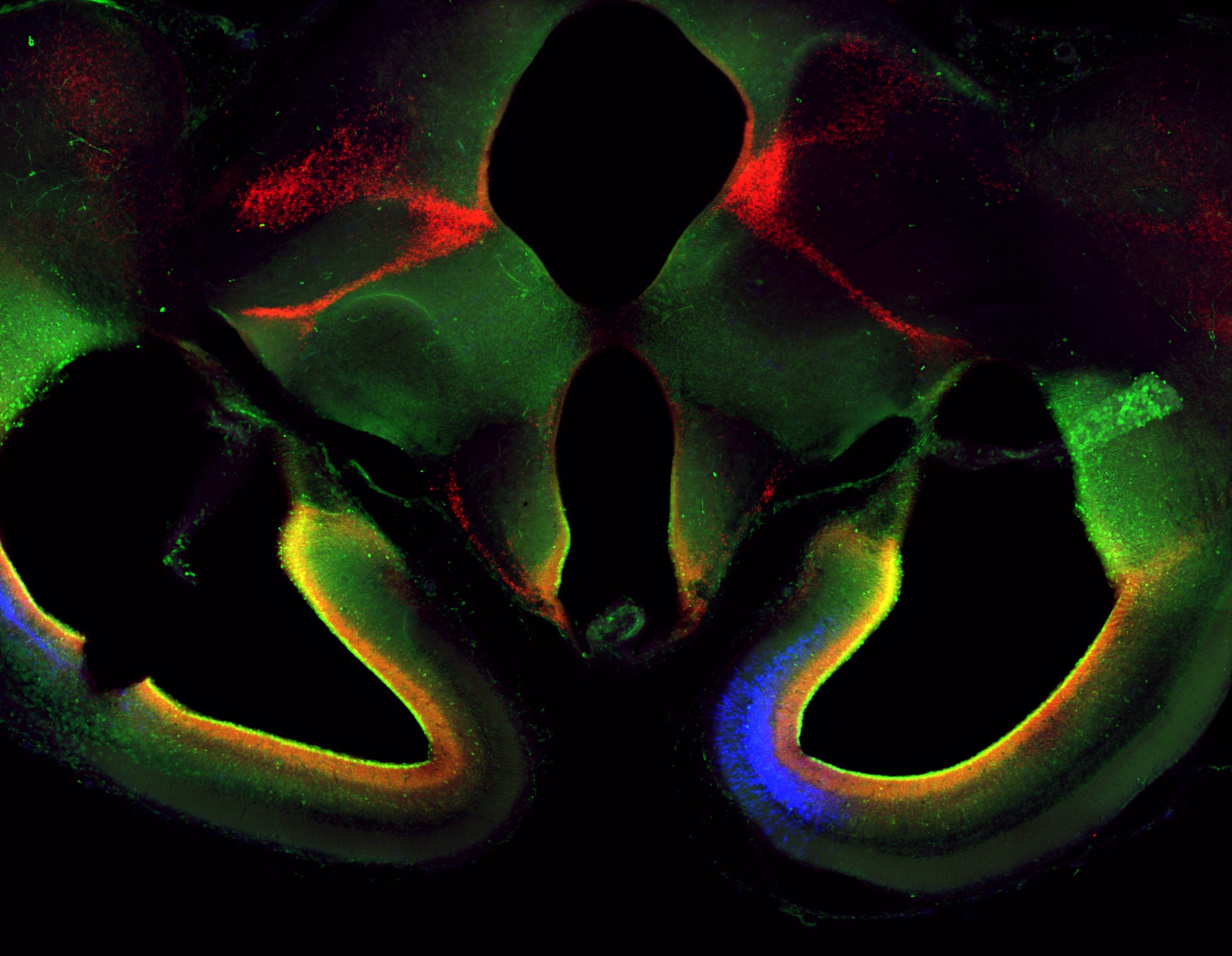
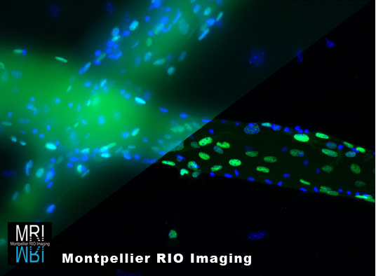
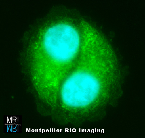
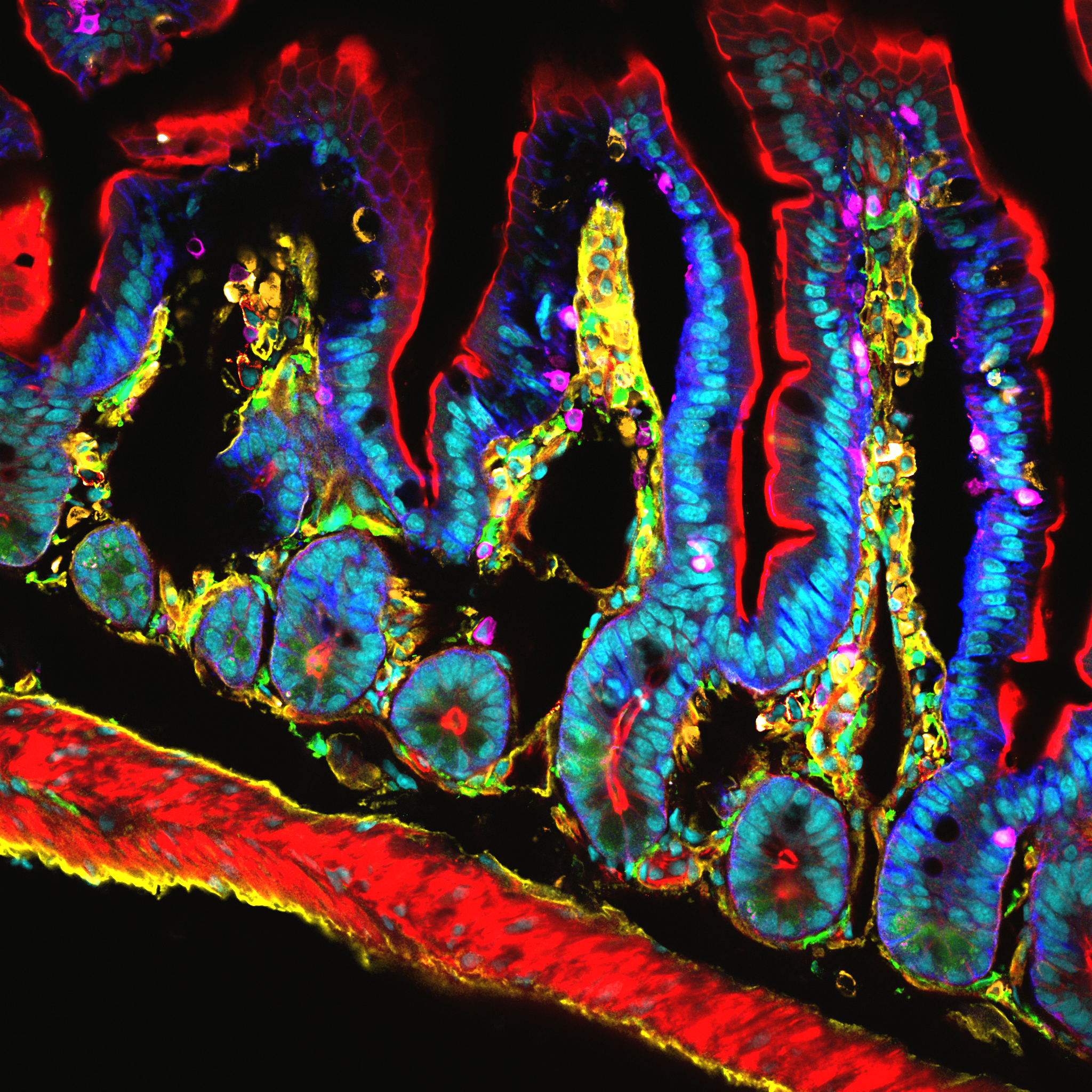
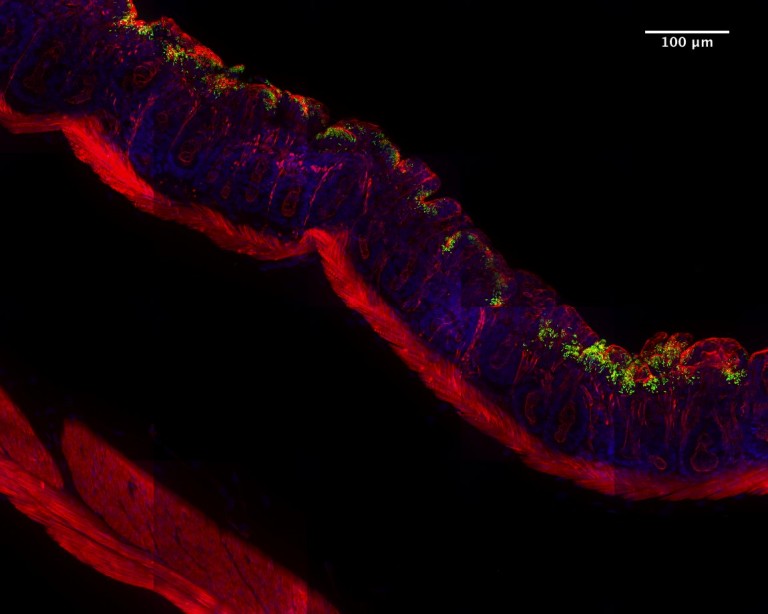
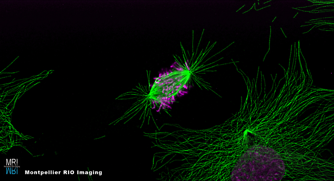
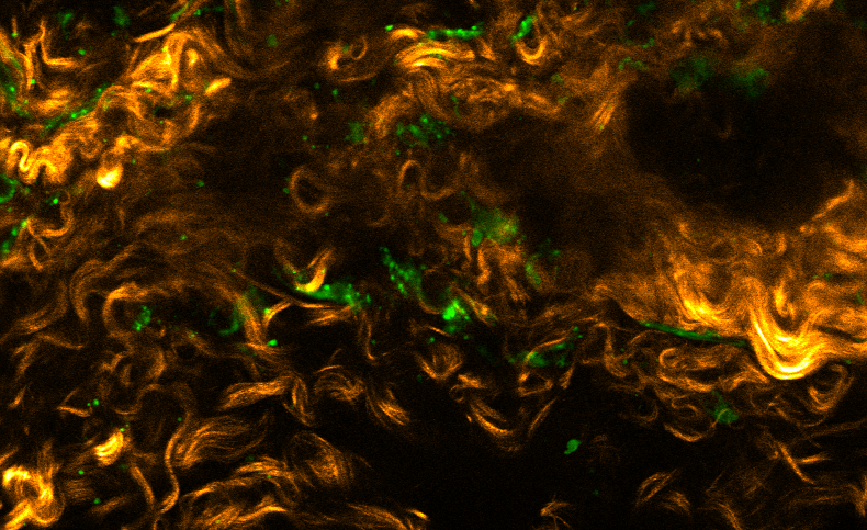
Biological imaging often lends itself to breathtaking displays of beauty under the microscope, blurring the frontiers between science and art. France BioImaging is proud to present below a sample of the artworks of its contributors, curated through personal initiatives and our Image Contest .
Do you have images you would like to share with us? Write to us: contact[at]france-bioimaging.org.
Image Contest 2017
Hairy Scan © Sébastien Mailfert, CIML
3D multicellular spheroids-the new world for discoveries on cancer research © Filippo Piccinini, IRST
Mickey Mouse © Lucie Sengmanivong, Laure Coquand, A. Baffet Lab, UMR 144, Institut Curie, Paris
Turquoise and Coral © Dario Donnarumma, Laboratoire Charles Coulomb UMR 5221 CNRS-UM
La petite Venise de la biologie © Carine Rossé, Emilie Lagoutte & Marie Irondelle, Institut Curie
FBI Logo © Mailfert Sébastien & Raphalen Morgane – CIML
Marvellous sea creature © Eve Gazave & Nicolas Rabet, Institut Jacques Monod-CNRS
Arabidopsis inflorescence 2 © Nathanaël Prunet, Caltech, Meyerowitz lab
Tree of Life © Mailfert Sébastien & Rossignol Rafaëlle – CIML
Fly with a broken face © Mathieu P. Dailly, CMAS
The Embryo Forest © Aude Nommick, IBDM – Marseille University
Hypnoblue © Anna Smirnova, University of Strasbourg – GMGM
Xmas Tree (NSIM in 2 colors) ® Lucie Sengmanivong, Olga Iuliano, B. Goud Lab, UMR 144, Institut Curie, Paris
Scream © Anna Smirnova, University of Strasbourg – GMGM
L’éléphant tricéphale © Lucie Sengmanivong, Laure Coquand, A. Baffet Lab, UMR 144, Institut Curie, Paris
Warhol’s Style SHG Muscle © Sébastien Marais, Bordeaux Imaging Center
J’y ai mis tout mon cœur ! © Marcello Delfini, CIML CNRS-INSERM-AMU
Nano Maze © Mathieu P. Dailly, CMAS
Glial cells in rat brain © Françoise Geffroy, CEA-DRF-NeuroSpin-UNIRS, Midas Team
Fly’s wings © Morgane Rabineau, Inserm
Les dents de la mer © Hugues Lelouard, Mathieu Fallet & Mailfert Sébastien – CIML
Vers quelle direction aller… © Lucie Sengmanivong, Olga Iuliano, B. Goud Lab, UMR 144, Institut Curie, Paris
Amphore © Mailfert Sébastien & Rossignol Rafaëlle – CIML
Microglia’s attack! © Françoise Geffroy, CEA-DRF-NeuroSpin-UNIRS, Midas Team
Nano-Horse © Jonathan Daniel, Institut des Sciences Moléculaires
Cannot see the nanorods for the blood cells © Marie Held, Biochemistry, University of Liverpool, Levy Lab
Virtuous Circle © Pierre-Olivier Strale, Interdisciplinary Institute for Neuroscience
Sunset on the mangrove © Mathieu P. Dailly, CMAS
Wood smoking muscle © Orestis Faklaris, Marianne Gervais-Taurel, Institut Jacques Monod
Arabidopsis inflorescence 3 © Nathanaël Prunet, Caltech, Meyerowitz lab
Poisson d’avril © Mailfert Sébastien & Rossignol Rafaëlle – CIML
Easter Egg © Mailfert Sébastien & Rossignol Rafaëlle – CIML
Une petite Sirène © Lucie Sengmanivong, Frederik Verweij, PhD, G.Raposo team, UMR 144, Institut Curie, Paris
Arabidopsis inflorescence 1 © Nathanaël Prunet, Caltech, Meyerowitz lab
Actin fireworks© Patrice Mascalchi, Bordeaux Imaging Center and Frédéric Saltel, INSERM U-1053, University of Bordeaux
Flower Time © Anna Smirnova, University of Strasbourg – GMGM
Monkey’s Head © Laurence Dubreil-APEX-UMR703 PAnTher INRA Oniris
Cat © Mailfert Sébastien & Rossignol Rafaëlle – CIML
Hanging on by a thread © Debora Olivier, LOB, Ecole Polytechnique for the use of the GE Deltavision OMX SR
The Tree of Life © Orestis Faklaris, Nicolas Chevalier – Institut Jacques Monod
Tracheids and surrounding tissues in Arabidopsis thaliana © Corrado Viotti, Institut de Biologie Moléculaire des Plantes, CNRS, Strasbourg – P. Genschik Lab
Cicata © Mailfert Sébastien – CIML
Fertility © Mailfert Sébastien & Mossadegh-Keller Noushin – CIML
The Black Swan © Orestis Faklaris, Nicolas Chevalier – Institut Jacques Monod
What is behind the heart curtain © Xavier Baudin, Véronique Monnier, Institut Jacques Monod
C. Simon, UMR8576
Superman flower © Nathanaël Prunet, Caltech, Meyerowitz lab
Going with the flow © Jérémie Teillon, INSERM U1034
Nanoparticles in brain tumor vessels © Françoise Geffroy, CEA-DRF-NeuroSpin-UNIRS, Midas Team
Fertility 3 © Mailfert Sébastien & Mossadegh-Keller Noushin – CIML
Mice Egg © Mailfert Sébastien – CIML-CNRS-Inserm-AMU
The Embryo Forest 2 © Aude Nommick, IBDM – Marseille University
Arabidopsis inflorescence 4 © Nathanaël Prunet, Caltech, Meyerowitz lab
Quand la fibre rencontre les FONs © Jonathan Daniel, ISM & Etienne Gontier, Bordeaux Imaging Center (BIC)
Star Pigments © Aude Nommick, IBDM – Marseille University
Tumour of Fire © Marcello Delfini & Mathieu Fallet – CIML CNRS-INSERM-AMU
Quand la chimie transcende la lignine ! 2 © Clémence Simon, Unité de Glycobiologie Structurale et Fonctionnelle, UMR8576
Growth can shorten distances © Valeria Davi, ImagoSeine – Institut Jacques Monod – CNRS – Paris
Main Gallery
Glowing grasp © Liu Zeng Zhen – Institut Jacques Monod – ImagoSeine
Fluorescent focal adhesion proteins in a stem cell, confocal microscope Zeiss 700
CNRS Photothèque
Magali Mondin, Daniel Choquet
UMR5091 – Physiologie cellulaire de la synapse (PCS) – Bordeaux
Un café agent J © PETREL Melina – Bordeaux Imaging Center – CNRS, Inserm, université Bordeaux Segalen
Cellules inflammatoire suite à introduction d’un corps étranger recouvert d’or.
© Noushin Mossadegh & Mailfert Sébastien – CIML, CNRS-INSERM-AMU
Coupe d’un testicule de nouveau-né de souris.
Marquage en immunofluorescence des noyaux cellulaires (DAPI).
C’est l’amour
© PETREL Melina – Bordeaux Imaging Center – CNRS, Inserm, université Bordeaux Segalen
Algues préparées par HPF.
Insitut des Neurosciences de Montpellier (INM)
Montpellier RIO Imaging
Montpellier
Le bouquet de vie © Noushin Mossadegh & Mailfert Sébastien – CIML, CNRS-INSERM-AMU
Coupe d’un tube séminifère de testicule de souris. On visualise les spermatozoïdes formant un « bouquet de fleurs ». Marquage en immunofluorescence des noyaux cellulaires (DAPI) représentés ici en cyan révélant la queue des spermatozoïdes. L’actine, représentée ici en rouge, révèle «la tête» des spermatozoïdes (phalloïdine marquée avec le fluorochrome Alexa-647). L’image représente 213×213μm sur 2048×2048 pixels. La coupe est d’une épaisseur de 20μm. Image de microscopie confocale sur Zeiss LSM780 ; lasers 405nm et 633nm ; objectif 40X, O.N. 1.4, immersion à huile.
Lionel Simmonneau, Institut des Neurosciences de Montpellier (INM) – Neurosphère
Alexandre Philips
Laboratoire Muscle et pathologies (ERI25)
Montpellier RIO Imaging
Montpellier
L’immunologie vue du ciel © Noushin Mossadegh & Mailfert Sébastien – Centre d’immunologie de Marseille Luminy (CIML) – CNRS /INSERM/AMU
Véronique Ripetti, Laboratoire Développement et amélioration des Plantes (DAP), Montpellier – Tige de pommier
Projections solaires © PETREL Melina – Bordeaux Imaging Center – CNRS, Inserm, université Bordeaux Segalen
Particules magnétiques encapsulées de polymère.
Jean-Michel Bellanger, CRBM, Montpellier – C. elegans RTM
Insitut de Génétique Moléculaire de Montpellier (IGMM)
Montpellier RIO Imaging
Julien Cau, Montpellier RIO Imaging – Fibroblaste de Xénope en division
ImageJ=1.47a
unit=micron
finterval=1
Michel Eybalin, Institut des Neurosciences de Montpellier (INM), Montpellier – Ganglion spiral de cochlée
Pierre Savagner, Unité Identité et plasticité tumorale (U868, CRLC), Montpellier – Glande Mammaire de souris- MRI 2006 Image contest 2nd Prize
The burning mesh © Patrice MASCALCHI – IINS, UMS3420 CNRS – University of Bordeaux, US4 INSERM – Bordeaux Imaging Center
This image is about tracking of single molecules on live neurons (uPaint technique). It is the superimposition of hundred of tracks (red) and instantaneous diffusion coefficient (gradient of yellow to white).
Alexandre Philips
Laboratoire Muscle et pathologies (ERI25)
Montpellier RIO Imaging
Montpellier
The Tracking Trip © Melina HEUZE – Institut Jacques Monod, CNRS, UMR7592 – ImagoSeine
Neuron © D. Choquet (CNRS)
Suzanne Vigneron – Centre de Recherche en Biochimie Macromoléculaire (CRBM) – Montpellier -Kinétochores dans un noyau de xénope
Bioimage analysis of Shigella infection reveals targeting of colonic crypts (PNAS, 2015) © Ellen T. Arena (Pasteur)
Institut des Neurosciences de Montpellier (INM)
Montpellier RIO Imaging
Montpellier
Véronique Ripetti, Laboratoire Développement et amélioration des Plantes (DAP), Montpellier – Tige de pommier
Incandescence © Noushin Mossadegh & Mailfert Sébastien – CIML, CNRS-INSERM-AMU
La chambre magmatique est en ébullition, les futures générations de spermatozoïdes se préparent à rentrer dans la vie de manière explosive. Coupe d’un tube séminifère de testicule de souris. De la périphérie à la lumière du tube, on visualise les cellules souches qui vont donner naissance aux spermatozoïdes. On distingue ces derniers grâce à leur queue. Marquage en immunofluorescence des noyaux cellulaires (DAPI). L’image représente 213×213μm sur 2048×2048 pixels. La coupe est d’une épaisseur de 20μm. Image de microscopie confocale sur Zeiss LSM780 ; lasers 405nm; objectif 40X, O.N. 1.4, immersion à huile.
Marie Mirouze, Laboratoire Biologie et Physiologie Moléculaire des Plantes (BPMP), Montpellier – Trichome d’Arabidopsis halleri
Laboratoire Guichet IJM
Frédérique Scamps, Institut des Neurosciences de Montpellier (INM), Montpellier – Neurone sensoriel
Adrien Demilly – Institut Jacques Monod
L’Art Confocal © Mailfert Sébastien – CIML, CNRS-INSERM-AMU
Mouse kidney section with AF 488 WGA in green, AF 568 Phalloidin in red, and DAPI in blue. From LifeTechnologies, F24630 FluoCells prepared slide. Confocal microscopy image obtained on a Leica SP5 ; 405nm and white light laser @488 and 568nm ; 63/1.4 oil immersion objective.
Le St Pierre Mediterranneen © Noushin Mossadegh & Mailfert Sébastien – CIML, CNRS-INSERM-AMU
R Gaudin
Frédéric Causeret
Génétique et développement du cortex cérébral
Institut Jacques Monod
Fly monster © Orestis Faklaris & Michael Lang – Institute Jacques Monod, CNRS UMR7592 – ImagoSeine
Multifocal confocal microscopy (spinning disk, Spinning CSU W1)
Drosophila third instar larval head, nuclear-RFP, neronal-GFP and green autofluorescence, 25x magnification, scale bar is 100 μm.
Second to the FBI Image Contest 2016
Link the dots! © Patrice MASCALCHI – IINS, UMS3420 CNRS – University of Bordeaux, US4 INSERM – Bordeaux Imaging Center
DNA-Paint technique performed to image triple-spot nanorulers (DNA origami from Gattaquant company). DNA probes is covalently attached to Atto 655 dye. Images were acquired on Leica GSD.
Steve Bourane, Institut des Neurosciences de Montpellier (INM), Montpellier – Colonne vertébrale de souris- MRI 2007 Image Contest 2nd prize
Florence Iral-Comeau, Laboratoire de Génétique Moléculaire et Chromosomique (IURC), Montpellier – Cellules épithéliales d’origine pulmonaire
Geneviève Conéjéro, Laboratoire Biologie et Physiologie Moléculaire des Plantes (BPMP), Montpellier – Anthère de Tabouret Bleuâtre (Thlaspi caerulescens)
Bioimage analysis of Shigella infection reveals targeting of colonic crypts (PNAS, 2015) © Ellen T. Arena (Pasteur)
© Théophile Déjardin – Institut Jacques Monod – ImagoSeine
3t3 cells expressing fluorescent Nesprin2 going through 5µm constrictions.
Alexandre Philips
Laboratoire Muscle et pathologies (ERI25)
Montpellier RIO Imaging
Montpellier
Gilles Carnac, Laboratoire Muscle Myotube humain
Dalton © Hugues Lelouard, Mailfert Sébastien & Mathieu Fallet CIML CNRS-INSERM-AMU
Confocal microscopy
Revealing sub-population of immune cells on small intestine by 10 colors spectral imaging
First Price of the FBI Image Contest 2016
Prendre De Bonnes Resolutions © Mailfert Sébastien – CIML, CNRS-INSERM-AMU
3D confocal microscopy image of a fluorescent calibration bead to quantify the resolution of the system
Willy Joly, Institut de Génétique Humaine (IGH), Montpellier – Embryon de drosophile
Galactic Love © Olga Nagy – Institute Jacques Mondod, CNRS, Drosophila Evolution Group – ImagoSeine
Immunostaing of Drosophila santomea pupal genitalia. Nuclei are in blue colour stained with DAPI. Neurons and glial cells are in red labeled with anti-HRP-Cy3 antibody. Hairs and chitin are in green autofluorescence. Image was taken at ImagoSeine by Zeiss LSM 710.
Geneviève Conégéro, Laboratoire Biologie et Physiologie Moléculaire des Plantes (BPMP), Montpellier – Feuille d’Arabidopsis thaliana
© Orestis Faklaris – Institut Jacques Monod – ImagoSeine
collagen SHG signal and autofluorescence (green) in the aorta
Dominika Sliwa
Equipe « Mitochondries, métaux et stress oxydatif »
Institut Jacques Monod
Frédéric Bernard
Equipe Génétique Moléculaire de la Différenciation
Institut Jacques Monod
Jean-Michel Bellanger, Centre de Recherche en Biochimie Macromoléculaire – C. elegans
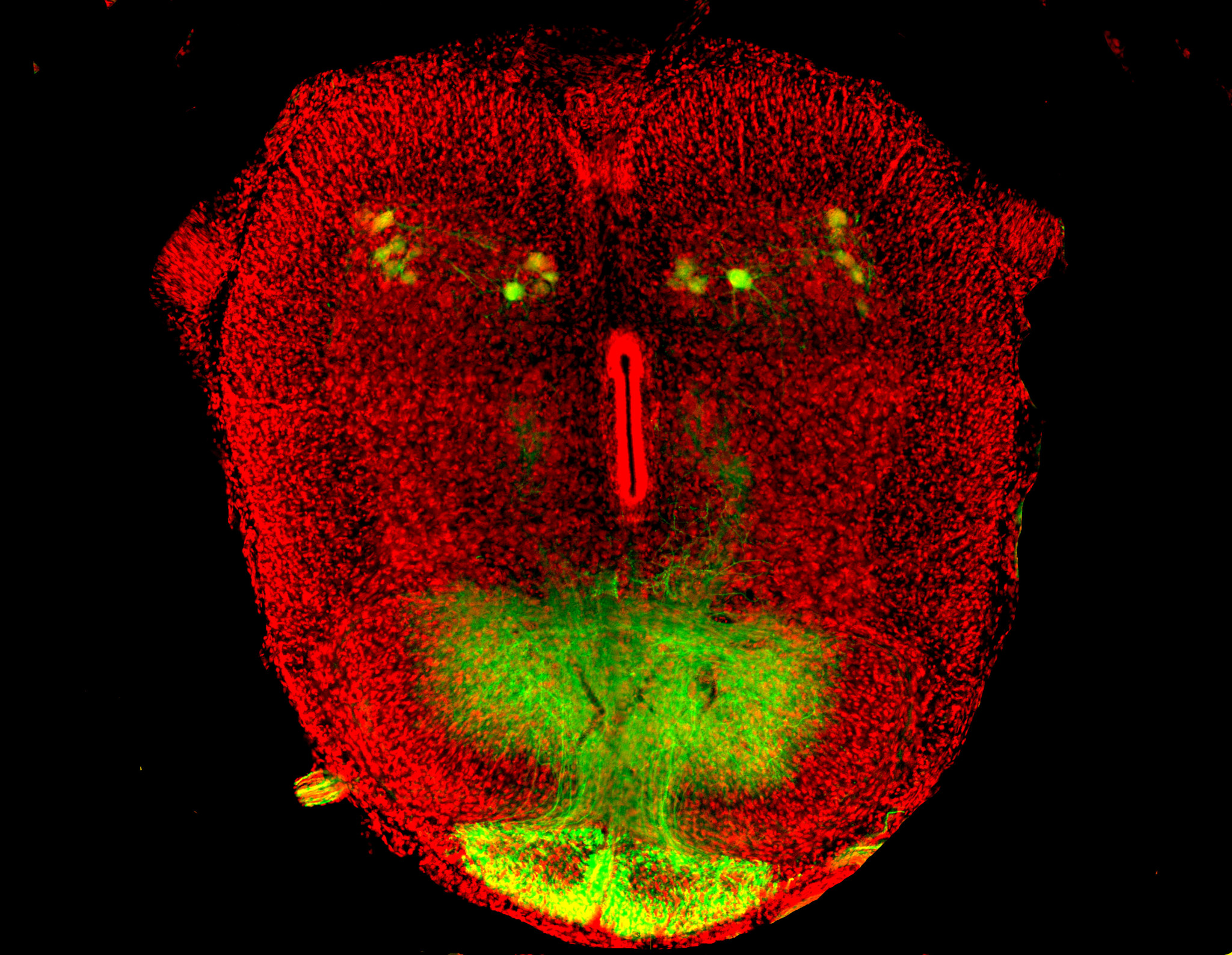
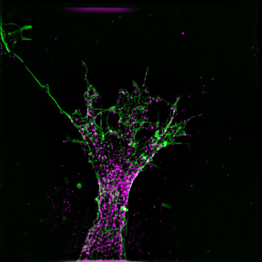
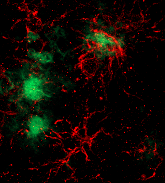
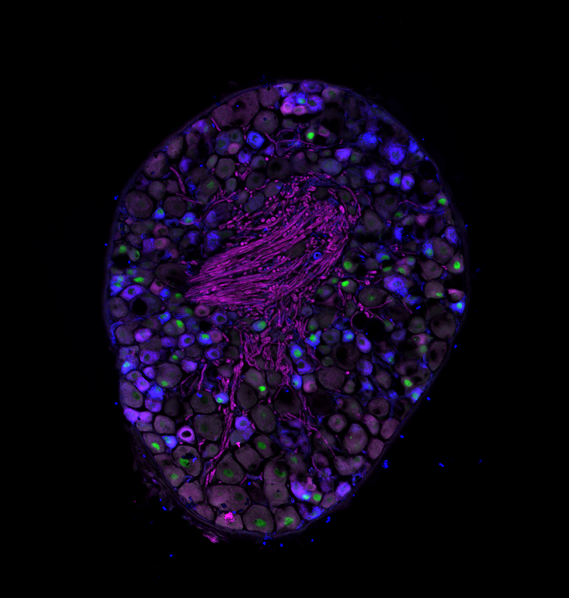
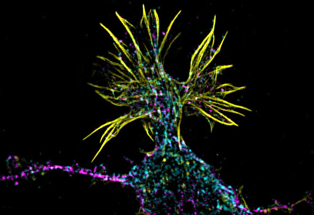
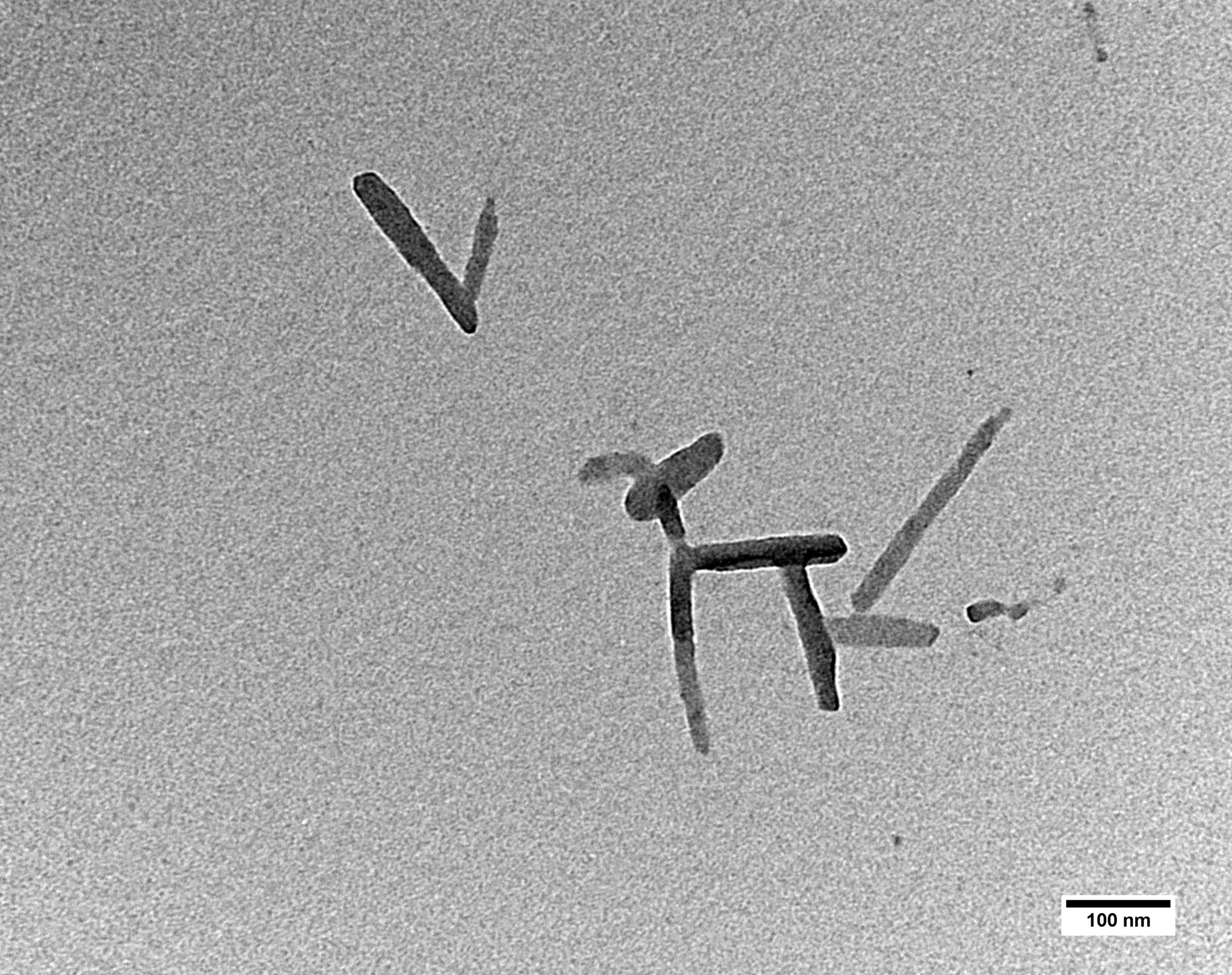
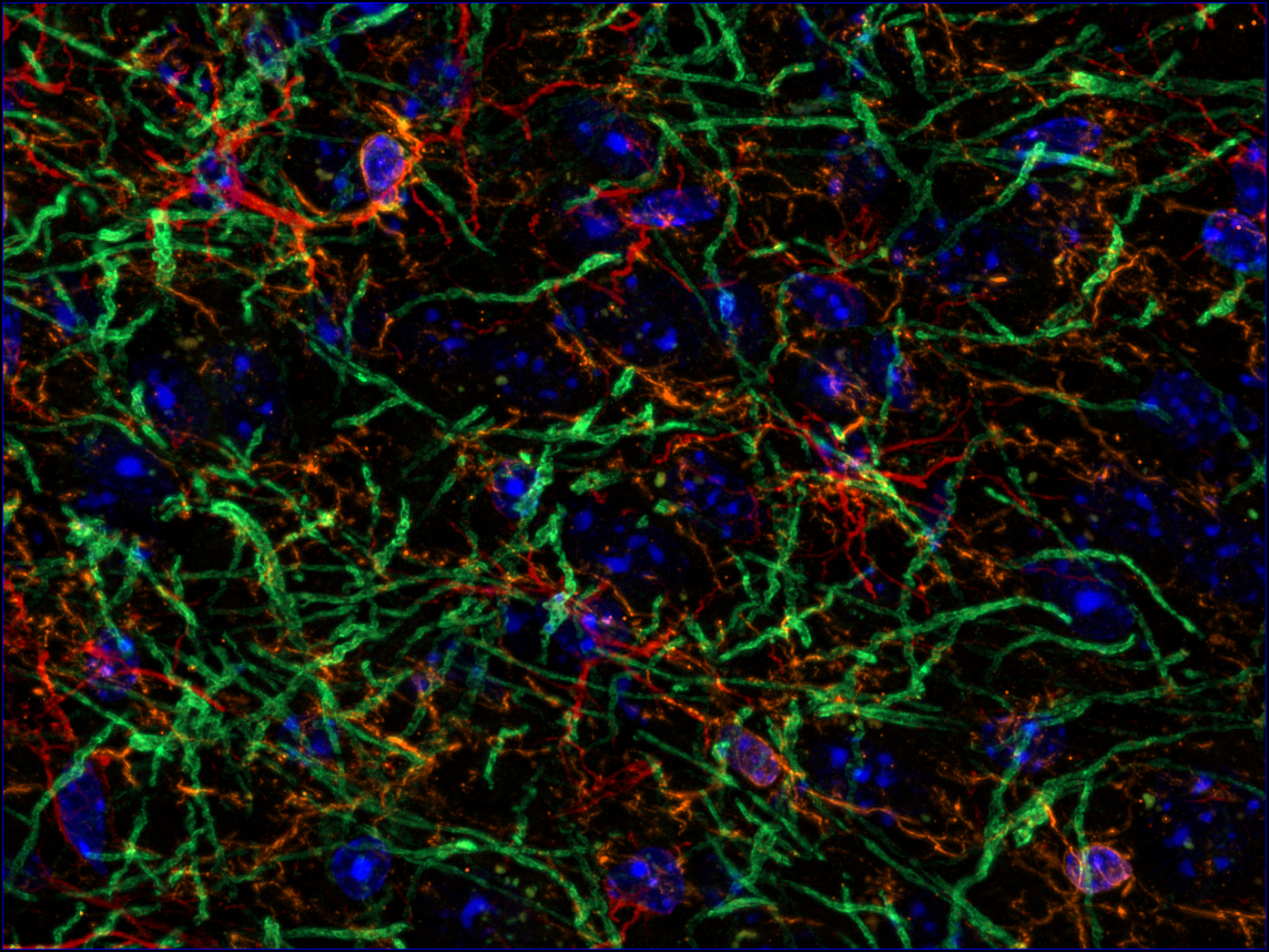
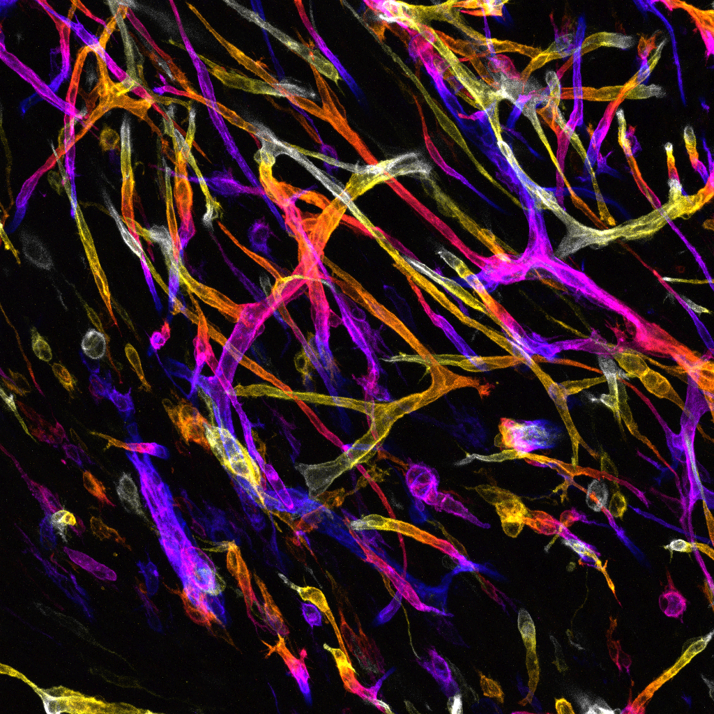
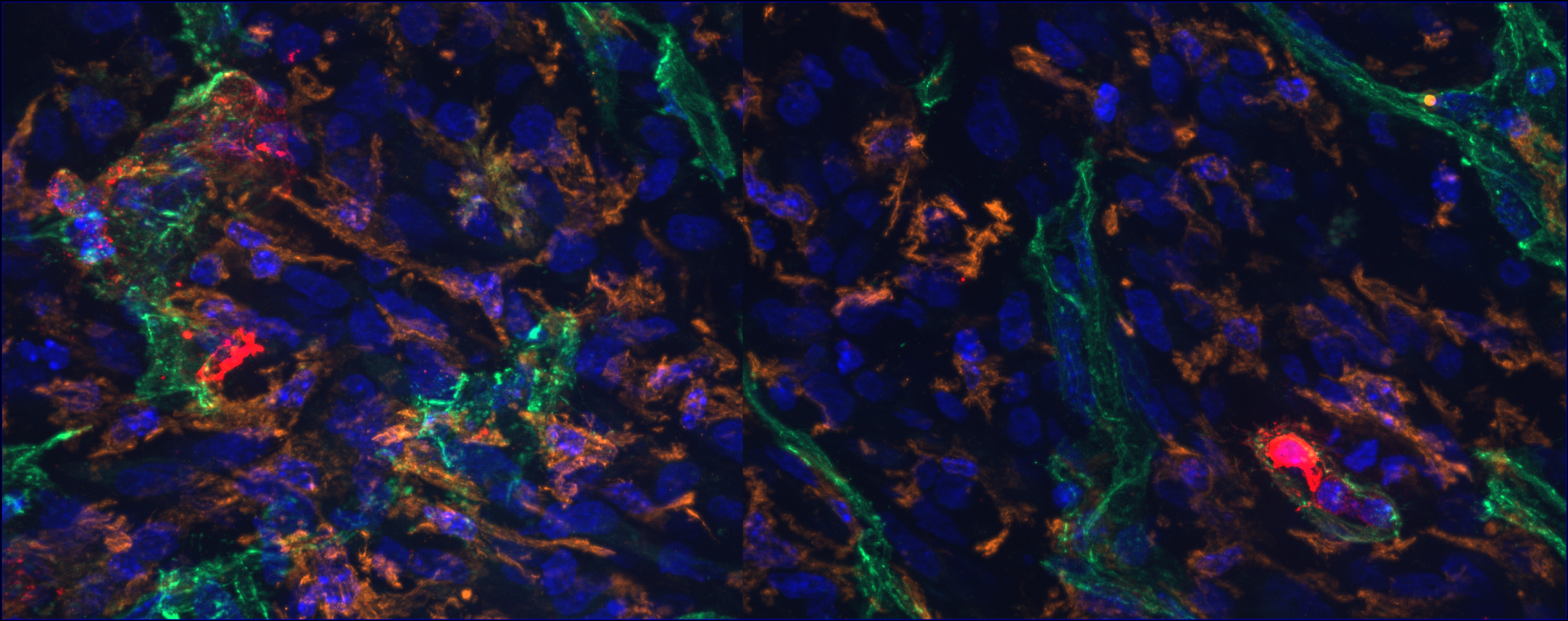
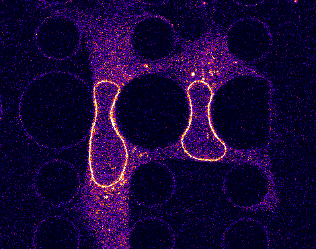
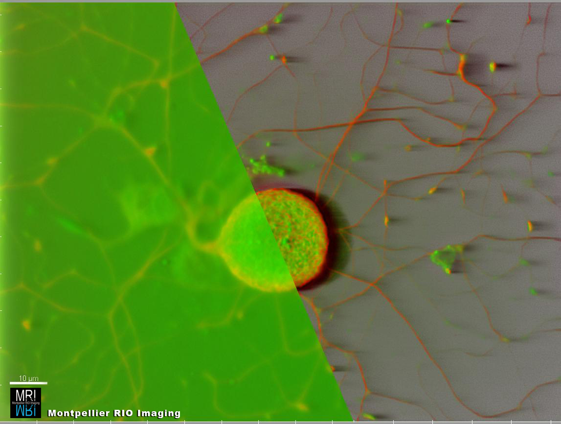
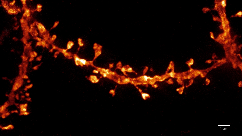
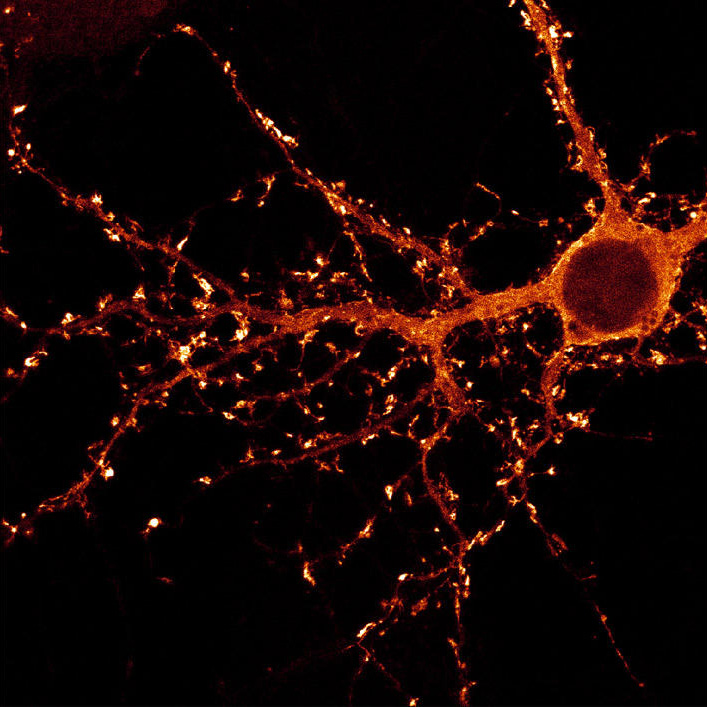
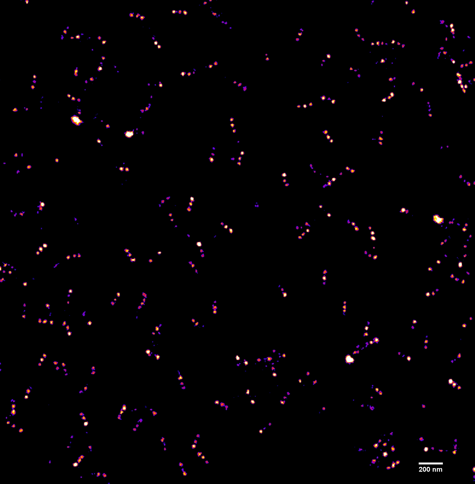
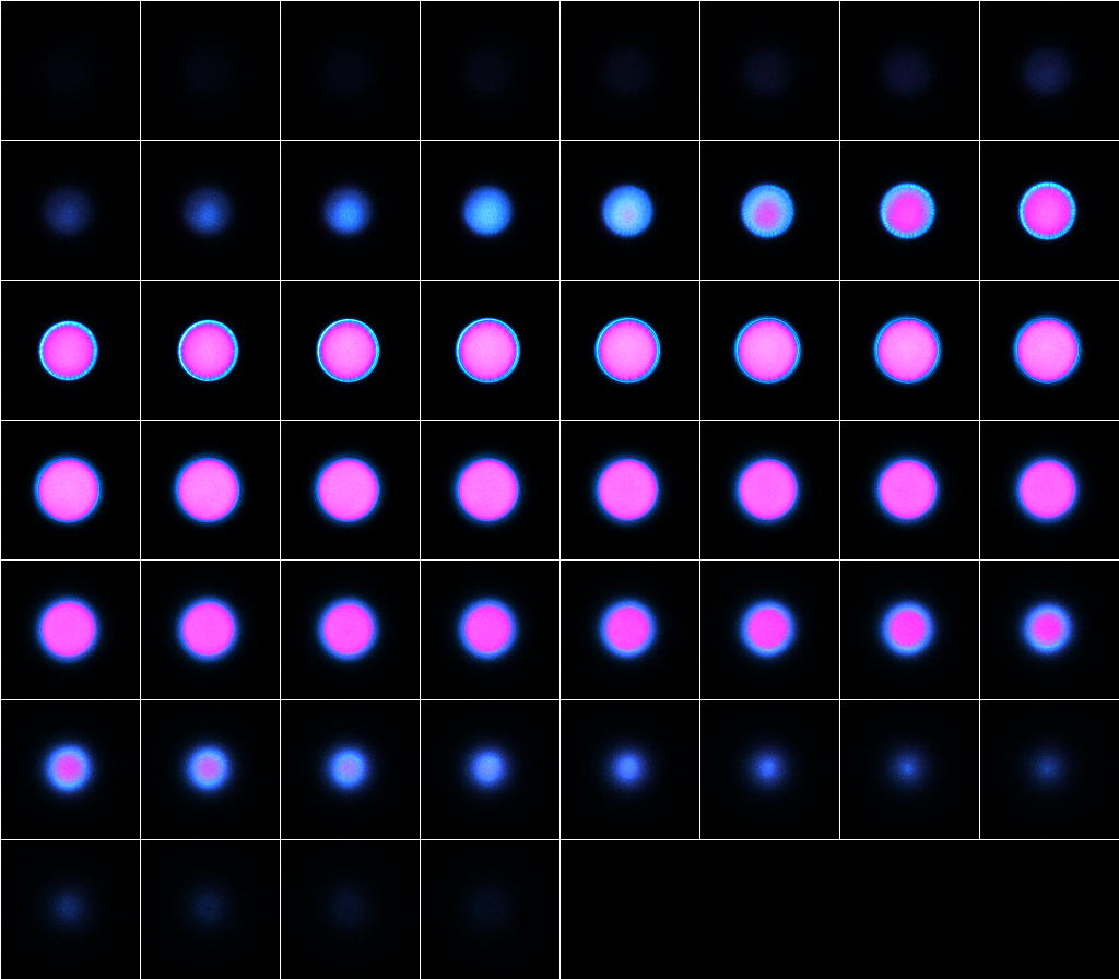
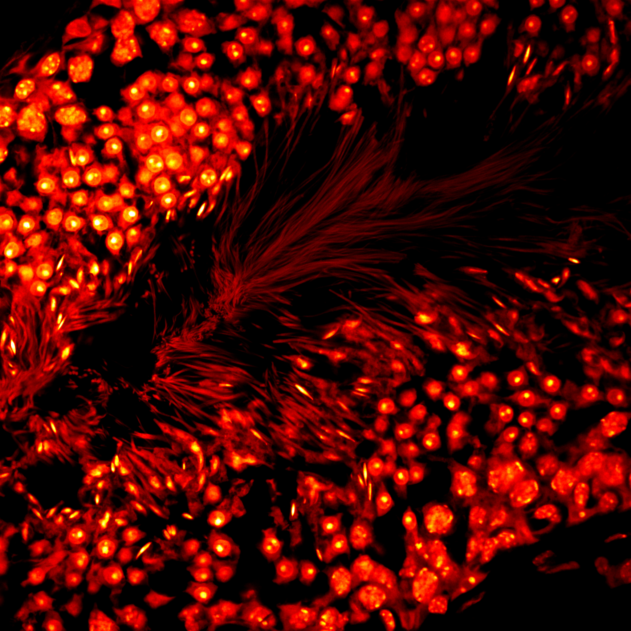
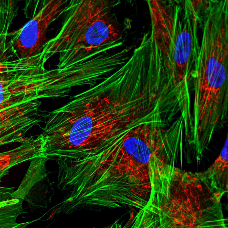
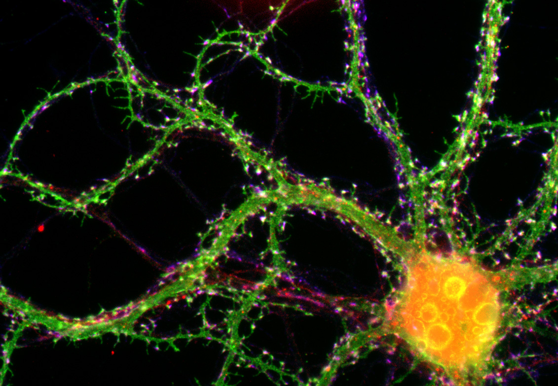
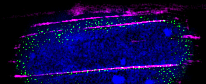
STED image of neuron spines acquired in a fixed mouse brain slice using Atto 647N immunostaining. Image is a the result of a maximum projection of a 12 plane z-stack.
Frédérique Scamps, Institut des Neurosciences de Montpellier (INM), Montpellier – Neurone sensoriel
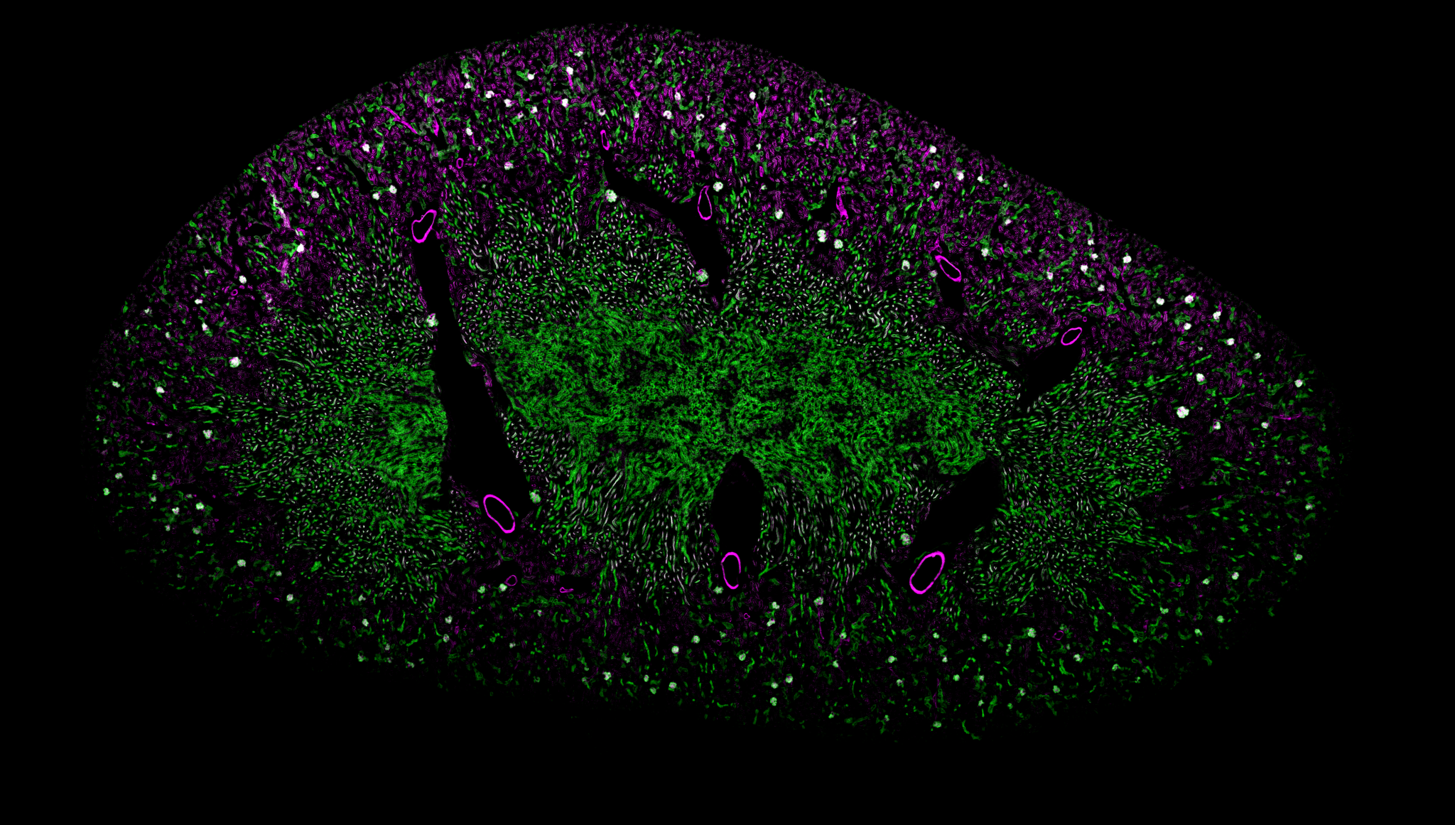
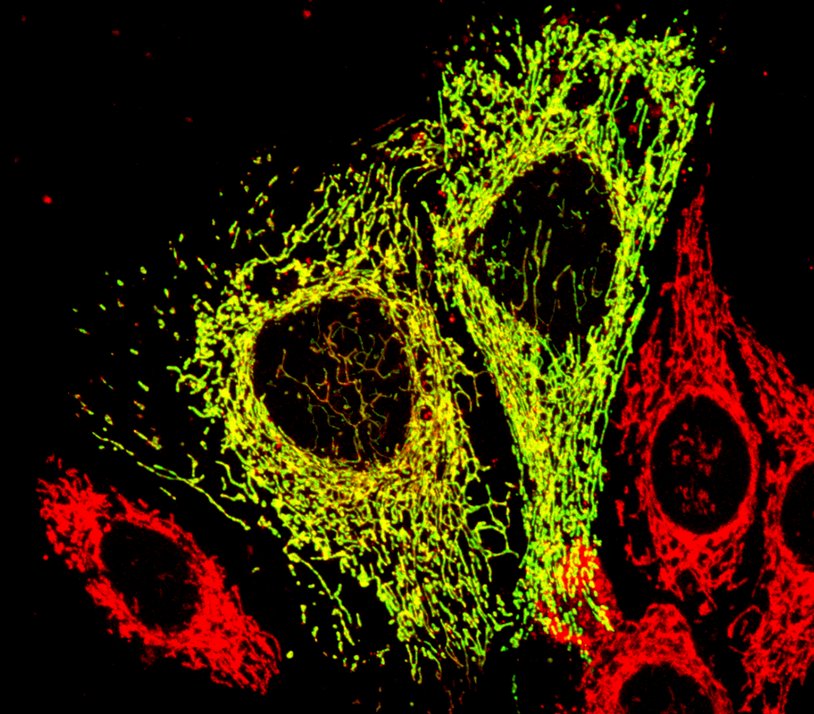
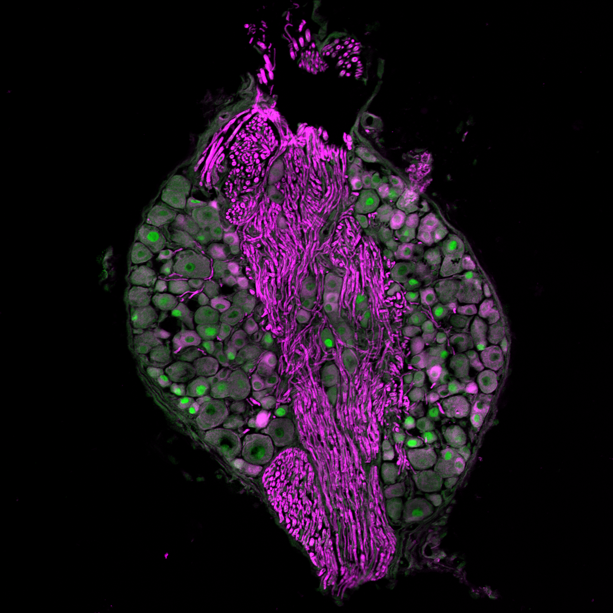
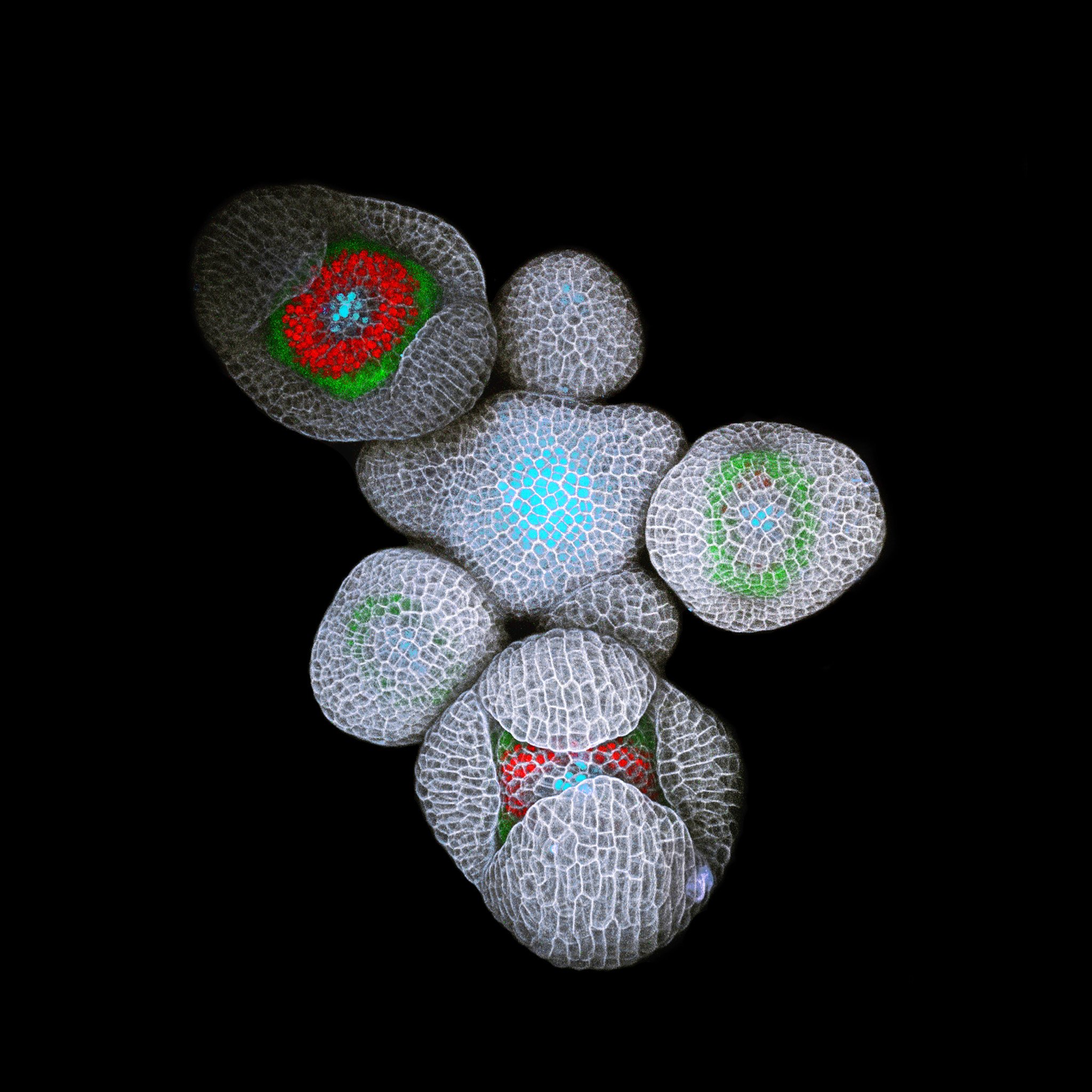
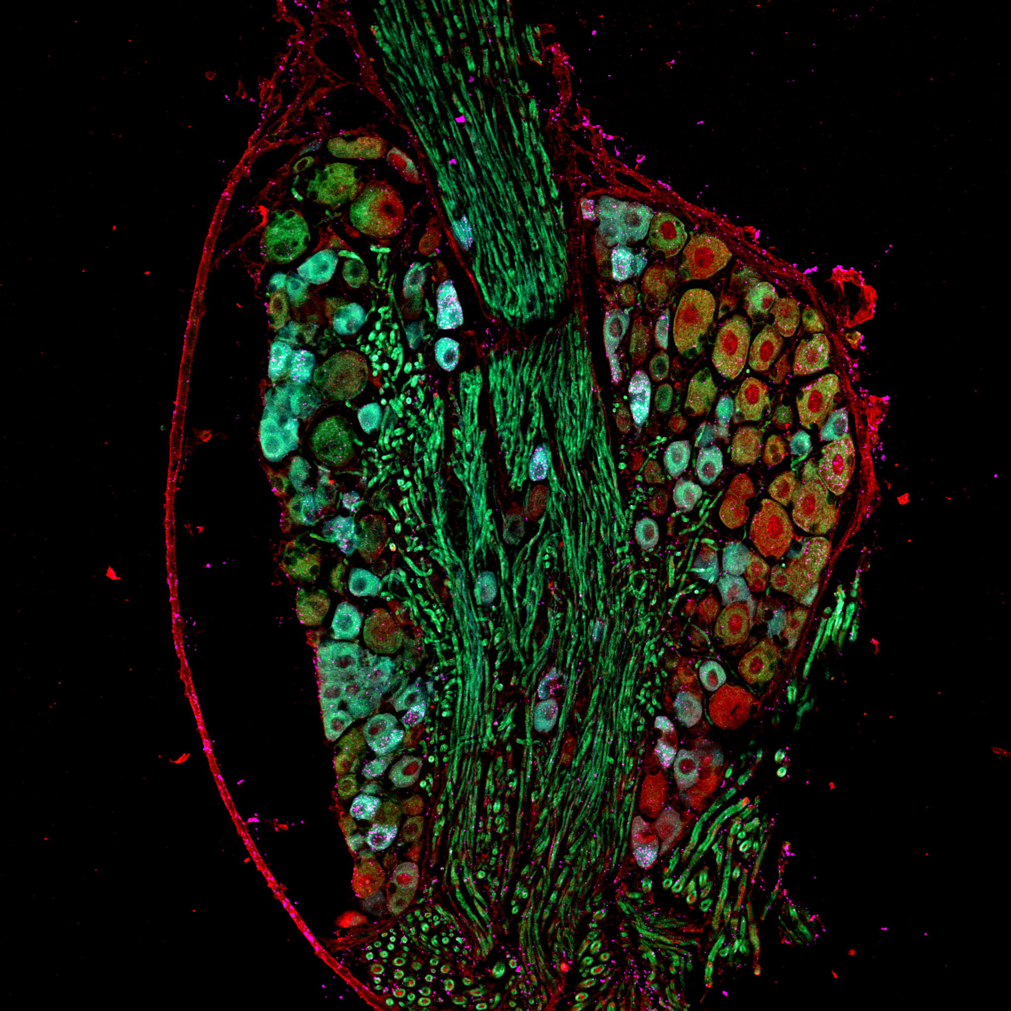
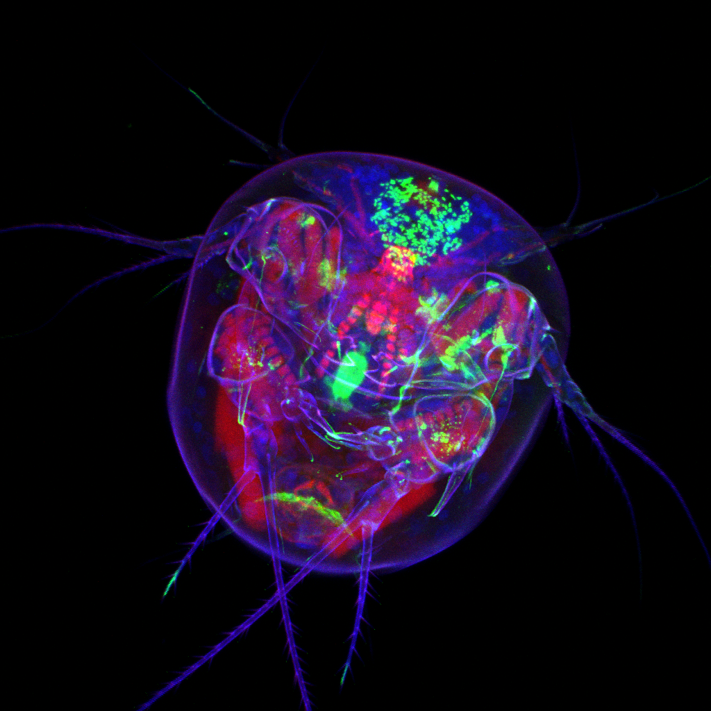
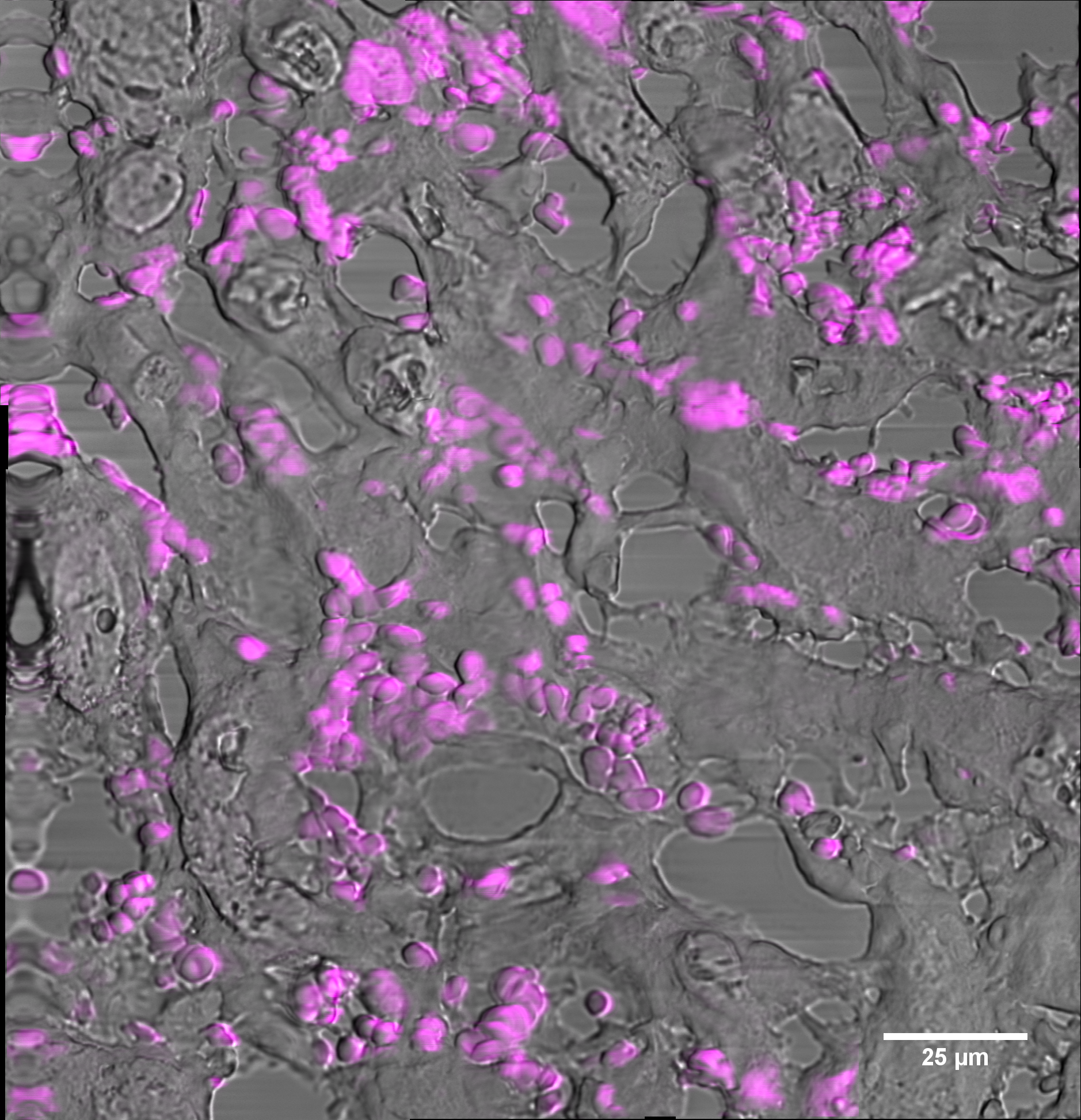
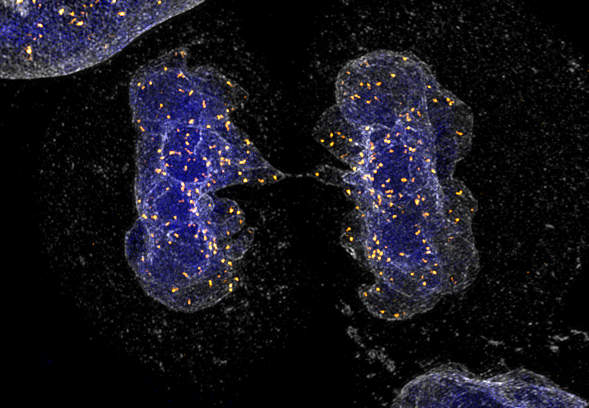
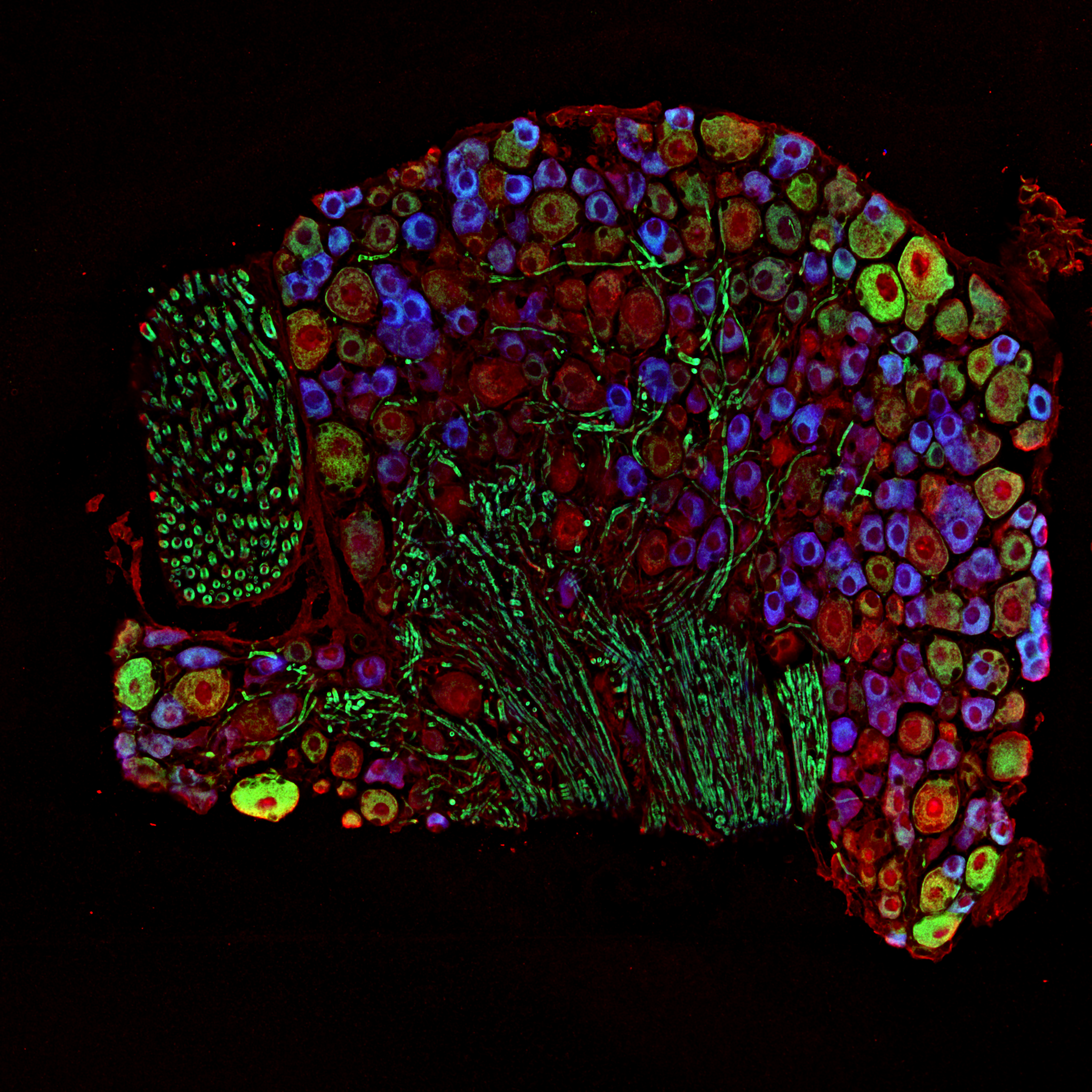
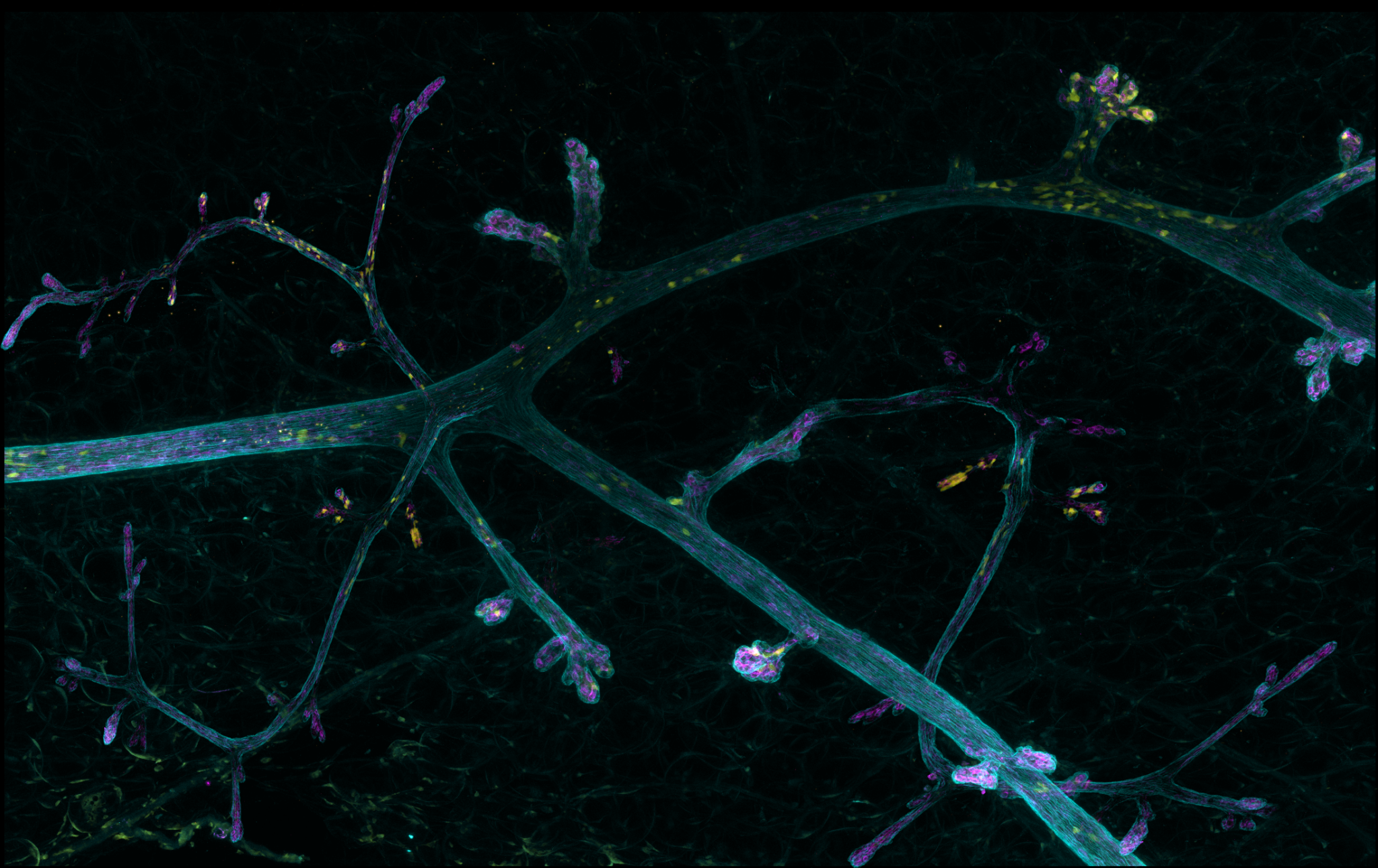
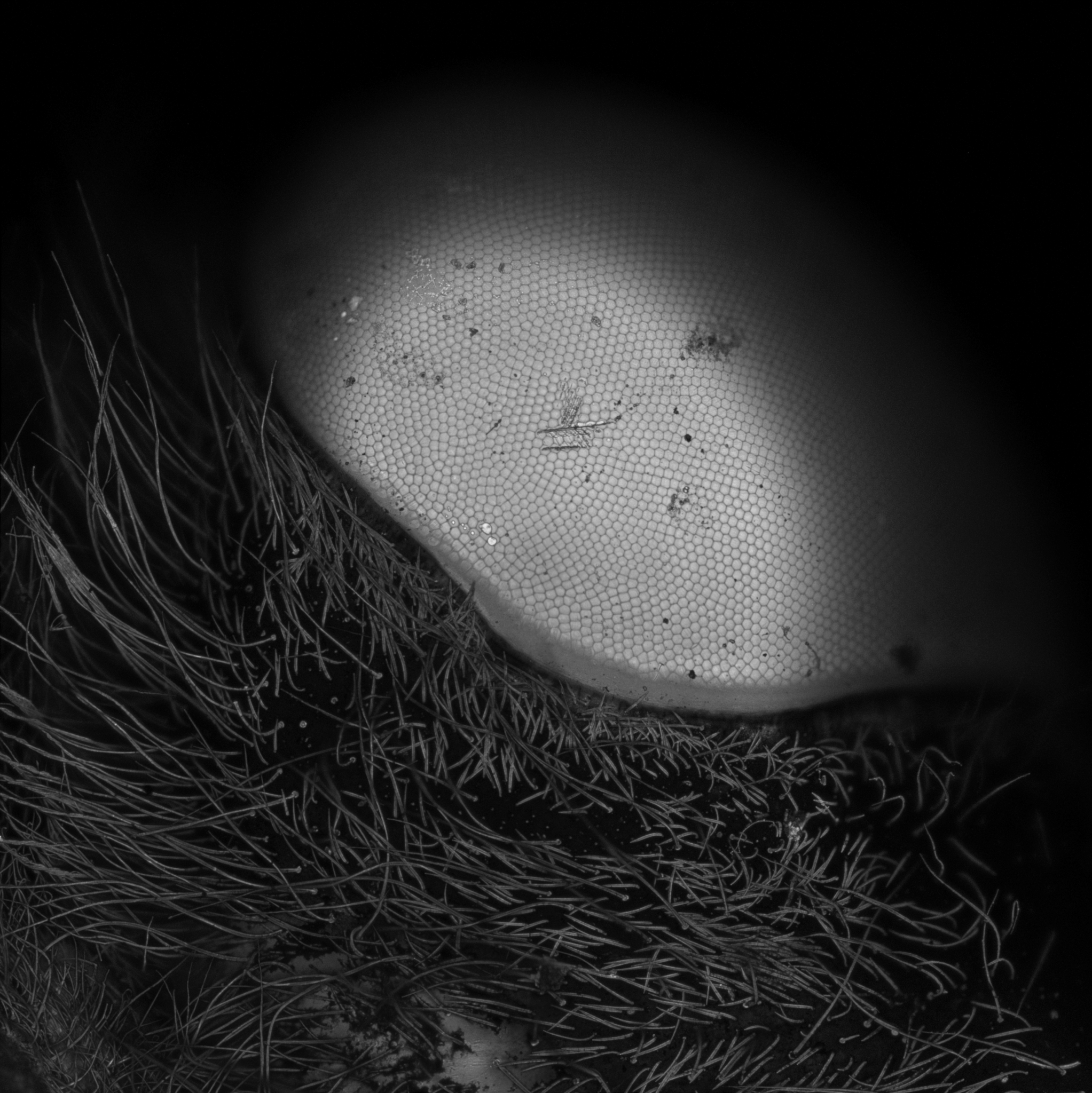
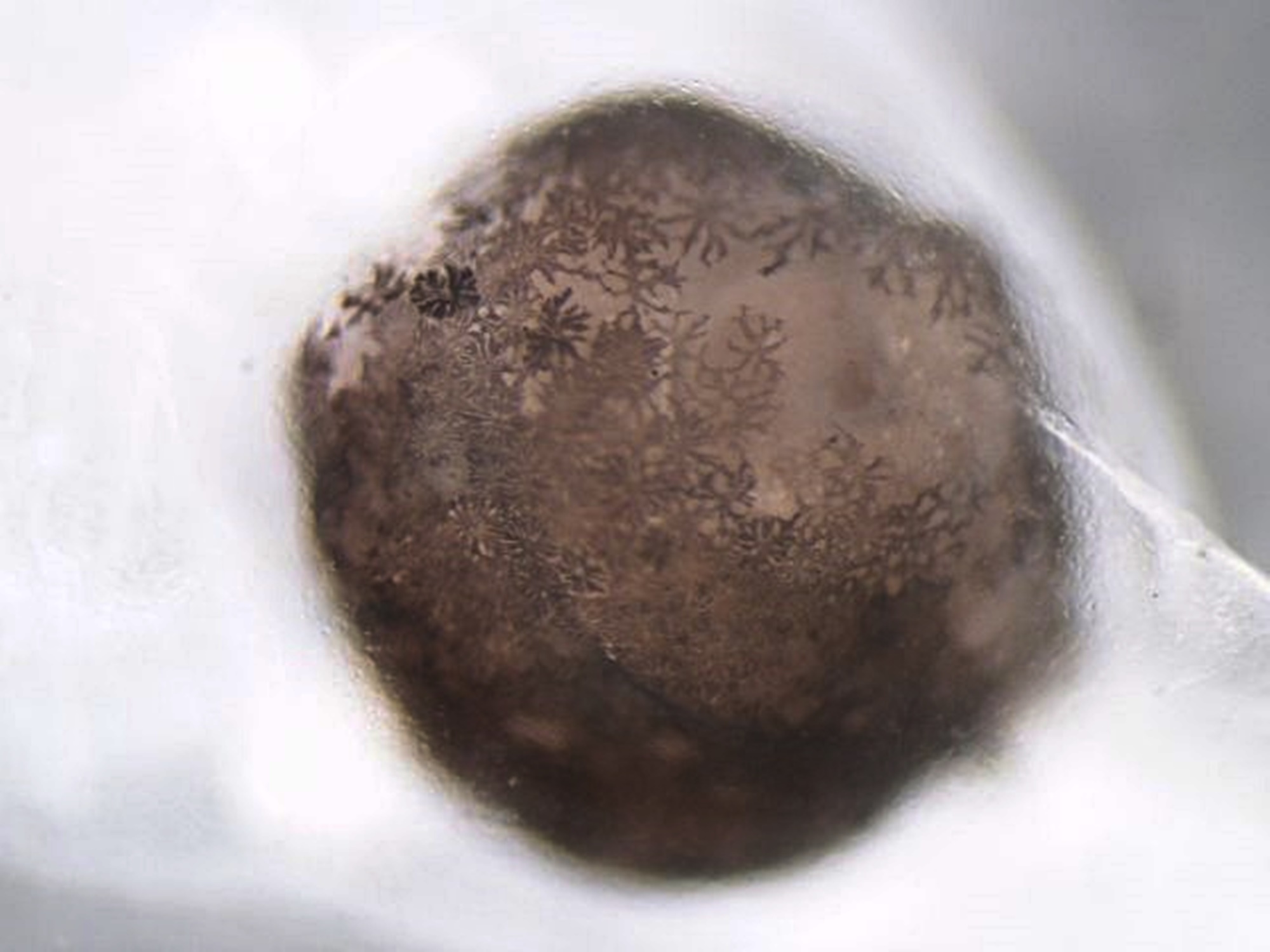
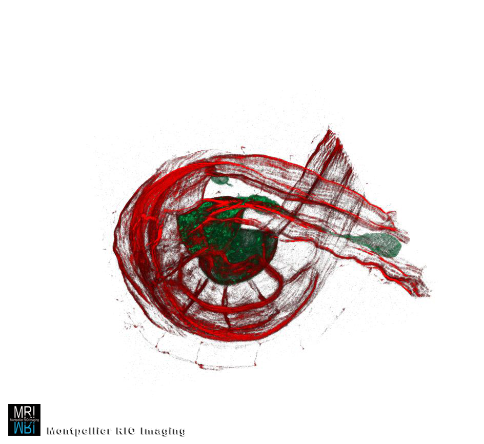
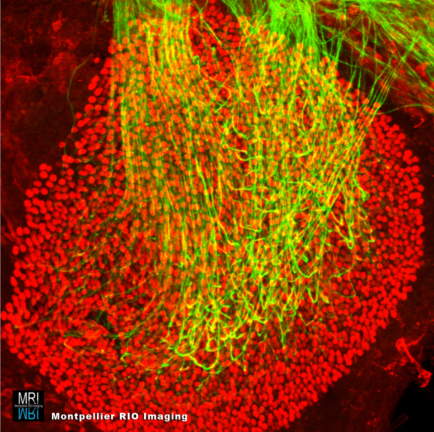
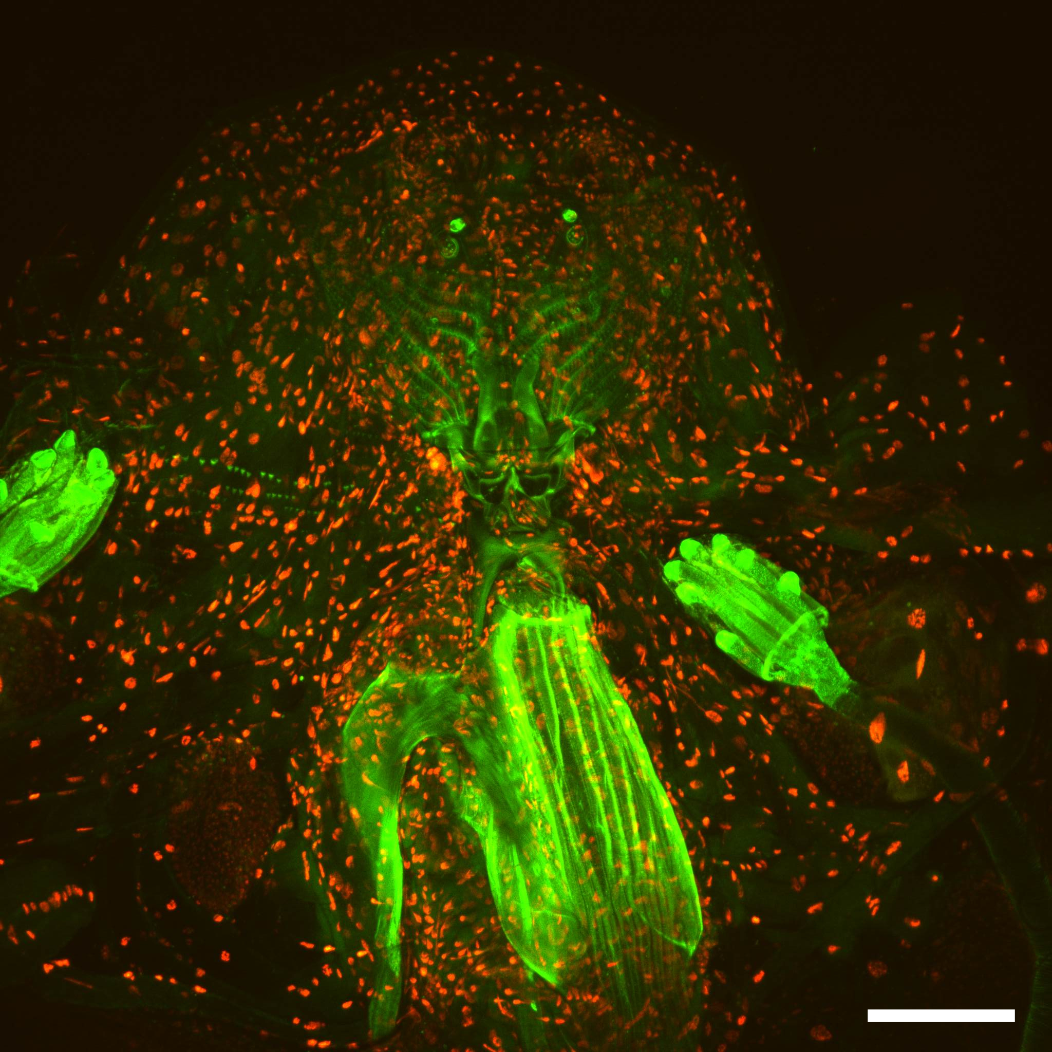
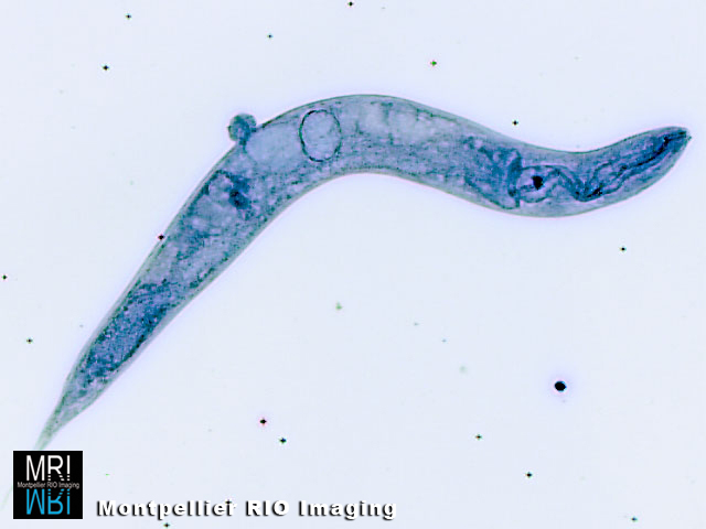
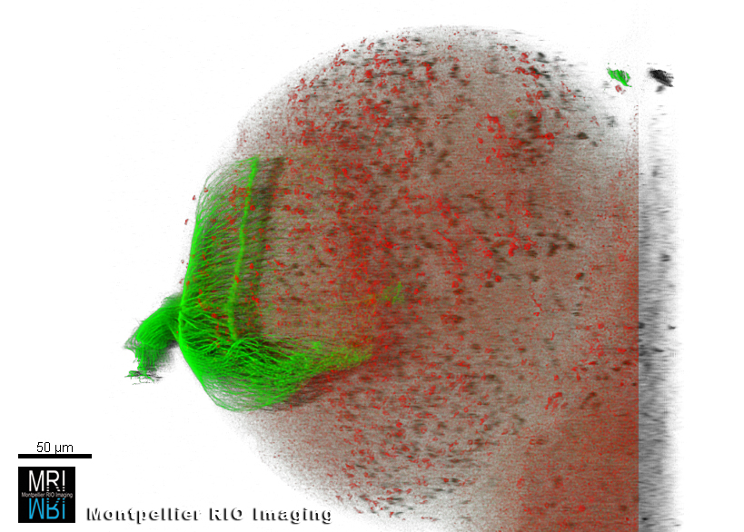
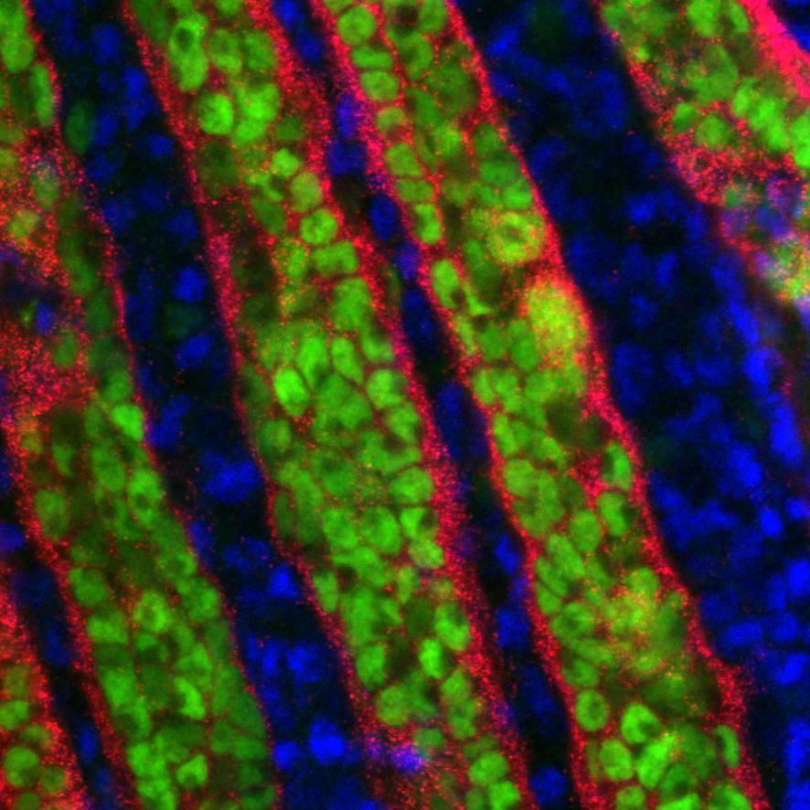
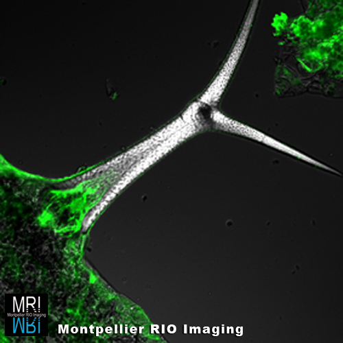
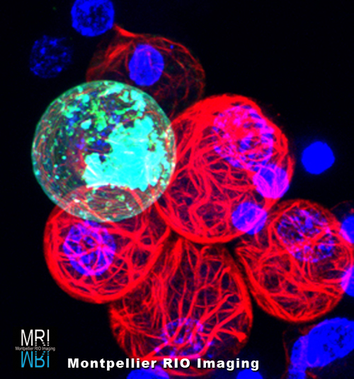
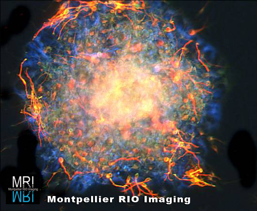
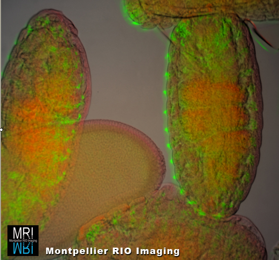
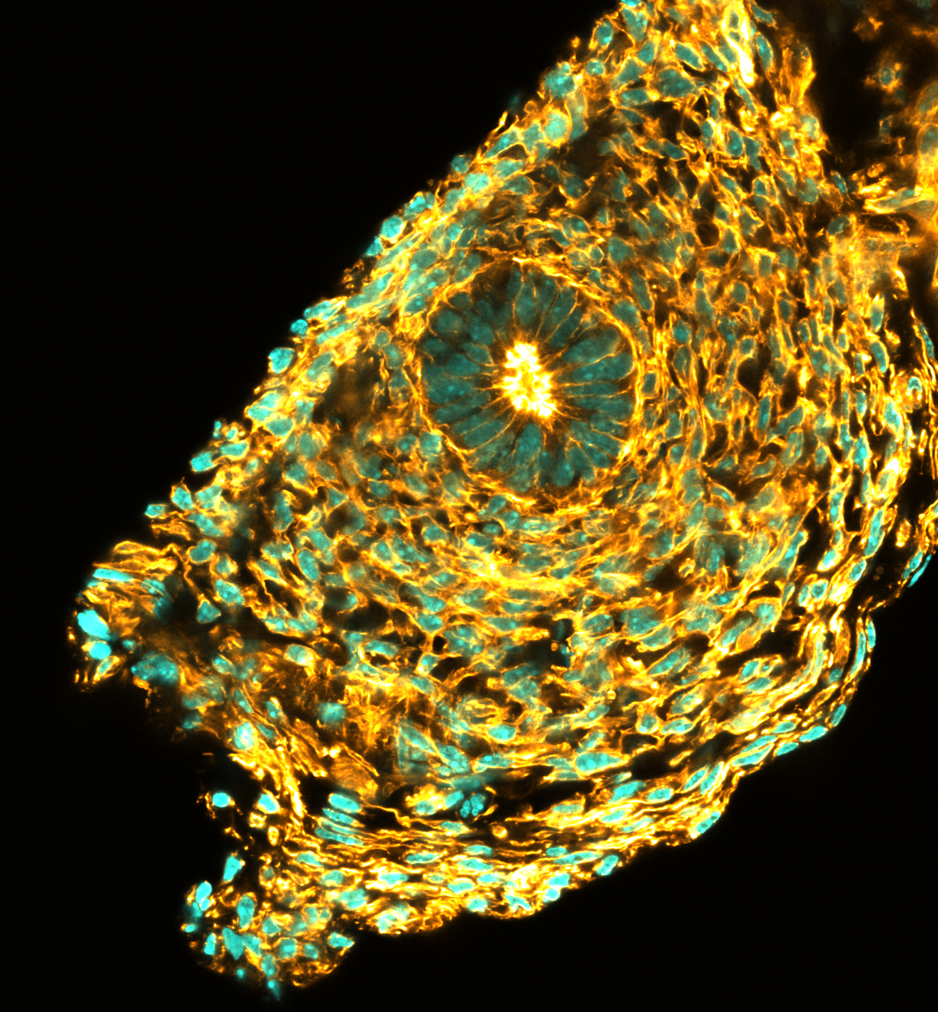
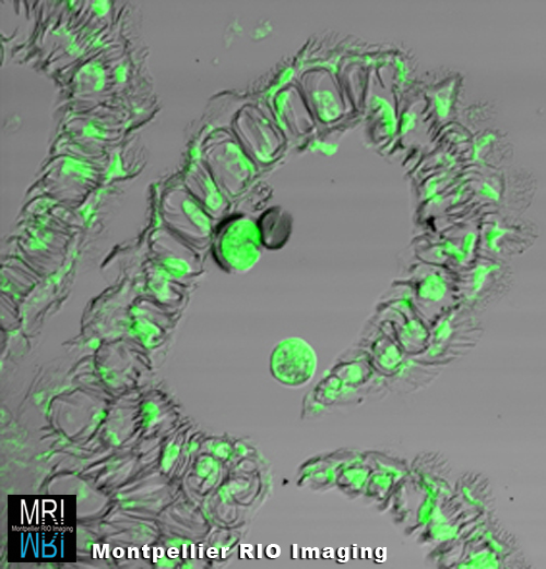
Mathieu Gabut, Institut de Génétique Moléculaire (IGMM), Montpellier – Disque imaginal d’oeil d’embryon de Drosophile
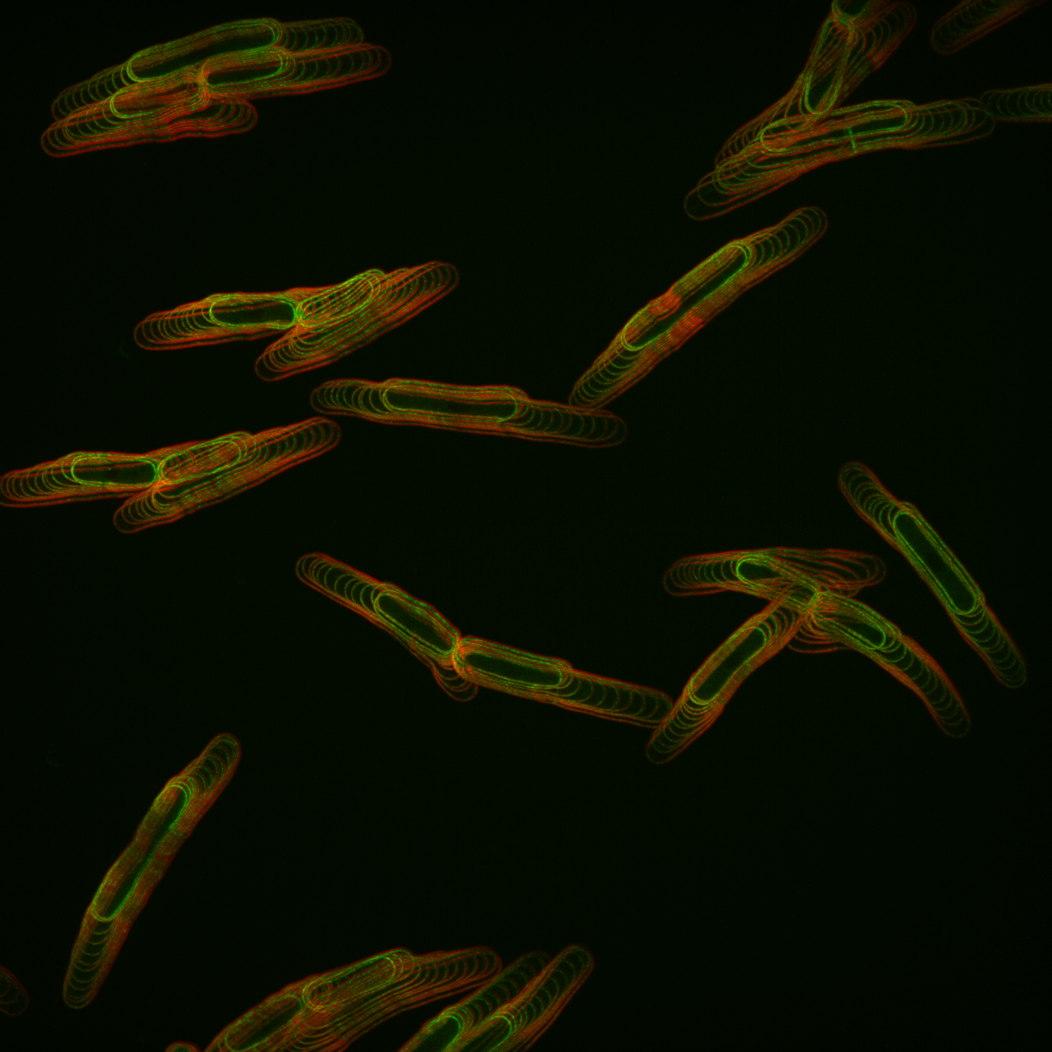
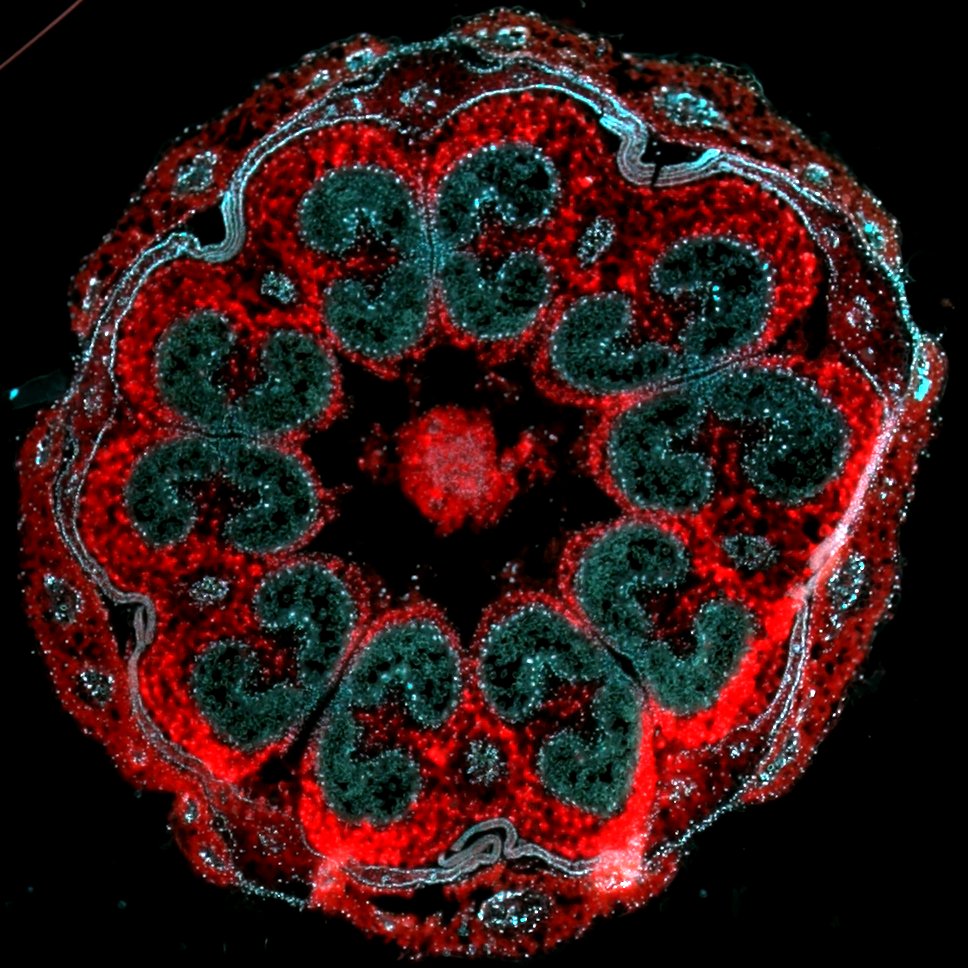
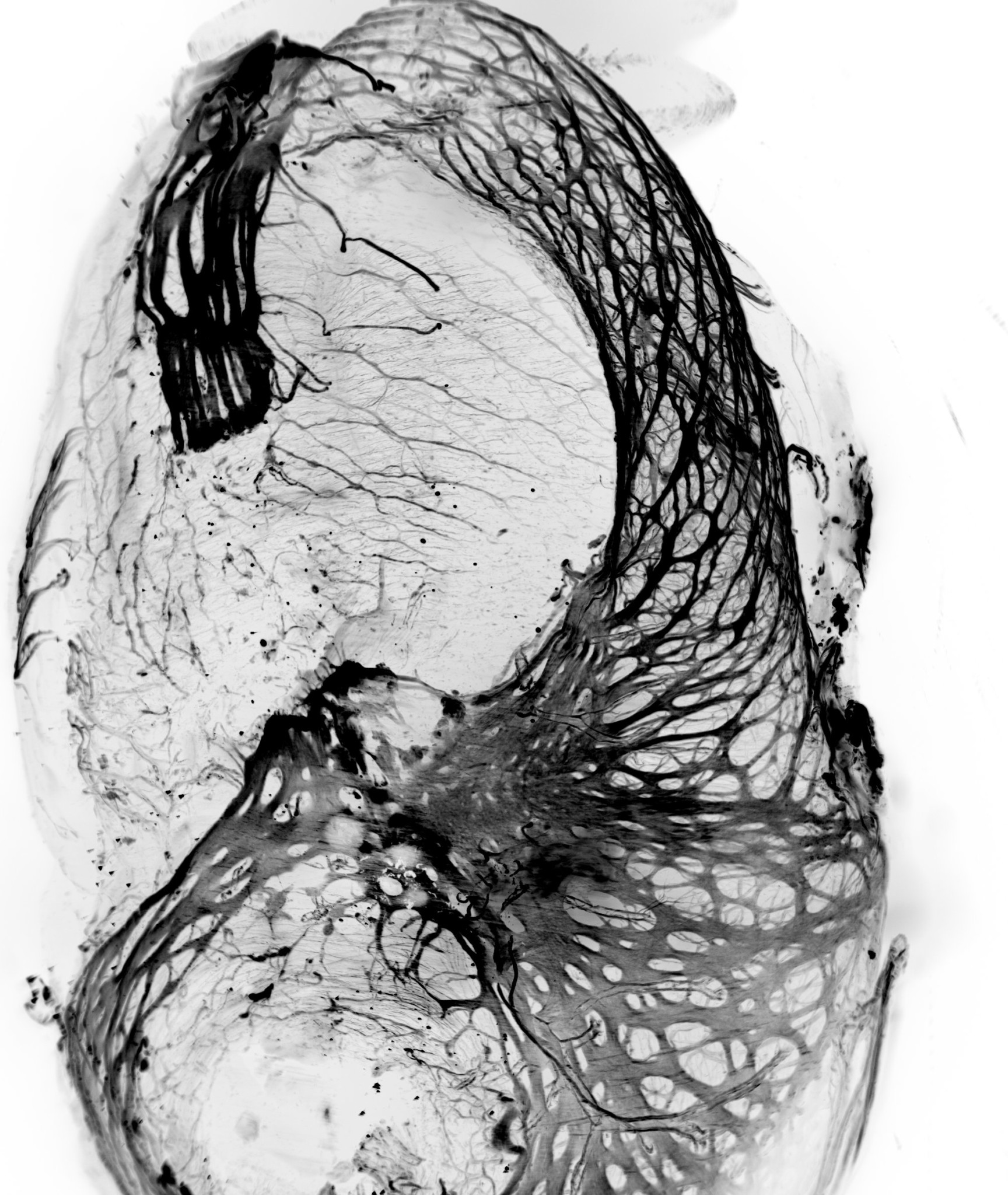
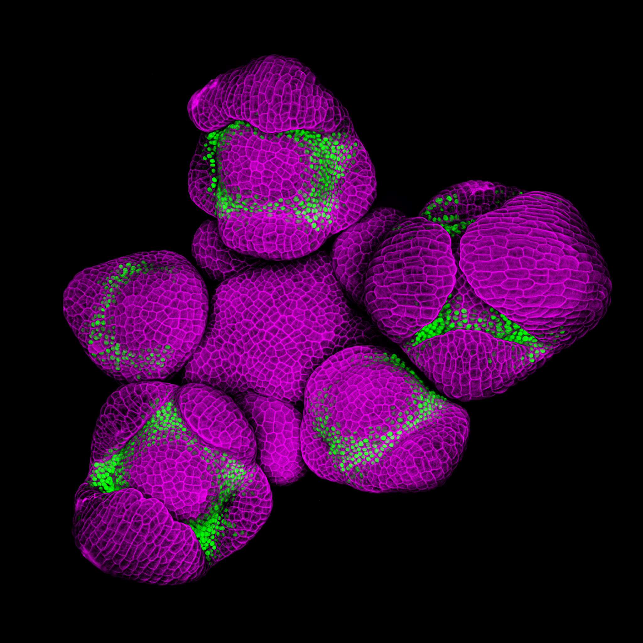
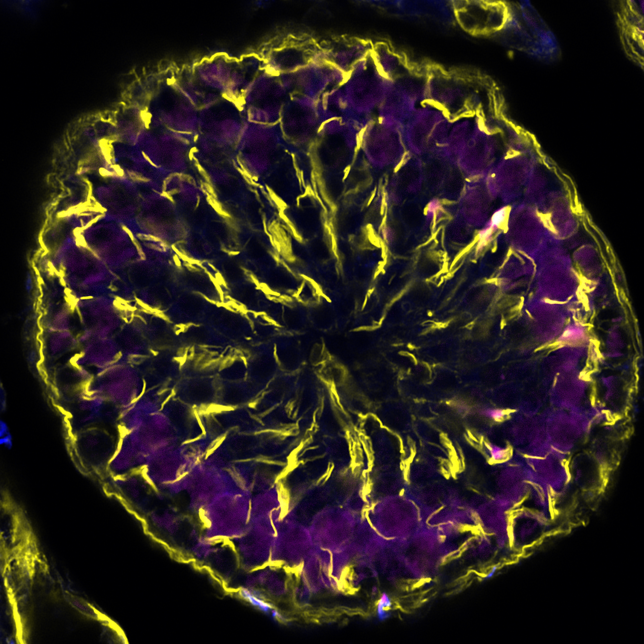
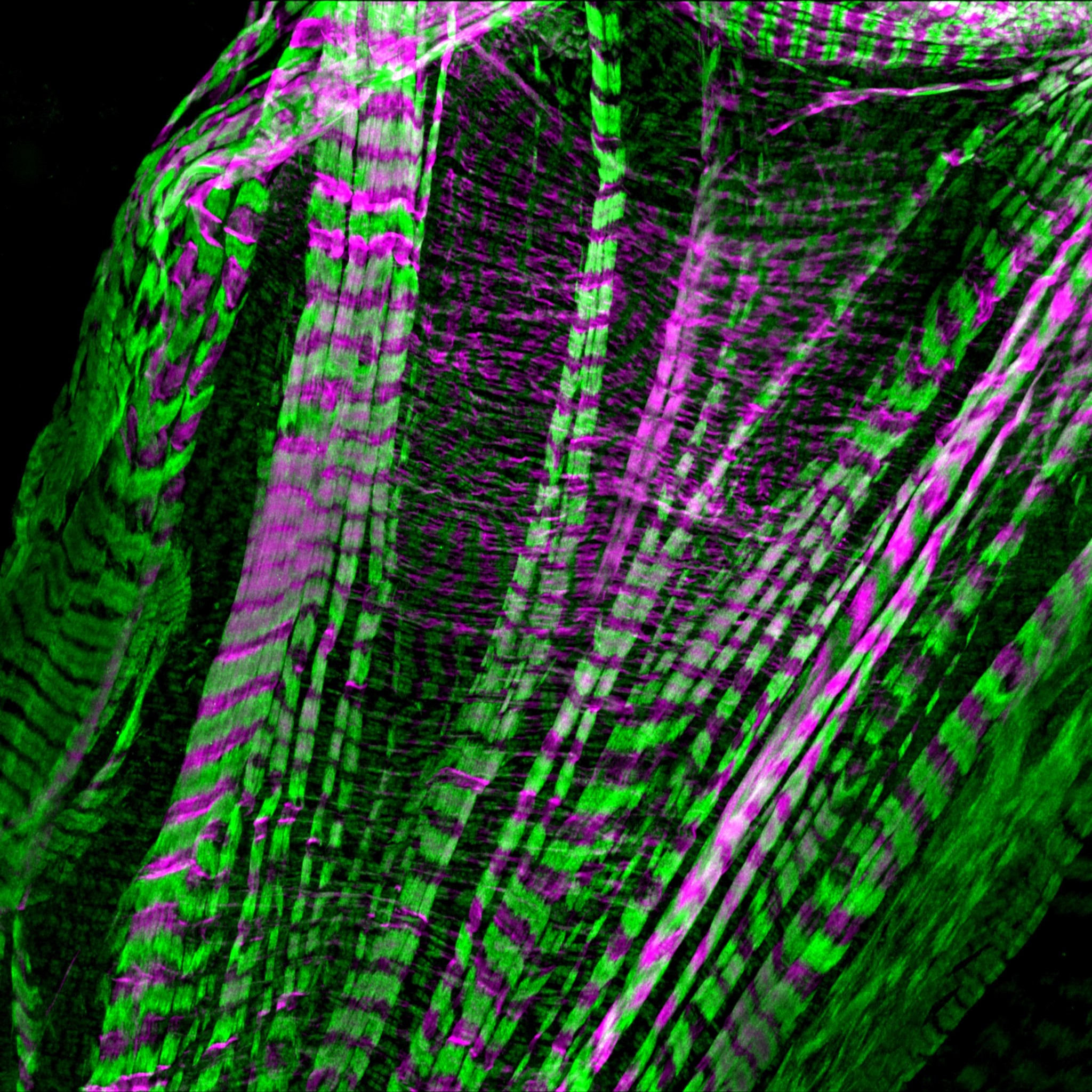
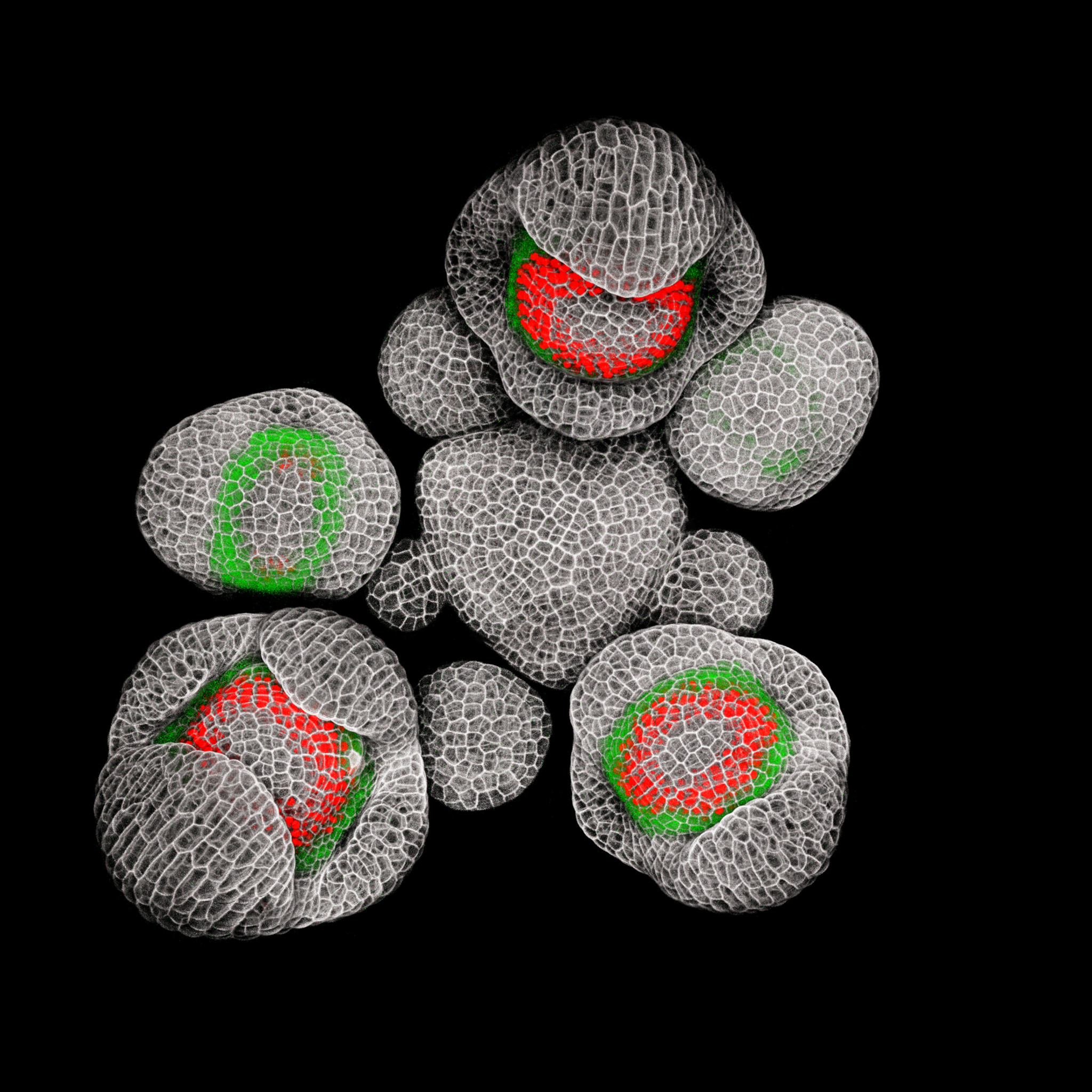
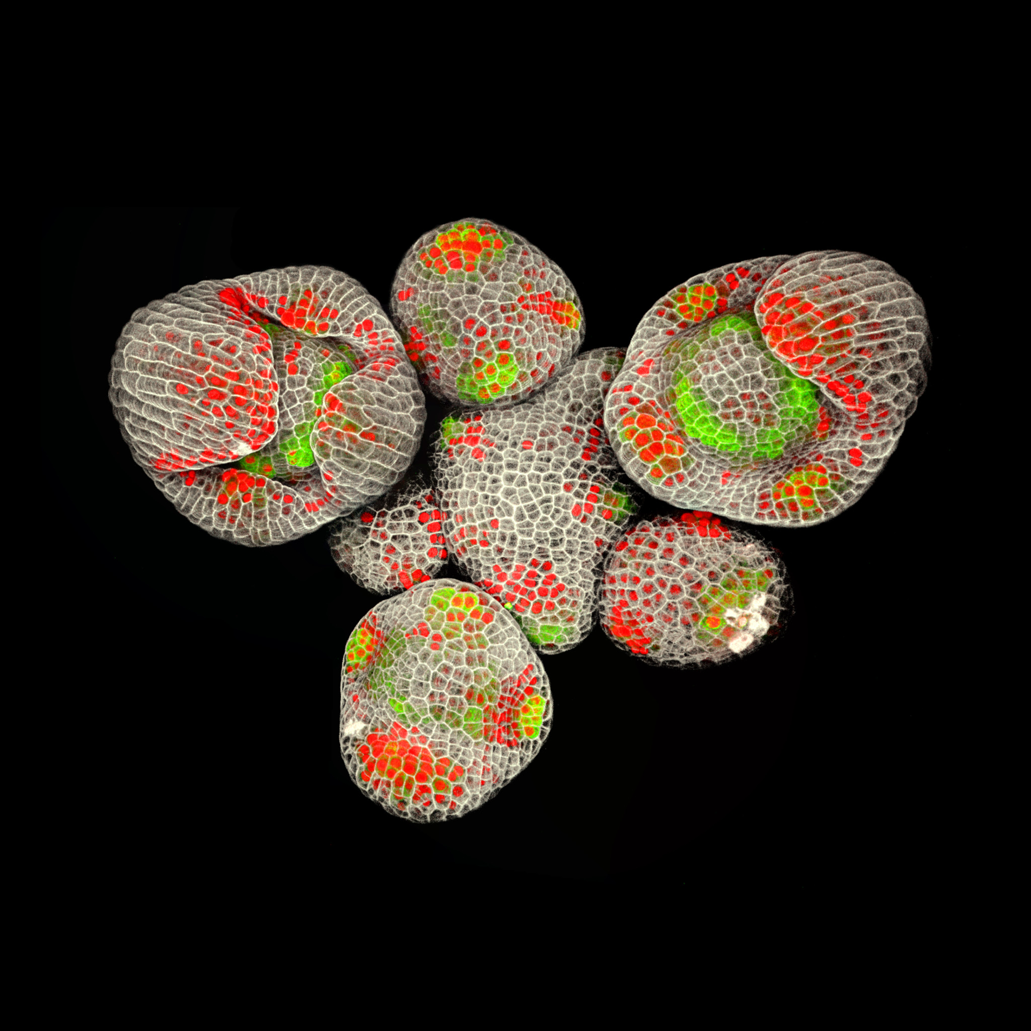
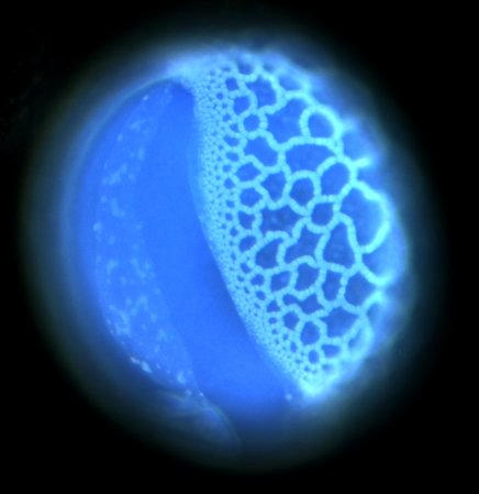
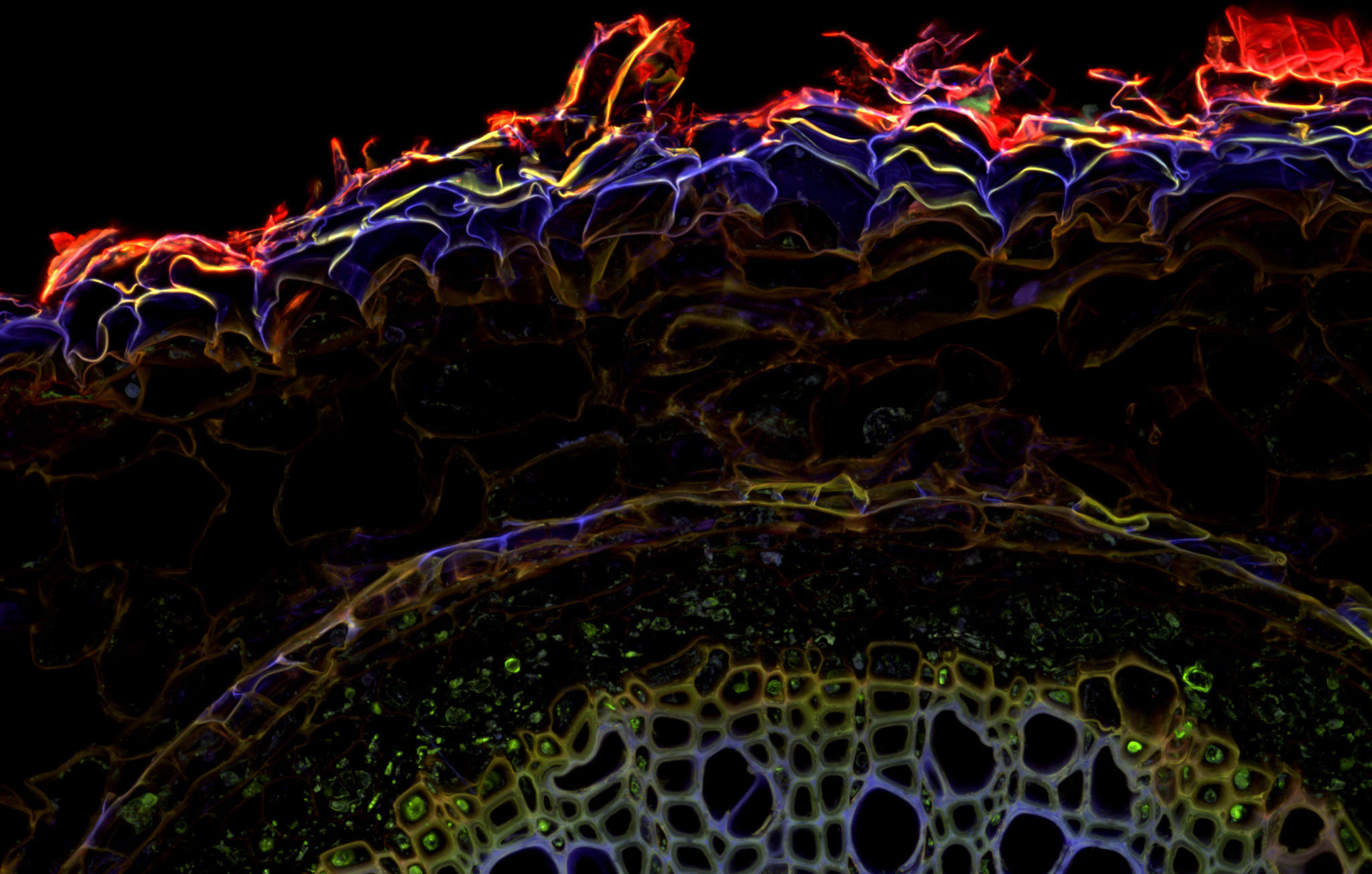
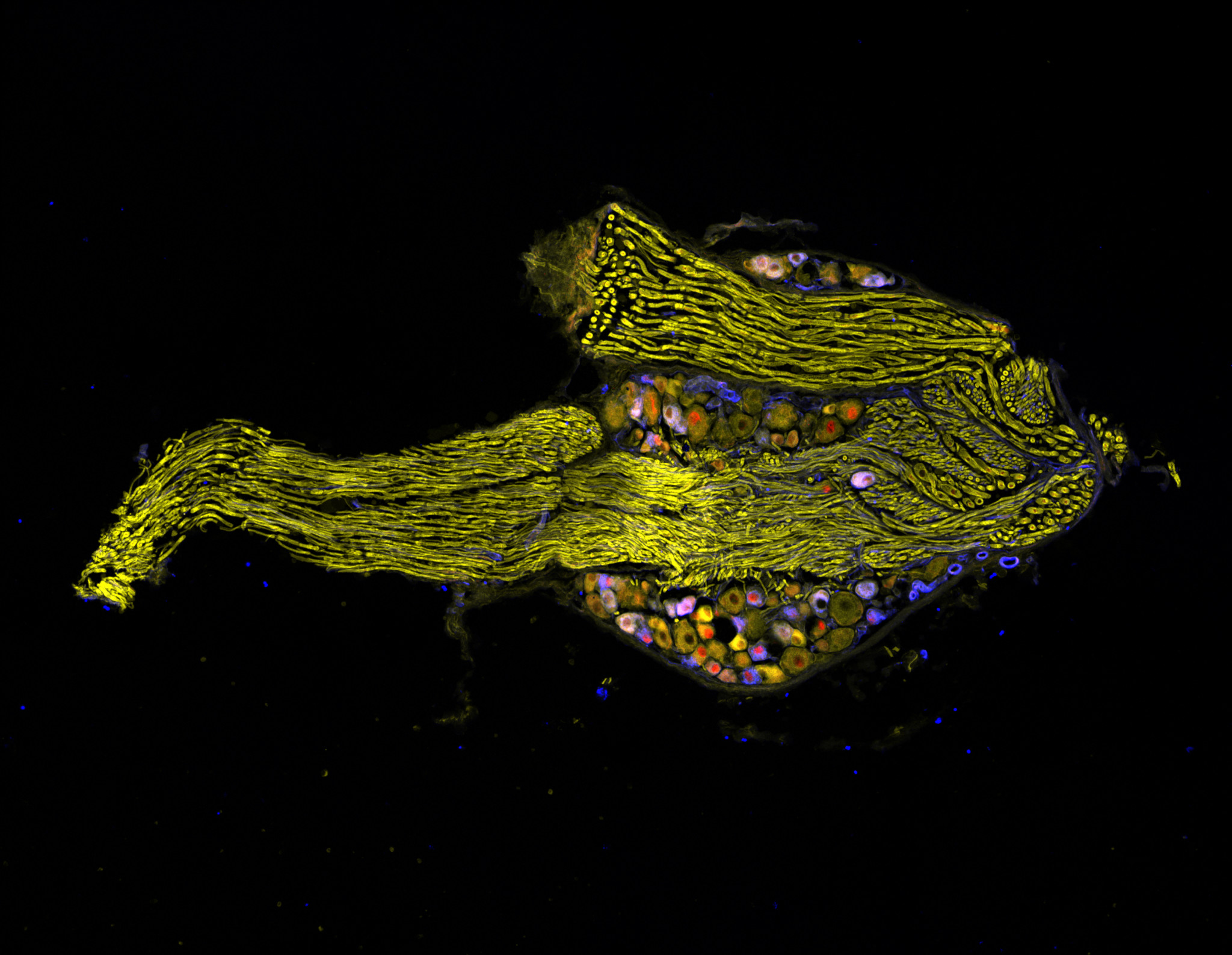
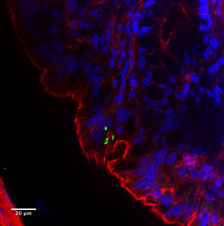
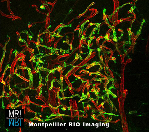
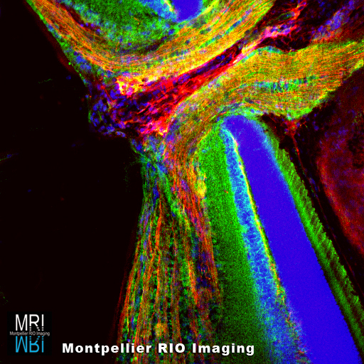
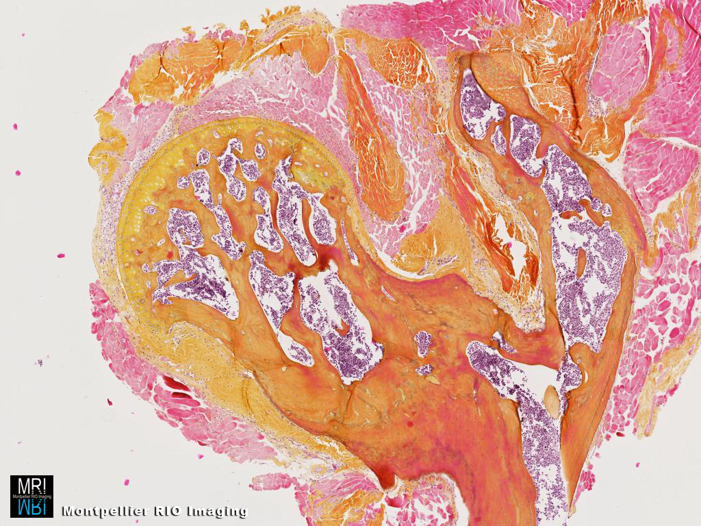
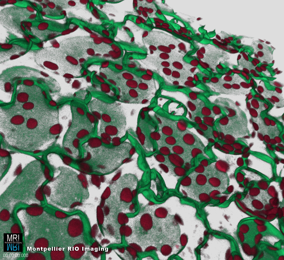
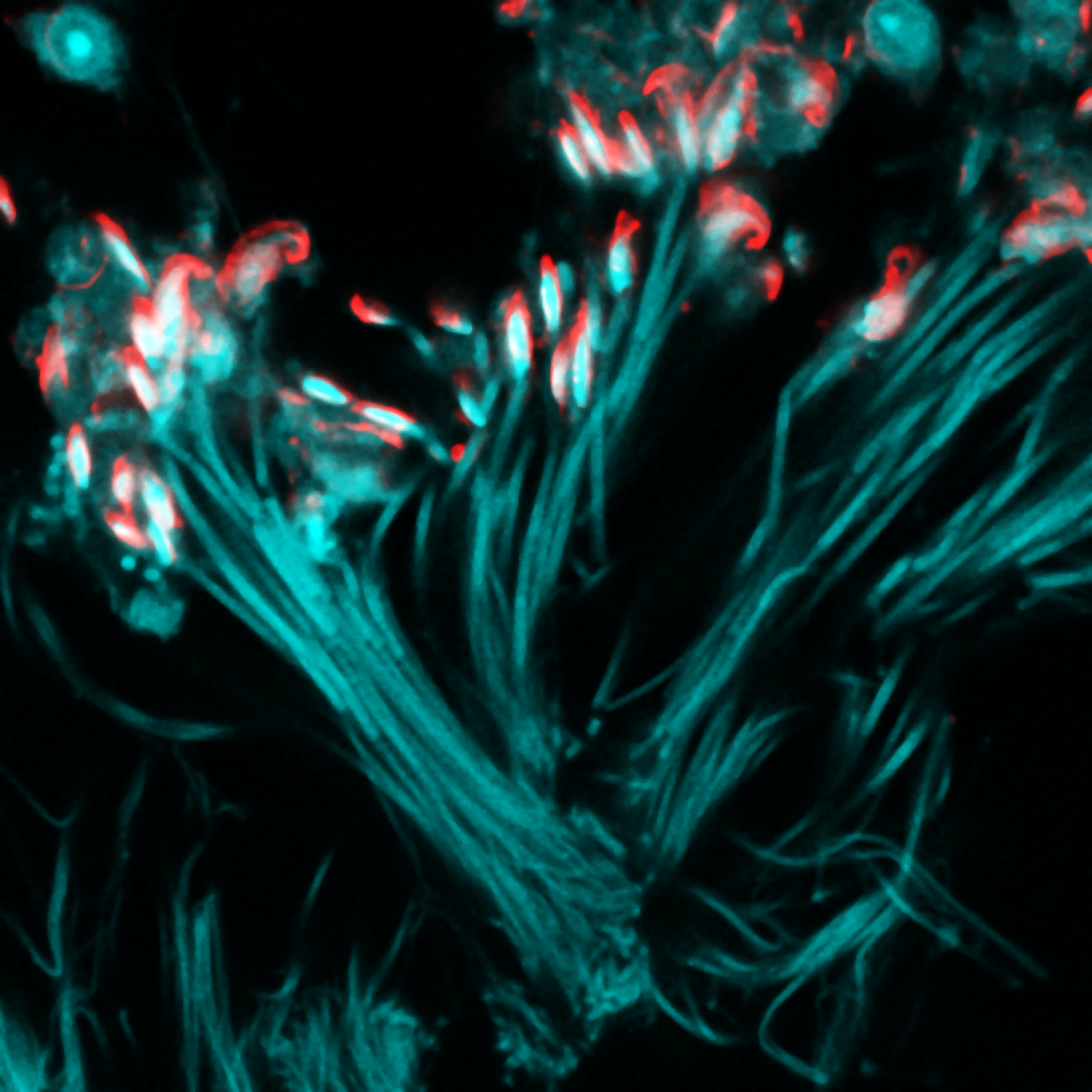
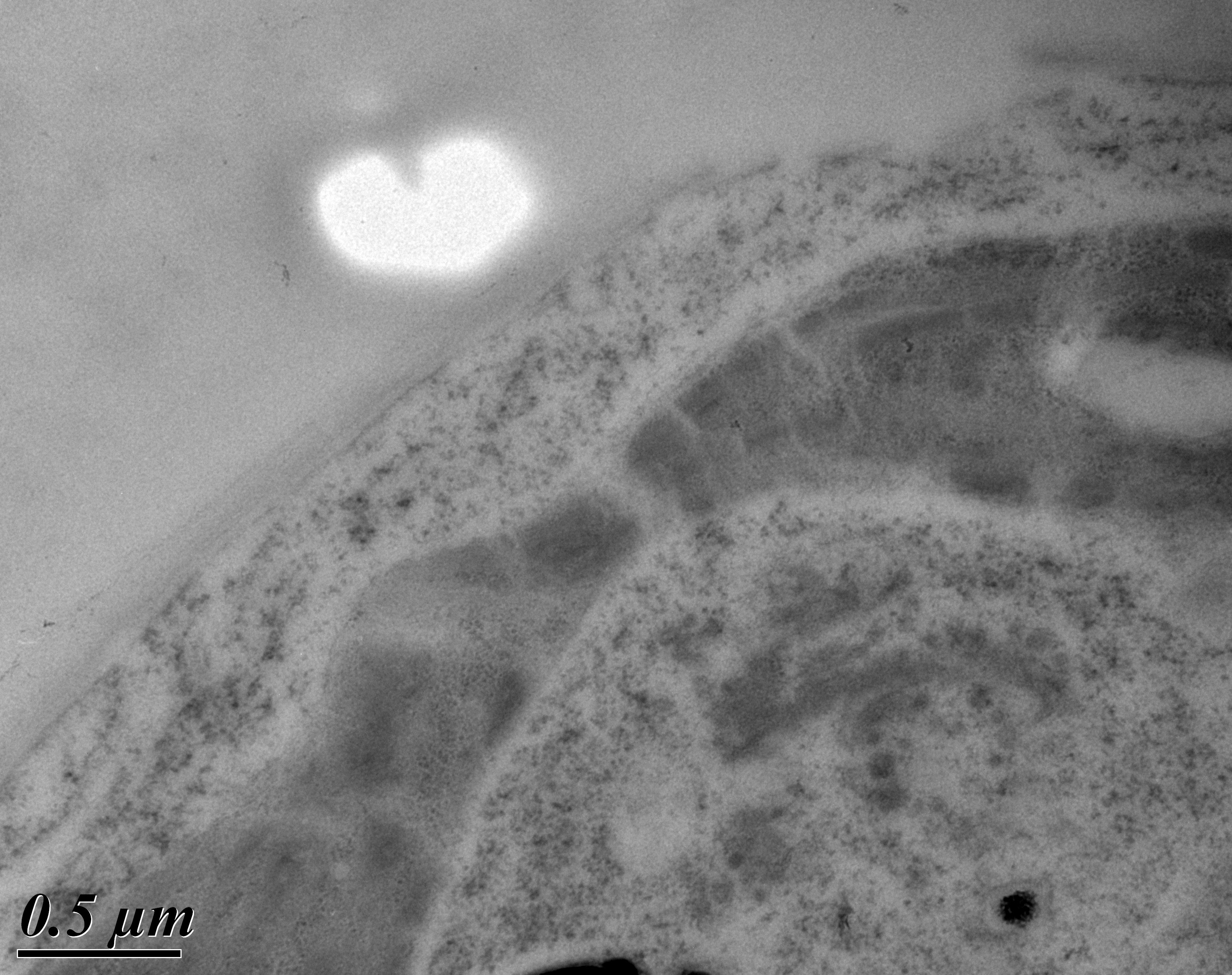
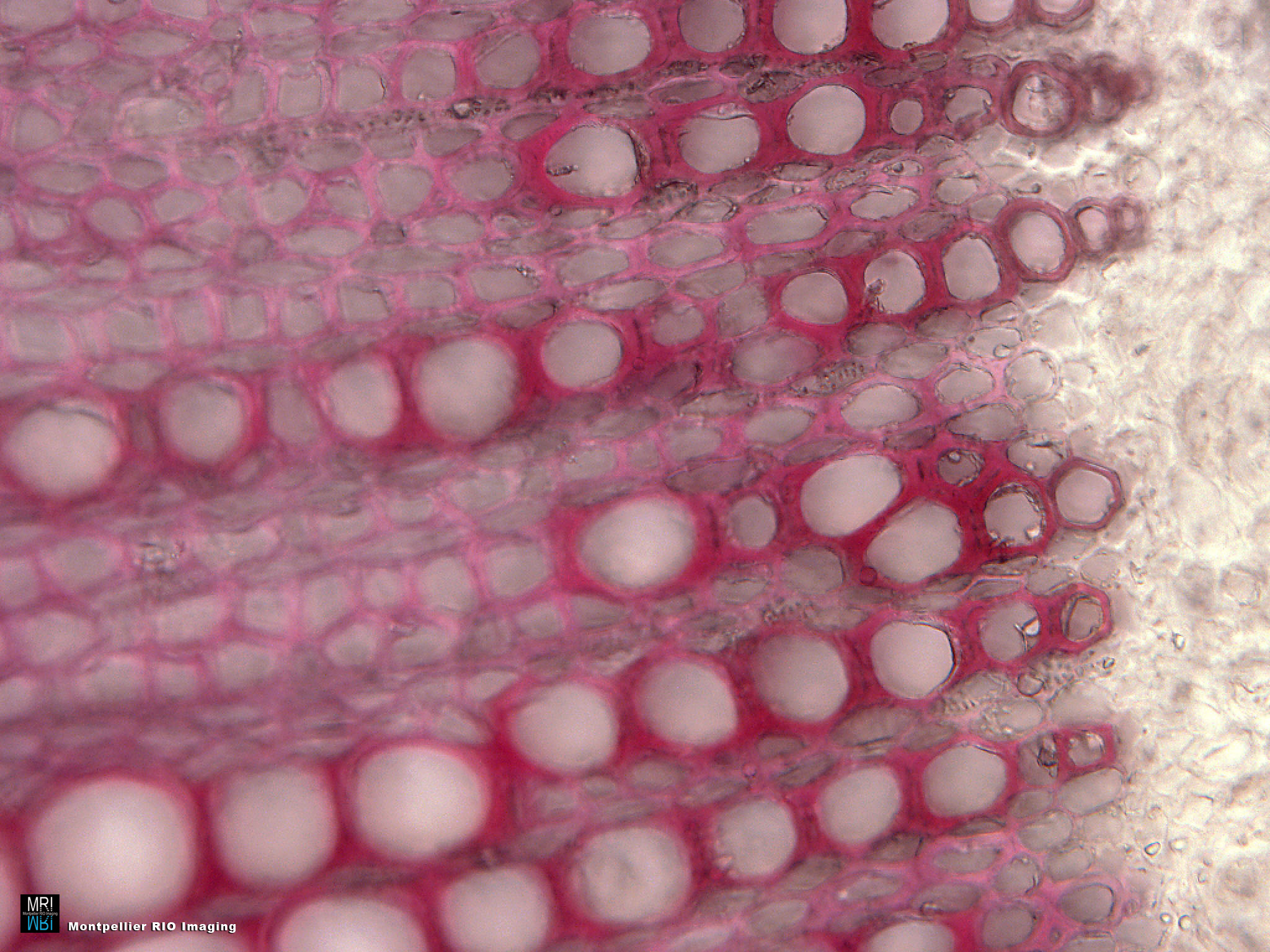
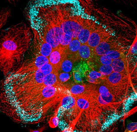
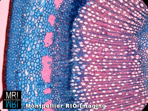
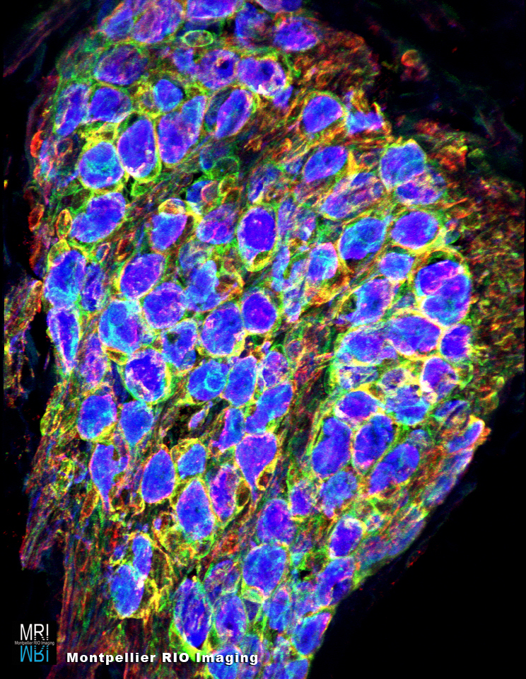
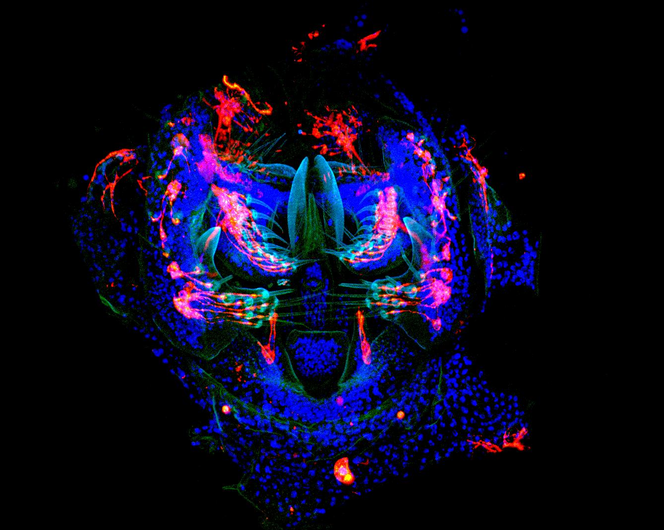
Gérard Alonso, Institut de Génomique Fonctionnelle (IGF), Département d’endocrinologie, Montpellier – Vascularisation de cerveau de rat
Pattern Face © Melina HEUZE – Institut Jacques Monod, CNRS, UMR7592 – ImagoSeine
MDCK cells seeded on fibronectin-coated micropatterns, fixed and stained for alpha-tubulin (green), gamma-tubulin (red) and DNA (blue).
Le caniche à sa mémère © PETREL Melina – Bordeaux Imaging Center – CNRS, Inserm, université Bordeaux Segalen
Particules magnétiques encapsulées de polymère.