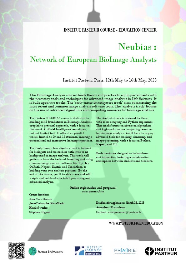
As the FBI Correlative Light-Electron Microscopy workshop is currently happening at the Bordeaux Imaging Center (FBI Bordeaux node), what a better occasion to highlight a correlative microscopy technique: The Array Tomography.

​Correlative Light and Electron microscopy (CLEM) increases our capacity of biological investigation. By combining light microscopy and electron microscopy, this complementary approach takes advantages of both techniques. In fact, light imaging provides valuable functional information thanks to its labeling power, whereas Electron microscopy excels at high resolution.

Where array tomography (AT) is special is that this technique is based on serial ultramicrotomy (cutting many sections less than a micrometer in thickness) of the sample, section collection onto support, and serial scanning EM (SEM) imaging.

An array of microscopy modes

Array tomography is a versatile microscopy method that offers opportunities to explore cell and tissues in three dimensions. This technique is well suited to image large tissue volumes of your sample with fine structural and molecular details.

Different modes are available, each having its own specificity and benefits:

  • The fluorescence microscopy AT mode (FM-AT) delivers volumetric resolution and molecular marker multiplexing highly superior to traditional fluorescence microscopies.
  • The electron microscopy AT mode (EM-AT) captures three-dimensional ultrastructure at size scales that would require prohibitive effort using traditional serial-section EM methods.
  • And of course, you can combine both modes in a unique one FM/EM-AT with three-dimensional light and electron images acquired in perfect volumetric data.

Why you should consider this technique next time?

The use of FM-AT should be considered for volumetric fluorescence imaging of fixed tissue specimens whenever there is need for very high resolution, high-order molecular multiplexing and/or rigorously depth-independent quantification of fluorescence signal intensities. Use of EM-AT offers perhaps the most convenient approach to volumetric electron microscopy available. Moreover, even though fields of applications are numerous, these attributes establish AT as an ideal choice for the most demanding analyses of diverse cellular architectures within mature and developing tissues such as brain tissue (neuroscientists, this technique is for you!).

Finally, while originally developed for EM, physical cutting of ultrathin sections was found to improve the axial resolution and provide accessibility to the sample for molecular labeling, beneficial for both, EM and light microscopy. Need another argument? The same or adjacent sections can be imaged with different modalities in correlative or even conjugate microscopy!

Get access to one of our services!

You need Array Tomography or another imaging technology or expertise that France-BioImaging provides? To get open access, please login via Euro-BioImaging website! You just have to choose the technology you want to use, then submit your proposal. All applications will be processed by the Euro-BioImaging Hub in close relation with France-BioImaging. And of course, all scientists regardless of their affiliation, area of expertise or field of activity can benefit from open access services! Users whose projects will be validated by Euro-BioImaging will benefit from a waiver for the access cost on France-BioImaging core facilities (https://france-bioimaging.org/access/).

Technical information: https://www.arraytomography.org/

Smith SJ. Q&A: Array tomography. BMC Biol. 2018 Sep 6;16(1):98. doi: 10.1186/s12915-018-0560-1

Launched earlier this year in coordination with the African BioImaging Consortium and Imaging Africa and within the framework of the Horizon Europe Programme, the Africa-France Joint Initiative for Biological Imaging aims at extending its partnership with colleagues in Africa that have interest in using advanced microscopy approaches for their own research programs and projects. With this in mind, we have previously designed two calls for funding: one for access to FBI’s bioimaging core facilities, the other as a twinning program.

Good news! Our first project has started! Granted by our second call, the Twinning program has begun between Stellenbosch University and FBI-Paris Node. A fantastic experience based on sharing practices, knowledge transfer and many fruitful discussions on image analysis and correlative approaches between light sheet and serial block face microscopy techniques. For the South African partner, Madelaine Frazenburg (Stellenbosch University), it is the opportunity to see how other microscopy laboratories in France works but also to learn more about cryo-SEM and to study new kind of sample preparation methods. From the French side, Ludovic Leconte (Institut Curie, FBI Paris-Centre node) is indeed very interested in gaining new experience in electron microscopy mainly in Serial Block Face, another tissue section imaging that is not available on his site and for which the Stellenbosch imaging platform has the mastery.

Our warmest thanks to Lize Engelbrecht, Professor Ben Loos and Janica Conradie for making this event possible and for the warm welcome they extended. The second stage of this “Twinning” project will take place at Institut Curie next spring. We look forward to welcoming Madelaine!

Atomic Force Microscopy (AFM) is a scanning probe microscopy technique that relies on measuring the interaction forces between a sharp tip and the surface of a sample to generate high-resolution images of its surface features and mechanical properties. A very broad range of sample types can be imaged with this technique at a very high resolution – at sub-nanometer level for some of them! Discover the AFM at the Montpellier node of France-BioImaging with Christine Doucet from Integrative Biophysics of Membranes team of the Centre de Biochimie Structurale.

Quickly visualize dynamic biological processes with High-Speed AFM

AFM provides images in physiological conditions, in liquid, over a length-scale ranging from few nanometers (single biomolecules) to tens of micrometers (living cells). In fact, the resolution depends on the tip radius and sample properties. For some of them, you can routinely obtain a nanometer lateral resolution and Angstrom axial resolution!

You want a video-rate version of the biological samples you are imaging? The High-Speed AFM, permits the acquisition of movies at approximately 10 images per second, enabling the visualization at nanoscale of dynamic biological processes involving biomolecular interactions, diffusion or conformational changes. It delivers nanometric resolved images typically at the same speed as conventional fluorescence microscopes!

Unravel the chemical information of your sample by combining AFM with…

AFM in ambient conditions and in liquids has a key limitation in that it does not directly provide chemical information about the sample being imaged. However, this limitation can be overcome by combining AFM with other techniques to obtain additional information about the sample’s composition. 

One commonly used technique in correlation with AFM is fluorescence microscopy. This combined approach of fluorescence labeling and AFM provides valuable insights into the chemical and biological properties of the sample. It was recently used on the Montpellier custom-made correlative AFM / fluorescence setup to observe the sublocalization of proteins in HIV-1 budding sites 1. They also used it to unambiguously attribute some unexpected configurations of the nucleoplasmic sides of Nuclear Pore Complexes 2. In these two cases, fluorescently-labeled proteins were imaged by dSTORM (direct STochastic Optical Reconstruction Microscopy). Of note, the lateral resolution of dSTORM and AFM are both in the 20 nm range with such samples, which makes their combination ideal!

In addition to fluorescence microscopy, AFM can also be correlated with other complementary techniques to obtain chemical information about the sample, such as Raman spectroscopy, Infrared Spectroscopy, X-Ray spectroscopy, microscopy and scattering.

Learn more about AFM applications

Here are 2 studies where Atomic Force Microscopy were essential: 

  • Structure and mechanics of the human nuclear pore complex basket using correlative AFM-fluorescence superresolution microscopy

Combining mechanical and superresolution measurements to reveal the plasticity of the Nuclear Pore Complexes

Nuclear pore complexes (NPCs) are the only gateways between the nucleus and cytoplasm in eukaryotic cells, facilitating the transport of selected cargoes of size from a few up to hundred nanometers. This versatility implies an important pore plasticity. Here, by combining atomic force microscopy (AFM) and single molecule localization microscopy (SMLM), a group led by France-BioImaging R&D team members Christine Doucet and Pierre Emmanuel Milhiet revealed that the NPC basket is very soft and explores a large conformational landscape: apart from its canonical basket shape, it dives into the central pore channel or opens, highlighting  how this structure can adapt, and let morphologically diverse cargoes shuttle through NPCs.

Vial et al., Nanoscale, 15, 5756-5770 (2023) 

  • The structure of pathogenic huntingtin exon 1 defines the bases of its aggregation propensity

Structural Biology meets Correlative Imaging

Huntington’s disease is a neurodegenerative disorder caused by an extended polyglutamine (poly-Q) tract in huntingtin. Here, using NMR, the team of Pau Bernado (CBS Montpellier) demonstrated that this poly-Q tract adopts long α-helical conformations. By adding correlative Atomic Force Microscopy and Fluorescence Microscopy data obtained in the FranceBioImaging facility PIBBS in Montpellier, they could demonstrate that the stability of this α-helix is a stronger signature than the number of glutamines, in defining the aggregation kinetics and the structure of the resulting fibrils, potentially linked to their pathogenicity.

Elena Real et al., Nature Structural & Molecular Biology, 30, 309–320 (2023)

How to use Atomic Force Microscopy at France-BioImaging?

Atomic Force Microscopy is open to collaborations under Proof-of-concept studies via Euro-BioImaging webportal (www.eurobioimaging.eu/service)! At the Montpellier node of France-BioImaging, you will be in contact with Dr Luca Costa (costa@cbs.cnrs.fr) with whom you will talk about the feasibility and the inherent experimental constraints linked to the technique. The collaboration procedure is discussed on a case-by-case basis, depending on the duration and technicity of the required experiments. Feel free to submit your project!

Thanks to Christine Doucet and Emmanuel Margeat for providing helpful information!

1. Dahmane, S., Doucet, C., Le Gall, A., Chamontin, C., Dosset, P., Murcy, F., Fernandez, L., Salas, D., Rubinstein, E., Mougel, M., et al. (2019). Nanoscale organization of tetraspanins during HIV-1 budding by correlative dSTORM/AFM. Nanoscale 11, 6036–6044.

2. Vial, A., Costa, L., Dosset, P., Rosso, P., Boutières, G., Faklaris, O., Haschke, H., Milhiet, P.-E., and Doucet, C.M. (2023). Structure and mechanics of the human nuclear pore complex basket using correlative AFM-fluorescence superresolution microscopy. Nanoscale 15, 5756–5770.

Age-related macular degeneration (AMD) affects more than 150 million people worldwide (early AMD) and 10 million of patients suffer from debilitating late stage AMD. Blurring central vision, this eye disease progresses over time, usually beginning when people are around their 50s or 60s by causing damage to the macula, in the retina. Researchers from the Institut de la Vision (Sorbonne Université, INSERM, CNRS, UMR_S 968) recently published about the AMD. Thanks to Serial Block-Face Scanning Electron Microscopy (SBF-SEM) experiments carried out at the ImagoSeine core facility (Institut Jacques Monod / FBI Paris-Centre node), they describe in this new study melanophages as a disease-progression marker.

Early or intermediate AMD is characterized by pigmentary changes and lipoproteinaceous debris accumulation between the photoreceptors and the melanosome-rich retinal pigment epithelium (RPE) or below the RPE. Later, AMD can be complicated by central choroidal neovascularization or by an expanding lesion of the photoreceptors. Even though patients with early or intermediate AMD can progress and develop late AMD, a large part of patients stay stable for years, underlining the potential usefulness of progress. 

AMD is associated with the appearance of hyperreflective foci, with reflectivity comparable to melanocyte-containing RPE cells. Thbs1 and CD47 are both important for the elimination of these cells. In the absence of either of them, melanocyte-containing RPE cells would then accumulate. The goal was to determine the origin of these cells in the retina, and the main question was: are these cells RPE migrating to the wrong place, or melanosome phagocytes cells having ingested melanosomes?

SBF-SEM: the key to answer this question

The Serial Block-Face Scanning Electron Microscopy (SBF-SEM) is a 3D electron microscopy imaging technique, where an ultramicrotome is placed inside a SEM. Biological samples are beforehand stained with heavy metals and embedded in a plastic resin block. Inside the microscope, a thin-section is cut at the surface of the block and discarded. Then, an image of the surface of the block – therefore inside the sample – is made, using back-scattered electrons. The process of cutting and imaging is repeated automatically as many times as necessary to produce a 3D stack of images inside the sample, as it is progressively imaged and destroyed. 

This technique allows 3D imaging of large samples for Electron Microscopy standards (up to several hundred microns in each of the X,Y,Z direction) at high resolution. This technique is often used to image whole cells, or even small pieces of tissues in 3D. The two major domains of application are to:

  • find a rare structure within a cell or tissue. The sample is imaged until the structure of interest is found.
  • understand the 3D spatial organization of organelles within cells, or of cells between them.

The benefits of bioimaging in this study

In the study, SBF-SEM was essential. As previously mentioned, AMD is associated with the appearance of hyperreflective foci, with reflectivity comparable to melanocyte-containing RPE cells. In the images produced by SBF-SEM, the retinal pigment epithelium (RPE) surrounding the melanophages in mice, where CD47 was inhibited, were markedly less pigmented and deformed compared to those where Thbs1 was blocked. This suggests that melanosomes have been transferred by phagocytosis from the RPE to nearby melanophages because they lack CD47. Finally, authors have shown that CD47 acts as a “don’t eat me” signal. The SBF-SEM was a great addition to this study where understanding the 3D spatial organization of the structure of interest was key.

Thanks to Jean-Marc Verbavatz for providing very helpful insights of the study!

Augustin, S., Lam, M., Lavalette, S. et al. Melanophages give rise to hyperreflective foci in AMD, a disease-progression marker. J Neuroinflammation 20, 28 (2023). https://doi.org/10.1186/s12974-023-02699-9

Get access to one of our services!

You need SBF-SEM or another imaging technology or expertise that France-BioImaging provides? To get open access, please login via Euro-BioImaging website! You just have to choose the technology you want to use, then submit your proposal. All applications will be processed by the Euro-BioImaging Hub in close relation with France-BioImaging. And of course, all scientists regardless of their affiliation, area of expertise or field of activity can benefit from open access services! Users whose projects will be validated by Euro-BioImaging will benefit from a waiver for the access cost on France-BioImaging core facilities (https://france-bioimaging.org/access/)

Massive intracellular accumulation of RPE-derived melanosomes in subretinal MPs of CD47−/−-mice causes subretinal melanophage formation and their clinical appearance as hyperreflective foci.

Correlative X-ray imaging and electron microscopy (CXEM) is the combination of X-ray imaging and electron microscopy. It is a correlative approach that makes it possible to characterise a sample of interest and locate a structure of interest in a non-destructive way. Nicolas BROUILLY is in charge of the Electron Microscopy Unit of the PICsL imaging facility on the Marseille node of France BioImaging, where CXEM is used for developmental biology studies. As part of Euro-BioImaging’s Proof-of-Concept study, his facility is now accepting applications from external users for CXEM projects. Learn more about how this approach works and what it can be used for in the interview below. 

We are today talking about CXEM imaging. Please provide a short summary of this type of imaging and tell us some applications:

Nicolas Brouilly: It is often very useful to combine 2 imaging modalities to take advantage of each while trying to lower their respective drawbacks. For example, by combining Light Microscopy and Electron Microscopy, we obtain the popular CLEM (for Correlative Light and Electron Microscopy). Visible light can then be used in combination with EM either:

  • To target a precise region of interest ;
  • To localize molecules within the ultrastructural information obtained by EM.

Using the same acronym building, CXEM corresponds to Correlative X-ray and Electron Microscopy. X-rays are photons of shorter wavelength than those from visible light, and can again be used to characterize a sample in 2 different ways:

  • To use their ability to easily go through tissues in order to record the 3D morphology of a sample: either by computed x-ray micro-tomography (or micro-CT) for micrometric resolution of big samples (mm to cm range) or by Soft X ray tomography for nanometric resolution of small samples (100’s of nm to um range);
  • To use a focused beam of high energy x-rays to analyse the localization of the elements of a sample: X-Ray Fluorescence microscopy (or XRF).

Both modalities can be used to complement the ultrastructural information obtained by electron microscopy. At the Marseille node of France BioImaging, in the Electron Microscopy Unit, we routinely use Correlative Micro-CT and Electron Microscopy to answer developmental biology questions.

What are some advantages of this technique that make it suited to addressing this type of question?

Nicolas Brouilly: The main advantage of Micro-CT (or Computed X-ray Tomography) is its ability to “see through” a sample and to reveal its overall organization in 3D without any labelling. The second advantage of Micro-CT is the fact that it is non-destructive. Thirdly, the contrast we usually give to samples for electron microscopy is compatible and even beneficial for X-ray imaging.

Altogether, this means that we can use X-ray tomography to map the microscale morphology of a sample in order to target a specific region of interest without having to go through the time-consuming and destructive collection of semi-thin sections.

We routinely use the micro-CT tool, not only to target a given organ or a given group of cells, but also to pre-orient the sample in order to cut it under a specific orientation. It is a timesaving tool within the frame of a 2D electron microscopy project, but it really is key within the frame of a 3D electron microscopy project given that Serial BlockFace and Focused Ion Beam techniques are destructive.

Tell us a bit more about a specific project that was done in your facility using this technology? What scientific questions were you addressing?

Nicolas Brouilly: Imagine that, first, you have a ball of yarn, second, you cannot untangle it, and third, you want to cut small bits of the thread at 24 cm from the end (not 22, not 26… 24 !). CXEM enabled us to do this on Drosophila gut. The micro-CT gave us the 3D map of the sample within the resin block. We could then use this map to find the best itinerary within the sample to make transverse sections of the portion of interest that was precisely indicated by the user on the micro-CT dataset. At the end of the day, the user was able to look at perfect transverse ultrathin TEM sections, at a precise position of this ball of yarn that Drosophila gut is. He could finally get precise metrics from this precise part of the gut in several samples. None of this could have been achieved without CXEM.

Like a ball of yarn… Above is an example of how CXEM can be used to find the best itinerary within a sample to make transverse sections of the portion of interest. On the left, the micro-CT provided a 3D map of the sample within the resin block. On the right, a transverse ultrathin TEM section of the drosphilia gut. Image courtesy of Nuno Luis (Schnorrer lab, IBDM) & Nicolas Brouilly (Electron Microscopy Facility, IBDM AMU/CNRS, France BioImaging).

For another example, you can have a look at the following paper where we used CXEM to map platelet aggregates within arteries in order to explore them by Serial BlockFace SEM, another example of “Find a needle in a haystack”. Have a look at movie S1, it is a wonder that we could not obtain without CXEM:

Estelle Carminita, Lydie Crescence, Nicolas Brouilly, Alexandre Altié, Laurence Panicot-Dubois, Christophe Dubois, DNAse-dependent, NET-independent pathway of thrombus formation in vivo, Proc Natl Acad Sci U S A. 2021 Jul 13;118(28)

Finally, below is the seminal paper from the Schwab lab at EMBL, which revealed the power of micro-CT to the Electron Microscopy community : 

Matthia A. Karreman, Luc Mercier, Nicole L. Schieber, Gergely Solecki, Guillaume Allio, Frank Winkler, Bernhard Ruthensteiner, Jacky G. Goetz, Yannick Schwab, Fast and precise targeting of single tumor cells in vivo by multimodal correlative microscopy, J Cell Sci (2016) 129 (2): 444–456.

Contact: nicolas.brouilly@univ-amu.fr

How to apply:

CXEM is part of the Euro-BioImagingProof-of-Concept study. The Proof-of-Concept study makes it possible to introduce exciting, new imaging technologies to our portfolio that were previously unavailable via our network. We are currently accepting applications to use these technologies at participating Nodes as part of the Proof-of-Concept study. Be part of this study – and contribute to community-wide continuous technological innovation!

All scientists, regardless of their affiliation, area of expertise or field of activity can benefit from Euro-BioImaging’s pan-European open access services. Potential users of these new technologies are encouraged to submit project proposals via our website. To do so, you can Login to access our application platform, choose the technology you want to use and the facility you wish to visit, then submit your proposal. All applications will be processed by the Euro-BioImaging Hub. As usual, users will benefit from advice and guidance by technical experts working at the Nodes, training opportunities, and data management services.

For more information: info@eurobioimaging.eu

From left to right: Nicolas BROUILLY (Head of facility), Aïcha AOUANE (Engineer Assistant), Fabrice RICHARD (Engineer)

Thanks to Nicolas Brouilly for the article!
Original article on: www.eurobioimaging.eu/news/cxem-finding-a-needle-in-a-haystack

An innovative technology to look at thick samples at high resolution? Marc Tramier, a group leader at the Institute of Genetics & Development of the University of Rennes/INSERM/CNRS, and scientific director of MRic (Microscopy Rennes Imaging Centre), is currently working with his team on Random Illumination Microscopy (RIM), a fast and easy to use microscopy technique with low phototoxicity. His facility, which is part of the Bretagne-Loire Node of France-BioImaging, offers RIM as a Euro-BioImaging Proof-of-Concept study, and is now accepting applications for projects. He explains the ideas behind RIM in the article below.

The idea of Random Illumination Microscopy is to use the speckle of the illumination laser in wide field to create a structured illumination pattern at the diffraction limit. By varying the pattern from image to image using a diffracting element (in our case a SLM), scientists are able to acquire a stack of images (around 100 images) on a camera which corresponds to a cumulative homogeneous illumination. By resolving the inverse problem, a super-resolved image is, then, reconstructed, at the focal plane with unprecedented optical sectioning. In comparison to conventional SIM, RIM is able to work in depth inside diffusive samples as the speckle is insensitive to diffusion. 

A transfer full of advantages

The method was first implemented by Thomas Mangeat – that we are happy to welcome in our new Toulouse node! – and collaborators in Toulouse (Mangeat et al., 2021. doi: 10.1016/j.crmeth.2021.100009). In the MRic, after the transfer of the prototype, the facility was able to image microvilli of intestine in c-elegans (depth > 50µm) having a spatial resolution of around 100 nm. This structure is impossible to be revealed by conventional confocal microscopy. Before the use of RIM, only the airyscan approach allowed us to resolve the microvilli but with higher illumination power (photobleaching of the sample) and longer acquisition time (around 10 times more). Now with RIM, we are able to follow microvilli in the living C–elegans at the second time-scale during several minutes.

RIM is one of the powerful methods to achieve super-resolved images in depth at high speed with very low phototoxicity. This makes a very nice compromise of z-sectioning and super-resolution with wide field illumination particularly adapted to thick live samples. Beside reconstruction and data analysis, MRic is offering a user-friendly system, with a complete set of microscopy methods for live sample investigation, from wide-field to light sheet including spinning disk, confocal and airyscan. And of course, this technology is available in open access through France-BioImaging and Euro-BioImaging!

How to apply to use RIM:

Random Illumination Microscopy is part of the Euro-BioImaging Proof-of-Concept study, in collaboration with our Nodes. The Proof-of-Concept study makes it possible to introduce exciting, new imaging technologies to our portfolio that were previously unavailable via our network. We are currently accepting applications to use these technologies as part of the Proof-of-Concept study. Be part of this study – and contribute to community-wide continuous technological innovation!

All scientists, regardless of their affiliation, area of expertise or field of activity can benefit from Euro-BioImaging’s pan-European open access services. Potential users of these new technologies are encouraged to submit project proposals via our website. To do so, you can Login to access our application platform, choose the technology you want to use and the facility you wish to visit, then submit your proposal. All applications will be processed by the Euro-BioImaging Hub. As usual, users will benefit from advice and guidance by technical experts working at the Nodes, training opportunities, and data management services. 

For more information:  info@eurobioimaging.eu 

Thank you Marc Tramier and Marianna Childress, communication officer of Euro-BioImaging, for the original article.

From February 6th to February 10th, France-BioImaging organised a group meeting on the project “FBI.data” in Bordeaux. For a week, participants focused on the architecture and the implementation of image data management tools. A user-friendly response to the challenges of never-ending data production. 

New imaging technologies are very greedy in terms of image processing and data management. Beside the image itself, biological imaging generates a huge amount of metadata. The FBI.data project, one of the key missions of France-BioImaging, addresses the questions related to the computational analysis and handling of image data. 

Speeding up the implementation of tools across the infrastructure

Although the distributed FBI.data team meets once per week, the FBI.data Sprint aims at only focusing on data management scenarii and accelerating the project. Two essential aspects have been discussed:

  • First of all, the data management system architecture must be simple for them to be implemented across France-BioImaging nodes. It also has to be compatible with long-term data storage and of course, to be user-friendly – we want to keep it easy for our users! 
  • The second point is all about anticipating as many data management cases as possible. Running through all the needs of bioimaging experts and users, the team lists the specific features of each case and considers the perfect solutions for all of them.

Working for the FAIRisation of data

The FAIRisation of data for Open Science is an initiative fully endorsed by France-BioImaging. Meaning that data are Findable, Accessible, Interoperable and Reusable, the benefits for the bioimaging community are numerous. It improves transparency and reproducibility, enhances quality of results, accelerates scientific progress and method development and finally boosts collaboration within the scientific community. 

OMERO, developed by the University of Dundee & Open Microscopy Environment teams, on which the FBI.data group is working, is one of the software making user data FAIR. Being a microscopy image data management decentralised platform, it helps organise, access and archive data. Besides, it combines image and metadata storage, a viewer and data analysis resources. Furthermore, OMERO is linked to the most valuable tools for bioimaging experts (ImageJ, Napari, QPath, etc.). And users can access their data from anywhere and keep them safe.

But much more has to be set up to have functional solutions: to ease user authentication and management, manage big data transfer, and have an adequate metadata scheme. Accompanying users is one of the mission of the FBI.data team, and the FBI.data Sprint is also the occasion to join efforts from the training working group led by the training mission officer Fabrice Cordelières (Bordeaux Imaging Center) to produce adequate training material on data management.

Sharing efforts and helping the community

The FBI.data working group is composed of:

By joining their skills and experience, they are working together on setting up tools and good practices for the management and FAIRisation of data inside France-BioImaging nodes but also for the entire bioimaging community. With this in mind, the project has collaboration with, among others, the Institut Français de Bioinformatique (IFB) and the Centre National de Ressources Biologiques Marines (EMBRC), and other infrastructure through the MUDIS4LS Equipex+ project. Moreover, the FBI.data project has an open GitLab, providing image data management codes in open source, and a blog with tutorials, recommendations and so much more!

Check the France-BioImaging OMERO web portal: https://omero-fbi.fr/

And its gitlab FBI data · GitLab (in2p3.fr)

Learn more about our missions and working packages: https://france-bioimaging.org/about/work-packages/

Being designed in response to imaging challenges, the Roboscope is the product of a collaboration between Marc Tramier’s team (FBI Bretagne-Loire node) with Julia Bonnet-Gélébart, research engineer, Jacques Pécréaux’s team of the Institut Génétique & Développement de Rennes (IGDR), and the Inscoper company, spin-off of the lab. This technology could become a great timesaver for fluorescence microscopy.

Allowing the automation of fluorescence microscope acquisitions, the Roboscope is an embedded technology based on a deep learning algorithm. To be precise, it is a predesigned event-driven acquisition (PEDA) based on a learning automatization of any cellular changes tracked by fluorescence. Catching rare and fast cellular events then becomes possible!

The use of the Roboscope would also save precious time of research, providing users with results without the need to stand by the microscope during acquisition. This technology goes beyond as they will be able to recover the data already classified and with only the specific points of illumination that they have previously triggered. 

A broad range of applications

The teams have almost finished to develop an entire algorithm monitoring the cell cycle progression in mitosis. These events specific to the cellular division correspond to major challenges in the control and treatment of cancer progression (Kops, 2005). As the cell cycle study is needed to understand several biological processes helping the development of targeted drugs, the technology aims to monitor efficiently and automatically simple cell models through their division cycle. 

And this is not its only benefit: this automatized fluorescence microscopy acquisition can be adapted in very diverse fields. From a cell cycle progression analysis to specific analysis, organelles, proteins and biological events can be tracked or activated within cells. A noteworthy advantage of the integrated device that – we hope – will be deployed widely in the future. 

Workflow of a Roboscope experiment. 1. The user annotate a bench of images with different class of interest to be detected. 2. The pre-trained Convolutional Neural Network is adjusted for the experiment by fine tuning and/or transfer learning. 3. The algorithm is transfered on embedded systems to perform real-time image analysis during microscopy acquisition. 4. The biological application with event-driven acquisition is defined and started by the user in order to start, interrupt and parametrize different acquisition sequence following real-time image analysis and event classification.

MicroPICell core facility offers access and services to a broad range of bioimaging technology and expertise, specialized in cell and tissue imaging. Based in Nantes, the core facility is now certified ISO 9001 and NFX 50-900, demonstrating their investment in providing quality services to its users.

The ISO 9001 :2015 and NFX 50-900 :2016 standards ensures good practices in terms of organization and management of a life science core facility. These standards are focusing, among other criteria, on the ability to fulfill its scientific and technological research missions, to consistently provide products and services that meet customer and legal requirements and finally, aims to increase the satisfaction of its customers through the effective application of its management system.

A supported process

To implement the first standard, experts from MicroPICell core facility have been trained in the management of the required quality system. The core facility staff have then been supported, in close collaboration with the GIS Biogenouest, by the head of the IBISA quality mission in order to build and implement the quality system according to both standards. This “Groupement d’intérêt scientifique” also has a quality network, Iquare, to which the core facility participates in, to exchange, share and be advised in the implementation of the quality system. The NFX 50-900 :2016 standard has been applied on R&D projects such as the establishment of a digital histology center delivering deep learning data processing, smart microdissection or the imaging of thick samples in collaboration with the company Kaer Labs.

Guaranteeing quality to users

This double certification is a recognition of the core facility’s quality approach, and allows MicroPICell to guarantee to their industrial and academic users that the implemented tools and procedures meet the requirements of the standards. This quality approach facilitates day-to-day management and internal communication at the platform and with the various parties concerned. Above all, it is a way of continuously improving bioimaging core facilities and ensuring that the missions are carried out efficiently.

Since 2019, the “Cristal collectif” medal rewards teams supporting research with their technical expertise, the collective dimension, their innovation and outreach. Both nationally and internationally recognised, the Bordeaux Imaging Center (BIC) from the France-BioImaging node of Bordeaux received this award for providing access to innovating technologies and for the quality of its training. The BIC was commended for its investment in training, especially for its partnership with the International School of Neurosciences, a unique partnership in Europe. The CNRS also has particularly highlighted the core facility’s activities of research and development in implementing new techniques and image analysis. Among its achievements, the BIC has succeeded to optimize a homemade Lattice Light Sheet, which has the benefit of being a good compromise between resolution, acquisition speed, imaging depth and low phototoxicity.

© Gautier Dufau

Laureates :

  • Lysiane Brocard, Plant Unit manager
  • Fabrice Cordelières, Image analysis manager
  • Mathieu Ducros, R&D Lattice Light Sheet Microscopy manager
  • Mónica Fernández Monreal, R&D CLEM manager
  • Étienne Gontier, Electronic Unit manager
  • Sabrina Lacomme, Transmission Electron Microscopy manager
  • Florian Levet, R&D software manager
  • Sébastien Marais, Confocal and Two-photon Microscopy manager
  • Magali Mondin, Super-resolution Microscopy manager
  • Melina Petrel, Cryo-preparation and immunomarking manager
  • Christel Poujol, Photonic Unit manager
  • Isabelle Svahn, Scanning Electron Microscopy manager
  • Jérémie Teillon, Clarification and Light-Sheet Microscopy manager

More information: www.cnrs.fr/fr/talent/index

Imagerie-Gif core facility, from our Ile-de-France Sud node, is pleased to announce the acquisition of a Scanning Ion Beam Electron Microscope (FIB-SEM) and a Lattice Structured Illumination Microscope (SIM) Elyra 7. For the occasion, the core facility is organizing “3D Res/volution“, a scientific event on high-resolution 3D imaging on December 15, 2022 from 2:00 pm to 5:00 pm at B21 amphitheatre. This event will be a great opportunity to introduce to you the possibilities of these 2 new systems available at Imagerie-Gif.

Free but mandatory registration: https://evento.renater.fr/survey/3d-res-volution-day-8pdzetbi

Initiated a few years ago, the Inria-IPL-NAVISCOPE (“Image guided NAvigation and Visualization data sets in live cell imaging and microscopy”) project aims at overcoming challenges of bioimaging observation. Virtual and augmented reality could become the new way to visualize and analyze microscope image renders.

Despite incredible progresses in microscopy, imaging biomolecular dynamics in cells remains a challenge. A lack of sensitivity, limited recording speed, photobleaching and phototoxicity associated have restrained, for a long time, our capacity to study biomolecules in their natural environments. As microscopy image is commonly observed on 2D screens, it can narrow human capacities to grasp volumetric, complex, and discrete biological dynamics. Following new modes of visualization including virtual reality (VR)/augmented reality (AR) approaches, the NAVISCOPE project allows more accurate analysis and exploration of large time series of volumetric images, such as those produced by the latest 3D + time fluorescence microscopy.

Why should cell biologists be interested in this project?

The project to which 4 FBI-teams from the BI-IPDM node participate, aims at engineering a technology made with and for biologists. For VR/AR approaches to be adopted by the broader bioimaging community, it is, indeed, important that they are evaluated by the biologists, on their own datasets. 

The potentials of VR/AR technologies for scientists are numerous: navigating into multidimensional, large data sets with another view angle or perception, interacting with these data especially by selecting subregions, quantifying features of interests, etc. New VR/AR approaches also provide specific quantification tools to show distances, angles, counting, local density, and histogram profiler or include a selection of regions of interest for further analysis such as the 3D Timelines. Moreover, because communication with analysis software coded in Java or Python is now integrated, more post-treatment analysis is possible on selected features, providing a multifaceted and accessible tool for biologists.

A promising future ahead

In practice, immersion of the user within 3D + time microscopy data still represents an acculturation challenge for the concerned community. Thus, to promote a broader adoption of these approaches by biologists, further dialogue is needed between the bioimaging community and the VR&AR developers. Nonetheless, future innovation can already be foreseen as there are multiple way to upgrade this technology. For example, using eye-tracking (Günther et al., 2020) or haptic interfaces (Petit et al., 2020) can improve human perception by providing local sensations, which would improve the selection of responses in a 3D + time space. Besides, a better integration of multiple channels with high pixel resolution or the addition of vector representations could add information about the orientation, movement of molecules or organization of structures such as cytoskeleton elements or membrane lipids. The prospects initiated by the NAVISCOPE projects are, as mentioned above, endless and could be a technology that reshapes the way we see biology at the hearth.

Full article on:

Challenges of intracellular visualization using virtual and augmented reality

Valades-Cruz Cesar Augusto, Leconte Ludovic, Fouche Gwendal, Blanc Thomas, Van Hille Nathan, Fournier Kevin, Laurent Tao, Gallean Benjamin, Deslandes Francois, Hajj Bassam, Faure Emmanuel, Argelaguet Ferran, Trubuil Alain, Isenberg Tobias, Masson Jean-Baptiste, Salamero Jean, Kervrann Charleseub

Front. Bioinform. 2:997082.
https://doi.org/10.3389/fbinf.2022.997082