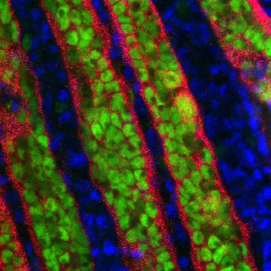
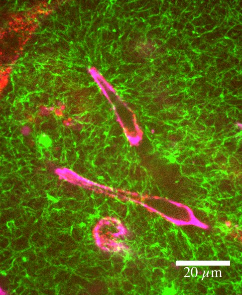
Embryonic murine lacrimal gland (E17), with fluorescent staining showing Actin, Fibronectin, phospho-MLC and nuclei. The acquisition was performed using a confocal microscope Zeiss LSM 980, with a 25X air objective.
Embryonic murine lacrimal gland (E17), with fluorescent staining showing Actin, Fibronectin, phospho-MLC and nuclei. The acquisition was performed using a confocal microscope Zeiss LSM 980, with a 25X air objective.
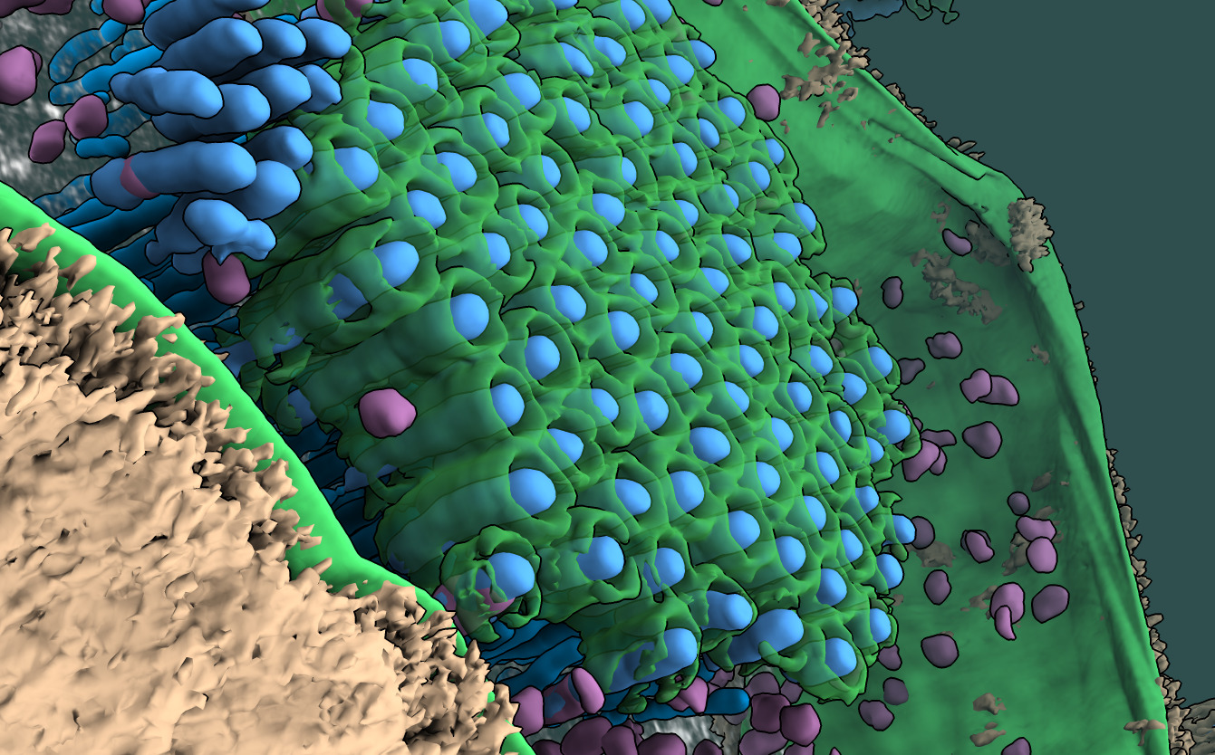
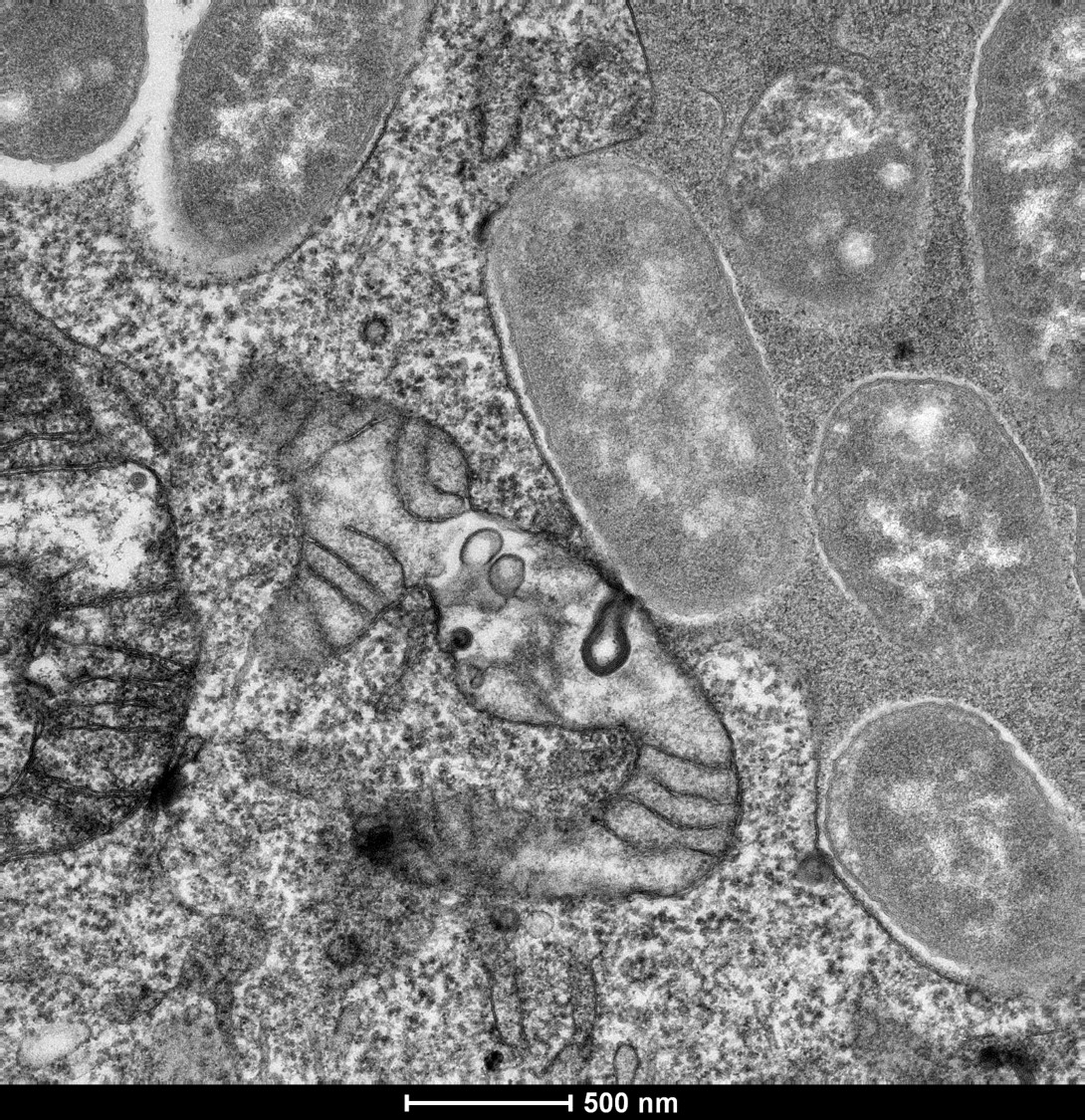
Sulfolobus islandicus filamentous virus (SIFV) inside its host (archeon). Although SIFV is an enveloped virus, it acquires its envelope and reaches its full maturity when inside the host cell cytoplasm! As SIFVs are twice in length than the diameter of their host cell they are tightly packed in twisted bent bundles. In the annotated volume: Membranes are displayed in green color (cell membrane and viral envelopes), viral nucleocapsid in light blue, ribosomes in magenta and S-layer in wheat (light gold).
Microscopy technique: Dual-axis electron tomography & Volume annotation using Convolutional Neural Networks
A triple-negative cancer cell navigating its path through a dense 3D collagen matrix network. Collagen is labeled in green; Actin in magenta, and plasma membrane structure, caveolae, component in red. The image was acquired through a spinning disc confocal microscope for a 3D migration time-lapse experiment.
Transmission electron microscopy observation of cells infected with Yersinia pseudotuberculosis in order to see their intra-vacuolar localization.
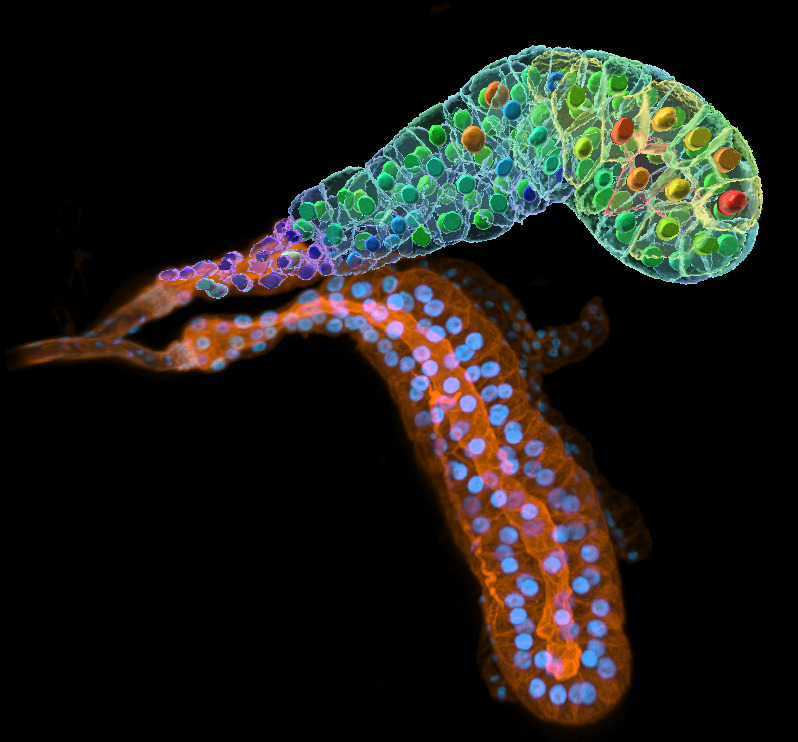
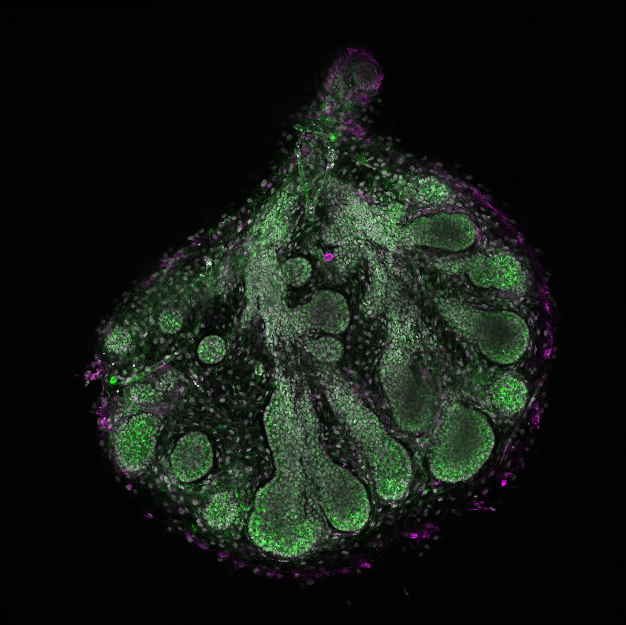
Two salivary glands from Drosophila simulans larva at L3 wandering stage. They have been stained with DAPI and rhodamine phalloidin. The upper gland is reconstructed in 3D on Imaris, nuclei and cells volumes are represented with a color scale from blue to red.
Microscopy technique: Microscope confocal Airyscan LSM 980
![]()
Ultramicroscope – light sheet microscopy
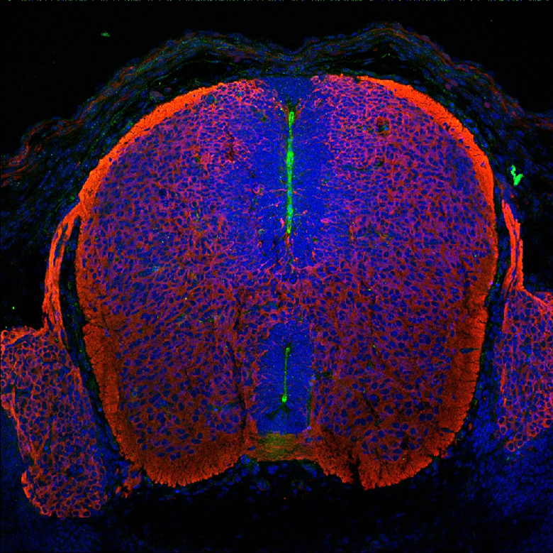
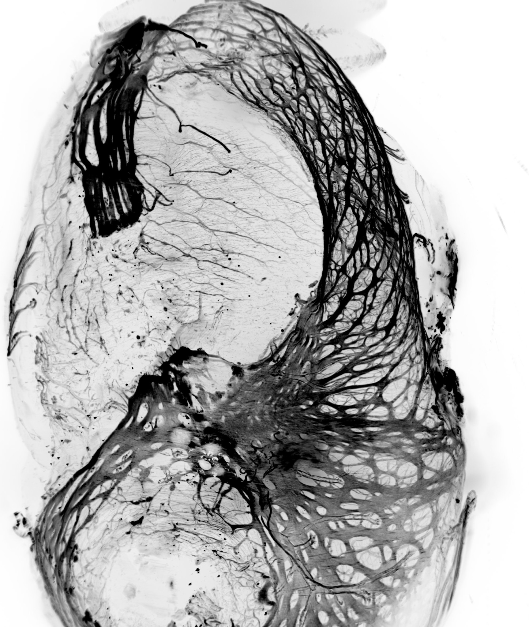
3D z-stack projection of transparised stomach and guts of chicken embryo. Label betaIII-tubulin – Alexa488.
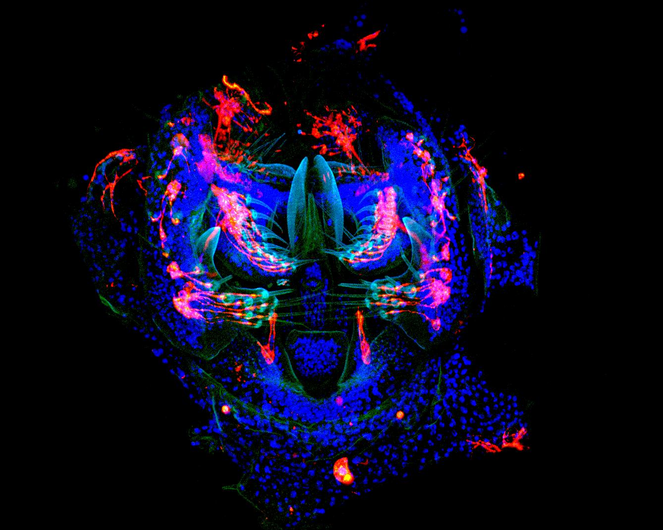
Fly monster © Orestis Faklaris & Michael Lang – Institute Jacques Monod, CNRS UMR7592 – ImagoSeine
Multifocal confocal microscopy (spinning disk, Spinning CSU W1)
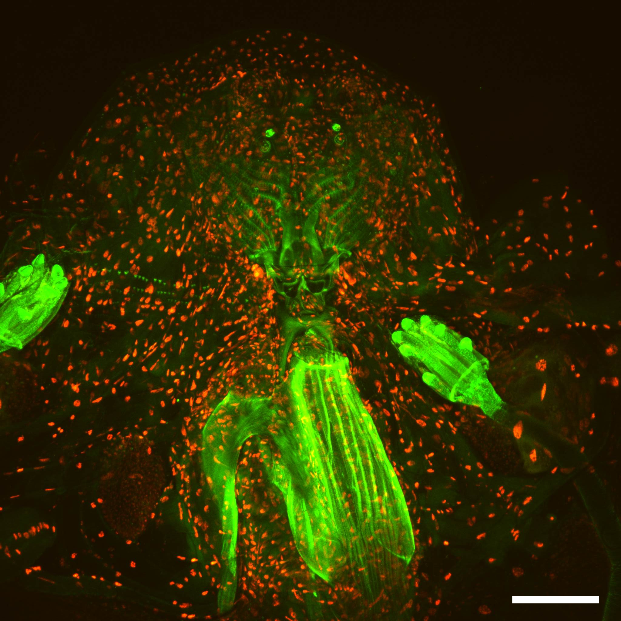
Drosophila third instar larval head, nuclear-RFP, neronal-GFP and green autofluorescence, 25x magnification, scale bar is 100 μm.
Second to the FBI Image Contest 2016