Initiated a few years ago, the Inria-IPL-NAVISCOPE (“Image guided NAvigation and Visualization data sets in live cell imaging and microscopy”) project aims at overcoming challenges of bioimaging observation. Virtual and augmented reality could become the new way to visualize and analyze microscope image renders.

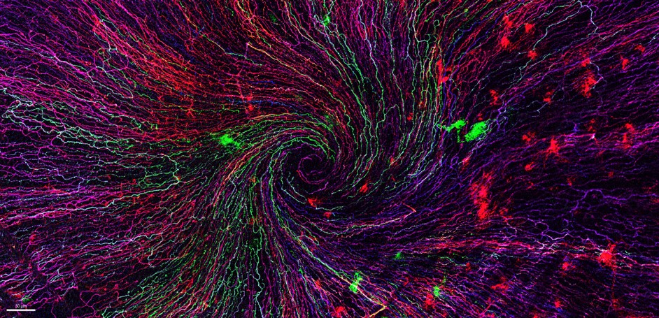
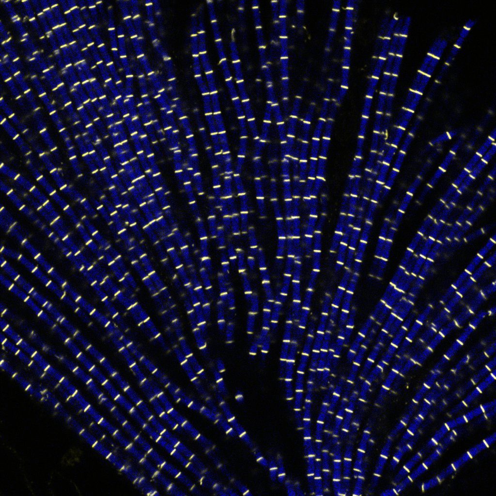
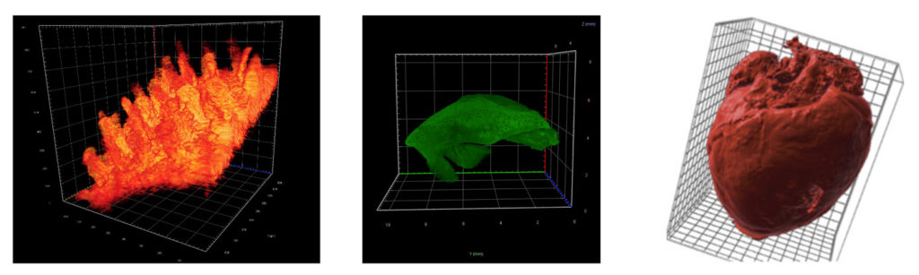
Despite incredible progresses in microscopy, imaging biomolecular dynamics in cells remains a challenge. A lack of sensitivity, limited recording speed, photobleaching and phototoxicity associated have restrained, for a long time, our capacity to study biomolecules in their natural environments. As microscopy image is commonly observed on 2D screens, it can narrow human capacities to grasp volumetric, complex, and discrete biological dynamics. Following new modes of visualization including virtual reality (VR)/augmented reality (AR) approaches, the NAVISCOPE project allows more accurate analysis and exploration of large time series of volumetric images, such as those produced by the latest 3D + time fluorescence microscopy.

Why should cell biologists be interested in this project?

The project to which 4 FBI-teams from the BI-IPDM node participate, aims at engineering a technology made with and for biologists. For VR/AR approaches to be adopted by the broader bioimaging community, it is, indeed, important that they are evaluated by the biologists, on their own datasets. 

The potentials of VR/AR technologies for scientists are numerous: navigating into multidimensional, large data sets with another view angle or perception, interacting with these data especially by selecting subregions, quantifying features of interests, etc. New VR/AR approaches also provide specific quantification tools to show distances, angles, counting, local density, and histogram profiler or include a selection of regions of interest for further analysis such as the 3D Timelines. Moreover, because communication with analysis software coded in Java or Python is now integrated, more post-treatment analysis is possible on selected features, providing a multifaceted and accessible tool for biologists.

A promising future ahead

In practice, immersion of the user within 3D + time microscopy data still represents an acculturation challenge for the concerned community. Thus, to promote a broader adoption of these approaches by biologists, further dialogue is needed between the bioimaging community and the VR&AR developers. Nonetheless, future innovation can already be foreseen as there are multiple way to upgrade this technology. For example, using eye-tracking (Günther et al., 2020) or haptic interfaces (Petit et al., 2020) can improve human perception by providing local sensations, which would improve the selection of responses in a 3D + time space. Besides, a better integration of multiple channels with high pixel resolution or the addition of vector representations could add information about the orientation, movement of molecules or organization of structures such as cytoskeleton elements or membrane lipids. The prospects initiated by the NAVISCOPE projects are, as mentioned above, endless and could be a technology that reshapes the way we see biology at the hearth.

Full article on:

Challenges of intracellular visualization using virtual and augmented reality

Valades-Cruz Cesar Augusto, Leconte Ludovic, Fouche Gwendal, Blanc Thomas, Van Hille Nathan, Fournier Kevin, Laurent Tao, Gallean Benjamin, Deslandes Francois, Hajj Bassam, Faure Emmanuel, Argelaguet Ferran, Trubuil Alain, Isenberg Tobias, Masson Jean-Baptiste, Salamero Jean, Kervrann Charleseub

Front. Bioinform. 2:997082.
https://doi.org/10.3389/fbinf.2022.997082

Ranging in size from 20 microns to 1 milimetre, Meiofauna is a crucial link between micro- and macro- marine ecosystems. Valentin Foulon, a research engineer in marine biology, is part of the Blue Revolution program that aims to develop a taxonomic identification protocol for these tiny creatures. He approached the Bretagne-Loire node of France-BioImaging, in Nantes, where a Single Plane Illumination Microscopy (SPIM) light-sheet available in open access helped him to further reach his goal.

Valentin Foulon loves imaging and microscopy – and he loves meiofauna. He works on the Blue Revolution program, whose goal is to accelerate the taxonomic identification of meiofauna using artificial intelligence algorithms. But while the confocal-microscopy-based protocol worked well for small plankton, the researchers quickly realized that it had to be adapted to work with meiofauna. 

Choosing the right microscopy approach

Identification of Meiofauna is usually operated manually on a classical microscope, which is “incredibly challenging” says Valentin. “And we only have one focal plane. To see important details, we need a 3D view of organisms.” explains Valentin. “That’s why we got curious about light-sheet imaging, because we would be able to see the organisms in 3D.” As France-BioImaging provides this type of microscopy in open-access in Nantes, “we clearly saw an opportunity there,” explains Valentin. His project proposal, which was submitted to the Euro-BioImaging pilot User Access fund, was fully funded by France-BioImaging, in a generous initiative introduced by France-BioImaging to fund all user projects to its facility received by Euro-BioImaging for the pilot User Access fund. 

“I had never tried light sheet microscopy before,” says Valentin, “And in Nantes, the lab didn’t have experience with marine organisms. Together, we found a compromise between different imaging parameters such as the resolution or the acquisition time.” Once the set-up was fine-tuned, he sent his samples, and the engineer at the lab did the imaging work. “It was a very nice experience,” expresses Valentin.  “I discovered a new technique, which is always enriching. I also think the lab was happy to work with a new sample type. And finally, we validated our proof-of-concept, proving that it is possible to image these organisms in 3D.

Image data management

But the project doesn’t end there. “The next step is data management. We imaged between 200-300 different organisms. It’s not enough for automatic classification, but nevertheless, we have 6-7 TB of data. Now we must process the data for this classification with machine learning. It’s an essential part of the project, to make the link between sample imaging, and data processing. We are working to close that gap.” 

“The outcome of this project is to validate a proof-of-concept, from the sample collection to the image classification. With this new taxonomic identification protocol, we will be one step forward in the comprehension of meiofauna, these mysterious marine organisms, who may hold the key to understanding the impact of human activity on marine ecosystems,” concludes Valentin. 

Thank you Marianna Childress, communication officer of Euro-BioImaging, for the original article.

As the 2022 edition of the France-BioImaging Image Contest admissions is coming to an end, we wanted to highlight our previous winners and their projects. Here is a quick throwback to our 2021 winners.

Before getting to the heart of the matter, we want to remind you that you still have time (before November 11th) to submit your best images and try to win your registration fees for one 2023 microscopy-related event! Please make sure you upload your images on the following link:

FBI Image Contest 2022 – Last days to submit your images!

Last year, we enjoyed the winning images submitted for their artistic take and their quality. Thanks to Léna Meneux, Eunice HoYee Chan, Camille Boutin et Nicolas Brouilly for their beautiful images!

1st place: Léna Meneux, Eye Team, Institut des Neurosciences de Montpellier

"The eye of the storm"

Sensory fibers of a mouse cornea imaged with a confocal microscope. The corneal nerves converge toward the centre forming a vortex. This particular transgenic mouse model allows stochastic expression of fluorescent proteins, unravelling the heterogeneity of the fiber origines inside the corneal epithelium.
Acknowledgements to Karine Loulier for the mouse model and Laetitia Hudececk for her help during the acquisition.

In the Institut des Neurosciences de Montpellier since 2020, Léna is a PhD student working in the team Eye lead by Dr. Frédéric Michon. This team is investigating the mechanisms related to the preservation and the integrity of the anterior part of the eye, including the lacrimal gland, the tears and the cornea. Léna’s project focuses on the cellular and molecular effects of the corneal innervation on the corneal homeostasis. The project goes further as they aim at highlighting new targets able to prevent and/or repair corneal damage. 

The image she submitted for the 2021 France-BioImaging Image Contest (The eye of the storm) represents the sensory fibers of a mouse cornea. This innervation follows a typical pattern where all the nerves converge toward the centre forming a vortex. This particular transgenic mouse model allows random expression of fluorescent proteins, unravelling the heterogeneity of the fibers’ origin inside the corneal epithelium. As cornea is the most innervated tissue in the whole body, this model shows the differences between fibers. In pathological context, for example wound injury, it is thus possible to follow a specific fiber during the healing process.

France-Bioimaging sponsored her participation to the FOM (Focus on Microscopy) 2022 congress where she presented her project through a poster. Even though the congress was online, it gave her the opportunity to share her results with experts and as a consequence, to gather advice on her ongoing experiments.

2nd place: Eunice HoYee Chan, Muscle Dynamics Team, Developmental Biology Institute of Marseille (IBDM)

Myofibrils isolated from Drosophila indirect flight muscle labelled with titin (yellow) and actin (blue). Image captured from confocal microscope. We are studying the role of titin protein in muscle mechanics and organisation during development

"Sarcomeric bouquet"

Myofibrils isolated from Drosophila indirect flight muscle labelled with titin (yellow) and actin (blue). Image captured from confocal microscope. We are studying the role of titin protein in muscle mechanics and organisation during development.

Research engineer in Frank Schnorrer's team at Institut de Biologie du Développement de Marseille (IBDM), Eunice focuses her research on Drosophila muscle dynamic and organisation during development using advanced biophysical and imaging techniques.

The image she submitted named “Sarcomeric bouquet" was from one of her very first muscle myofibrils isolation experiment. She dissected flight muscles from flies and labelled the individualised myofibrils with Llama nanobodies conjugated with different epitopes. Those labelled myofibrils were then subjected to various imaging methods including standard confocal microscopy, super resolution microscopy and cryo electron-tomogram. Using these novel labelling tools and imaging techniques, her team could study the dynamic and organisation of muscles during development in details.

France-BioImaging sponsored her registration to the 49th European Muscle Conference in Prague (22-26 September 2022). As she is new to the muscle field, this conference offered a great opportunity to have a broad view on different kind of state-of-the-art imaging techniques. Besides, she gave a presentation during the conference, highlighting her work and initiating discussion.

3rd Place: Camille Boutin, Biology of multiciliated cells Team, Developmental Biology Institute of Marseille (IBDM) & Nicolas Brouilly, PICsL Imaging facility, Electron Microscopy department

Lamellar structure in a differentiating multiciliated cell observed by transmission electron microscopy with a Tecnai G2 200kV FEI.

"Clown"

Lamellar structure in a differentiating multiciliated cell observed by transmission electron microscopy with a Tecnai G2 200kV FEI.

Camille is a researcher in Laurent Kodjabachian’s group at the Institut de Biologie du Développement de Marseille (IBDM). She develops projects as a principal investigator on the compartmentalization and sizing of multiciliated cells. With this in mind, she routinely uses confocal and super-resolution microscopy but also scanning and transmission electron microscopy and tomography.

Nicolas is in charge of the Electron Microscopy Unit of the Plateforme d’imagerie commune du site de Luminy (PICsL). In addition to the routine sample preparation and 2D TEM imaging, this imaging facility offers, to internal and external users, advanced sample preparation (cryo-methods, immunolabelling...) and advanced imaging (tomography, CLEM, serial blockface…).

To understand the production of multiple centrioles in multiciliate cells, they focused on the deuterosome, a membrane-less organelle that has been described 50 years ago but whose composition, organisation and function remain unknown to this day. In this context they have developed an inducible multiciliated cells line. This image was taken during the initial characterisation of this cell line by transmission electron microscopy.

Thanks to the France-Bioimaging Image Contest, Nicolas participated to the COST COMULIS Conference that was held by the Cyprus Institute in Nikosia. It was a great opportunity to exchange with the people at the cutting edge of the multi-modal imaging field. The program covered subjects such as the sample preparation for multi-modal imaging, image analysis and integrated industrial partners.

Published on August 23rd, 2022 in EMBO reports, this article questions the way that core facilities should be recognized in the scientific literature and their key contributions to data lifecycle. An initiative endorsed by France-BioImaging.

Core facilities are an integral part of the life science research landscape as providers of centralised access to technological resources and expertise. This article’s working group has estimated that between 40 and 80% of imaging, proteomics and genomics data at their institutes are generated at core facilities. The contribution of core facilities to scientific research and innovation must thus be accordingly recognised. In that respect, the most straightforward way is an acknowledgement. Unfortunately, the lack of formal rules still leaves core facilities being inadequately recognised. 

This article proposes that the recognition of core facilities should be deployed via two actions and implemented in two phases: first, with the systematic acknowledgement of core facilities in all scientific publications, and second, by including core facilities and their staff in data citations (Cousijn et al, 2018).

The first step can be accomplished at the manuscript-submission stage by asking the corresponding author to confirm if any data (and associated metadata) used in the manuscript originated from a core facility, and if yes, to identify the associated core facility. EMBO Press has recently included a question in the author checklist to confirm whether the work in the publication “benefited from core facilities” and that the core facility be acknowledged accordingly.

The next step would be to make it compulsory for authors to respond to such a query and explicitly identify the core facility and relevant data (and associated metadata). The MDAR (Materials, Design, Analysis, Reporting) form (Macleod et al, 2021), wherein one needs to provide information about data availability in the Analysis section, could likewise include a question to explicitly identify core facilities involved. Eventually, the information in the author checklist could be automatically fed into the acknowledgement section.

Acknowledging will have two key positive consequences: on the sustainability of core facilities and on their staff careers. In the absence of a high number of publications, particularly as lead or corresponding authors, acknowledgements are used as a measure of a core facility and its staff’s output and impact. Second, it further motivates and incentivizes core facility staff to actively contribute to scientific research. 

The acknowledgement of a core facility goes beyond professional courtesy: identifying the origin of data (and associated metadata) is essential for data traceability and reproducibility particularly since core facilities are major generators of data in life science research. 

Thanks to Jean SALAMERO, our “Action inter-infrastructures” mission officer, for contributing to this article. 

Full article on:

Acknowledging and citing core facilities

Katja Kivinen, Henri G A M van Luenen, Myriam Alcalay, Christoph Bock, Joanna Dodzian, Katerina Hoskova, Danielle Hoyle, Ondrej Hradil, Sofie Kjellerup Christensen, Bernhard Korn, Theodoros Kosteas, Mònica Morales, Krzysztof Skowronek, Vasiliki Theodorou, Geert Van Minnebruggen, Jean Salamero, Lavanya Premvardhan

EMBO reports (2022) 23: e55734
https://doi.org/10.15252/embr.202255734

The France-BioImaging Image Contest is back for its 4th edition!

This image contest is open to all within the imaging community: core facility staff and users, R&D labs teams and co-workers, students… Submit your best microscopy images for a chance to showcase your skills, research and creativity to the French bioimaging community and beyond, allowing people to see the visual appeal of the life sciences. Images from the contest will be featured on France-BioImaging communication tools, online and in print.

France-BioImaging and all the French community aims to develop and promote innovative imaging technologies and methods. But microscopy images can also take an artistic, creative look and make the invisible world beautiful.

We are all eager to see your work !

Prizes

1 to 3 images will be awarded depending on the quantity and quality of the entries submitted. France-BioImaging will cover the registration fees for one 2023 microscopy related event of the winners’ choice (FOM, ELMI, EMC, COMULIS conference, etc.).

Important: Only French or foreign participants affiliated to a French institution can enter the contest. Foreign participants non-affiliated to a French institution can submit images and will be featured in the gallery, but will not be evaluated as part of the contest.

Submission deadline: Friday, November 11th, 2022, 23h59 UTC+2. 

Click here to consult the terms and conditions of the contest. When you are ready, submit your entry by filling the form below. You can check out last editions’s entries for inspiration. One participant can submit several entries (up to 3).

(If you have any issues when submitting your image, please contact communication@france-bioimaging.org)

This form is currently closed for submissions.

Our three winners have been announced! Please discover all the submitted images on this following link: https://france-bioimaging.org/announcement/winning-images-fbi-image-contest-2022/

On December 13th and 14th 2022, we will have the pleasure to invite you to our Annual Meeting, to be hosted by FBI Bretagne-Loire Node at the Health Research Institute of the University of Nantes (Nantes, France).

2022 is an important landmark for France-BioImaging and its community, as the infrastructure is celebrating 10 years of operation and scientific advances. We will be happy to celebrate this milestone with all the members of the bioimaging community (within and outside the France-BioImaging community).

The Annual meeting will highlight France-BioImaging’s development as a research infrastructure and its node community accomplishments during these last 10 years, and the role they play in boosting innovation in bioimaging. Imaging scientists and users from the infrastructure’s nodes will present their key projects and demonstrate how they have profited from France-BioImaging and its community.

We look forward to meeting you there!

Gallery

Videos

Programme

Registration

Deadline to register: 04/12/2022 at 23:59

This form is currently closed for submissions.

Poster

INFOS PRATIQUES:

Adresse: Bâtiment IRS-UN 8 quai Moncousu 44000 Nantes

Plan d’accès https://sfrsante.univ-nantes.fr/medias/fichier/plan-acces-irs-un-1_1561112224023-pdf?ID_FICHE=1562669&INLINE=FALSE 

(arrivée directement à l’accueil, toutes les salles sont au rez de chaussé et seront fléchées)

Comment venir?

Arrêt de tram le plus proche: ligne 2 ou 3 du tramway Aimé Delrue

En train: 

Gare de Nantes à 20 minutes à Pied (préférez la sortie sud pour venir à pied). En bus ou tram compter 15 minutes.

Ligne de bus accessibles depuis arrêt sortie gare Sud C2, C3, 54 arrêt Hotel Dieu

Ligne de tram depuis la sortie Gare Nord : prendre la ligne 1 direction François Mitterand/Jamet et descendre à Commerce, continuer à pied (10 minutes de marche environ) 

En avion:

Navette aéroport  Navette aéroport Nantes Atlantique : horaires, tarif

Le départ et l’arrêt se font à cause de travaux de Hotel Dieu ( plus proche de l’IRS) et non pas Commerce. 

Taxis Nantais:

02 40 69 22 22 http://www.taxis-nantes.com/ 

02 40 85 40 85. https://heptaxis.com/ 

VTCs: 

LocalCab 09 80 66 62 82   TAXI / VTC – Réservez votre Chauffeur au Meilleur Prix ! (localcab.fr) 

Quelques Hôtels conseillés :

L’Hôtel  L’HÔTEL NANTES – Votre Boutique Hôtel idéalement placé centre-gare (nanteshotel.com) (135 à 195 euros la nuit avec petit déjeuner) 

Hôtel Amiral Hotel Nantes centre-ville – Hotel 3 étoiles – Hôtel Amiral | Nantes (hotel-nantes.fr) (128 à 150 euros avec petit déjeuner)

Hotel ibis Nantes Centre Gare Sud   ibis Nantes Centre Gare Sud – Hôtel 3 étoiles à Nantes – ALL (accor.com)  (110 à 130 euros)  ibis Styles Nantes Centre Place Royale 

Hôtel  ibis Styles Nantes Centre Place Royale Hôtel à Nantes – ibis Styles Nantes Centre Place Royale – ALL (accor.com) (85 à 95 euros) 

Prise en charge des missions: 

Se rapprocher de votre noeud FBI (fonds mission), sauf pour les intervenants qui seront directement contactés pour la prise en charge de leur missions. 

Advancing multimodal microscopy of thick samples for preclinical studies

The new France-BioImaging preclinical microscopy Working Group, whose aim is to advance multimodal microscopy of thick samples for preclinical studies, is organizing its first meeting! The event will take place on Monday December 12th 2022 in Nantes.
The event will present three aspects of preclinical microscopy:

  1. Imaging technologies (correlative microscopy, thick tissue or organoid microscopy, light sheet microscopy, intravital imaging and label free imaging)
  2. Image analysis (whole slide, correlative and 3D analysis)
  3. Regulatory frameworks (animal testing ethics and best practices in preclinical development)

Preliminary program

Registration Form

Deadline to register 04/12/2022 at 23:59

This form is currently closed for submissions.

The meeting will cover three aspects of preclinical microscopy with presentations of 30mn and a final round table:

  1. Imaging technologies
    • Correlative microscopy
    • Thick tissue imaging
    • 3D whole organ macroscopy
    • Intravital imaging (imaging chambers, exposed organs in living animal..)
    • Label free imaging
  2. Image analysis
    • Whole slide imaging analysis
    • Correlative image analysis
    • 3D reconstruction (distortion, alignment problems…)
  3. Regulatory frameworks
    • Animal testing ethics (3R, Ethics Committee…)
    • Preclinical developments (GMP, 21 CFR Part 11 Compliance…)
  4. Closing
    • Round table
    • Synthesis

Poster

France-BioImaging PreClinical Working Group

The PreClinical topic is usually connected to animal studies, but one needs to take into account the following aspects :

  1. Preclinical development, also termed preclinical studies or nonclinical studies, is a stage of research that begins before clinical trials (testing in humans) and during which important feasibility, iterative testing and drug safety data are collected, typically in laboratory animals.
  2. Preclinical imaging usually refers to whole animal imaging techniques allowing longitudinal studies such as high-frequency ultrasound, magnetic resonance imaging (MRI) and computed tomography (CT) which are usually used for anatomical imaging, while optical imaging (fluorescence and bioluminescence), positron emission tomography (PET), and single photon emission computed tomography (SPECT) which are usually used for molecular visualizations. This macro imaging scale is proposed within the infrastructure France Life Imaging.
  3. Multimodal imaging including the microscopy scale is largely developing. Technologies developed within FBI could benefit to the preclinical community
  4. The communities of cell, tissue and intravital microscopies need to know each other better.

Our Goal

The new France-BioImaging Working Group wants to focus on the microscopic aspect of the “preclinical imaging”. It aims to develop microscopy technologies for preclinical studies used to explore highly resolved information at the organ level. It includes, for example, whole slide quantitative tissue histology, correlative microscopy, 3D from sections as well as Light sheet fluorescence microscopy for 3D whole organ imaging, intravital organ imaging or multi photon microscopy. The goal is to bridge the gaps between communities dealing with either fixed sections or living organs.

Call for speakers

The Working Group would like to call on speakers interested in presenting one of the several aspects that will be highlighted during the meeting and contributing to the Working Group (open to everyone). If you are interested, please get in touch:

This form is currently closed for submissions.

Open-Neuroscience in collaboration with the Neuroscience department and the Photonic BioImaging Core of the Institute Pasteur and the University of Sussex neuroscience department is organising a FENS satellite event on Open Hardware Microscopy DevelopmentThe aim of this event is to bring into focus excellent open projects that permeate education, ease of access and data collection in advanced research environment.

The symposium will take place on July 8th at the Pasteur Institute campus in Paris (28 rue du Dr Roux). It will be divided into a morning workshop session and an afternoon series of talks. Registration is necessary to access the campus :

 REGISTER

The workshops will be led by specialists in the field, which will also participate in the afternoon session. By the end of the session, participants will be invited to discuss what they have learned, how they could implement Open-Science microscope in their research, and/or how they could improve/modify their own setup by implementing Open tools on it and how their research protocol could benefit from it.

The afternoon session will be dedicated to a series of talks by our invited specialists. It will be split into two distinct topics/sub-sessions:

  1. Open-Source microscopy for education and improving ease of access to research equipment.
  2. Open-Source microscopy for advanced research.

It is our goal that by the end of the day, participants get a full picture of the state of the art for Open-Source Microscopy. It is hoped that development and implementation of such tools and techniques in modern research units promote science accessibility and improve data reproducibility. Furthermore, an earlier and more systematic access to advanced science tools in education could only support shaping the research minds of tomorrow.

Open Microscopy is sponsored by EMBO, TReND in Africa and France-Bioimaging.

The first edition of “Digital Spaces for Research and Medicine” will take place at Institut Curie on July 7th, 2022. This event will be dedicated to providing scientists and clinicians insight into Virtual Reality, Augmented Reality and Mixed Reality based tools that offer novel and powerful ways for visualizing and analysing data in research and medicine. This day will include presentations of the tools followed by hands-on demonstrations. The day will close with an open forum for questions and discussing future perspective. The following tools will be presented:

  • MorphoNet-VR (Serpico-STED, Inria-UMR144 Institut Curie and LIRMM): Visualizing live microscopy data (3D+T) in VR
  • Genuage (Team LOCCO, UMR168, Institut Curie): visualization and analysis of multidimensional point cloud data, such as single molecule data, in virtual reality
  • DIVA (Institut Pasteur, Institut Curie): 3D reconstructions of raw experimental image stacks that are integrated in virtual reality for volumetric analysis
  • Tamed Cloud (ENSADLab): immersive VR experience to interact with cloud of images
  • HoloTracks (TEAM INRAE-Unité MaIAGE / AVIZ Inria, Paris Saclay): Immersive (AR) visualization of dynamic compounds in living cells
  • UnityMol A&VR (UPR 9080, IBPC) : deep inside molecules – digital twins at the nanoscale

Presentations of the tools will be at amphitheater Burg and online (click here to join)

Registration is mandatory for hands-on demonstrations : register here to book your slot!

Program

9 – 9.15 am Welcome coffee

9.15 – 11.30 am Presentations of the tools

11.30 – 1 pm Demonstrations part 1

2 – 5 pm Demonstrations part 2

5 – 6 pm Discussion “Future and perspectives”

6pm Cocktail

France-BioImaging, with its partner the GDR IMABIO, organizes the 4th edition of the FBI-AT: an advanced microscopy workshop to be held in Paris from November 21st to 25th, 2022.

The aim of this France-BioImaging-Advanced Training is to train microscopy users on the most advanced imaging techniques that will allow them to perform molecular studies at the cellular level as well as in thick samples. In particular, recent developments on fluorescent probes will be highlighted. The workshop will benefit from state-of-the-art equipment available on several of the Parisian Node Imaging facilities.

This year’s edition will have plenary lectures given by experts in the microscopy development field. These seminars will be advertised as a series and will be broadcasted for a large audience.  In addition, specific techniques will be introduced.

Hands-on practicals will train attendants on these techniques every afternoon in different sites in Paris including Institut Curie, Institut Pasteur, Institut Cochin, Institut Jacques Monod, Institut de Psychiatrie et Neurosciences de Paris and ENS Paris. Access to this part of training will be restricted to selected and registered trainees.

To guaranty access to set-ups and proper training, each practical session will host only 3-4 persons. The sessions will be run in parallel.

Apply now, attendance will be limited to 25 participants! 

FBI-AT is ideal for researchers with a basic training in microscopy willing to become familiar with advanced techniques to answer their specific biological questions, or to be exposed to new developments that will allow them to tackle new questions in their project. We will consider applications from early career researchers (PhD students, post-docs), technical staff members and more senior scientists.

AT A GLANCE

The workshop contains plenary lectures and specific training sessions. Plenary lectures will be on hybrid mode and largely open.

Invited Speakers

Emmanuel Beaurepaire

Giulia Bertolin

Joerg Bewersdorf

Peter Dedecker

Claire Deo

Marie Erard

Ricardo Henriques

Christophe Leterrier

Sandrine Lévêque-Fort

Gustavo Quintas

Gaelle Recher

Jean-Baptiste Sibarita

Lothar Schermelleh

Practicals on

  • Combining micro UV-irradiation and Single Particle Tracking in living cells
  • SMLM multi-color: from sample preparation to quantification
  • FRET-based molecular tension sensors and FLIM
  • Imaging of cellular ultrastructures with expansion microscopy
  • SIM, STED or STORM ? : from sample prep to 3D imaging
  • 3D STED : Comparing flat cells vs thick samples
  • Culturing and imaging multicolour 3D live brain organoïds
  • Combining fast live 3D imaging with Z resolution preservation
  • Light sources for optogenetics
  • Non-classical genetically modified fluorescent probes for biological imaging
  • Imaging biological structures in 3D using double helix-STORM and 3D-SIM

Organizers

Florence Niedergang, Lydia Danglot, Chloe Guedj, Mickael Lelek, Pierre Bourdoncle, Audrey Salles, Xavier Baudin, Nicolas Borghi, René-Marc Mege, David Geny, Ludovic Jullien

Poster

Sponsors

Euro-BioImaging is organizing a third online User Forum on Tuesday, April 5, 2022 from 14:00-17:00 CEST. The topic is “At the Forefront of Neuroscience.”

This event will highlight the importance of cutting-edge imaging technologies in support of brain research and showcase the specific expertise available at our Nodes across Europe through case studies presented in tandem with the research community. 

In addition, keynote presentations from Hervé Boutin, University of Manchester, and Selma Ding, Max Planck Institute for Animal Behavior, will further reveal the potential of biological and biomedical imaging technologies to boost brain research. 


Register here: https://us02web.zoom.us/meeting/register/tZ0ucuytpj8iE92A-JjvPbvpNnmTF4Z7iRaC

More information: https://www.eurobioimaging.eu/news/join-us-for-the-third-euro-bioimaging-user-forum-at-the-forefront-of-neuroscience/

Programme