Most mammals can maintain a relatively constant and high body temperature. This is considered to be a key adaptation for theses species, enabling them to successfully colonize new habitats and survive harsher environments. Scientists from the Institut des Sciences de l’Evolution de Montpellier (ISEM) investigate the possible correlation between the maxilloturbinal in the anterior nasal cavity and the body temperature maintenance by using Micro Tomography (or MicroCT) at the MRI core facility (FBI Montpellier node). This technique was essential in this study as it rebuilt the hypothesis around body temperature maintenance. Here is what they found.

MicroCT: Image in a non-destructive way

First of all, what is Micro Tomography? Micro Tomography, or Micro-CT, is a 3D imaging technique using X-rays to see inside biological material, at a small animal or body part level. Slice by slice, this technology scans the object in a series of 2D images that are reconstructed in a 3D model. Micro CT is, thus, non-destructive. This means that it can be used to image a sample without having to cut it! Not only your material is still in one piece but you can use it for further experiments.

Phylogenetic studies as an example of application

In this study examining the correlation between skull structure and the stabilization of body temperature, MicroCT was the key. The presence and the relative size of the maxilloturbinal has been proposed as a hypothesis that reflects the endothermic conditions and basal metabolic rate in extinct vertebrates. Among bony structures, respiratory turbinals (e.g., maxilloturbinal) are interesting anatomical structures that may offer important insights to the origins of endothermy, in other words to the origin of warm-blooded animals. Indeed, respiratory turbinals are highly vascularized, which amplifies the surface area and offers an effective mechanism to avoid loss of internally-produced and costly heat.

You probably figured it out: scientists needed to compare the structure of the maxilloturbinal in order to take conclusion. This is when Micro Tomography was very useful. They scanned 424 individuals from 310 mammal species using high-resolution X-ray micro-computed tomography, with approximatively half of the samples imaged at MRI, part of our Montpellier node. Using the obtained comparative 3D µCT dataset, they explored the anatomical diversity of the maxilloturbinal based on relative surface area, morphology and complexity. They specifically tested the relationship between multiple parameters such as the size-corrected basal metabolic rate (cBMR), the relative surface area of the maxilloturbinal (Maxillo RSA) or body temperature.

And the results surprisingly showed that…

…there is no evidence to relate the origin of endothermy and the development of some turbinal bones! Even though scientists used a comprehensive dataset of Micro CT-derived maxilloturbinals spanning most mammalian orders, they demonstrate that neither corrected basal metabolic rate nor body temperature significantly correlate with the relative surface area of the maxilloturbinal. These results challenge the hypothesis of thermal regulation being linked to respiratory bone structure.

So, what could be linked with the thermoregulation of mammals? Researchers proposed 3 more hypothesis. First of all, environmental conditions could have a bigger role: “the maxilloturbinal function could have a more prominent heat/moisture exchange role in species that face harsh environmental conditions, thus helping to limit spurious heat and moisture loss”. Another major role of the maxilloturbinal is water conservation. As an example, the naked mole-rat avoid breathing through the mouth when performing energy intensive digging because the lips close behind the digging incisors and this species has the lowest value of predicted Maxillo RSA of the entire sample. But most of all, the factor could be a multifactorial physiological question. What is the relation of the maxilloturbinal with the overall nasal cavity? Do other functions play a role in the evolution of this body part, such as its protective role against toxic elements? Is it linked with brain cooling?

Well, imaging will certainly give them an answer in the future!

Detailed view of the maxilloturbinal in selected mammalian species with peculiar thermal and metabolic conditions or that undergo different forms of heterothermy (https://doi.org/10.1038/s41467-023-39994-1)

Get access to one of our services!

You need Micro-CT or another imaging technology or expertise that France-BioImaging provides? To get open access, please login via Euro-BioImaging website! You just have to choose the technology you want to use, then submit your proposal. All applications will be processed by the Euro-BioImaging Hub in close relation with France-BioImaging. And of course, all scientists regardless of their affiliation, area of expertise or field of activity can benefit from open access services! Users whose projects will be validated by Euro-BioImaging will benefit from a waiver for the access cost on France-BioImaging core facilities (https://france-bioimaging.org/access/).

Martinez, Q., Okrouhlík, J., Šumbera, R. et al. Mammalian maxilloturbinal evolution does not reflect thermal biology. Nat Commun 14, 4425 (2023). https://doi.org/10.1038/s41467-023-39994-1

Explore the beauty of the invisible world through the 2024 FBI digital calendar!

Enjoy the diversity of microscopy techniques, models and applications represented, one image at a time. All 12 images used for this calendar were submitted to France-BioImaging Image Contest 2023.

A big thank you again to all the participants!

You can download the A4 print version (one month per page) 2024 FBI digital calendar here:

If you wish to use it as your computer desktop, you can download a PNG version of each month here:

On December 13th and 14th 2023, we have the pleasure to invite you to our Annual Meeting, to be hosted by our brand new FBI Toulouse Node at the Centre de Biologie Intégrative of the Université Toulouse 3 – Paul Sabatier.

We will be happy to celebrate yet another year of achievements and developments in bioimaging with all the members of the community.

This year, the Annual Meeting will focus on “Multiscale mechanobiology of cells and cell systems“, a topic specially selected for being one of Toulouse node’s expertise.

Mechanobiology aims to apply biophysical approaches to measure and perturb forces in complex physiological systems, and to develop in vitro systems including different types of organoids, enabling the controlled manipulation of cells and tissues and the measurement of mechanical forces to study cell mechanics and morphogenesis. 

This “mechanobiology” theme is including (i) how cells generate and transmit forces, (ii) the impact of forces on cell/tissue dynamics, (iii) the impact of extrinsic forces on cells and tissues, and (iv) the development of new tools for manipulating and measuring forces in cells and tissues in a controlled, non-invasive way.

We invite the France-BioImaging Community to present their mechanobiology-related projects during the second day of the Annual Meeting around a dedicated session with selected talks. We strongly encourage you to submit an abstract for a talk or a poster presentation during your registration!

The winner of the best talk and the best poster presentation will win their registration fees for one 2024 microscopy related event of their choice!

Core facilities will also have an opportunity to present a poster.

We look forward to meeting you there!

Gallery

Videos

Registration

Deadline: November 24, 2023

This form is currently closed for submissions.

Preliminary program

INFOS PRATIQUES:

Adresse: Centre de Biologie Intégrative, 169 Rue Marianne Grunberg-Manago, 31400 Toulouse

Télécharger le plan du campus

Comment venir?

Par la route

En venant de Bordeaux, contourner Toulouse par l’Est (direction Montpellier) ; avant le péage prendre la sortie no 23 direction Rangueil ou la no 19 direction Ramonville, puis suivre Université Paul Sabatier.

En venant de Montpellier, après le péage, prendre la sortie no 19 direction Ramonville, puis suivre Université Paul Sabatier.

En avion

Horaires aéroport Toulouse Blagnac

Une navette et le tramway relient l’aéroport et le centre ville de Toulouse (Transports en communs)

De là, il est possible de prendre le métro (lignes A ou B) pour rejoindre la station « Ramonville St Agne » (ligne B, direction Ramonville) : environ 1h de trajet

Le CBI est à 500 mètres à l’Ouest de la station et accessible à pieds ou en bus.

Il est également possible de prendre un taxi depuis l’aéroport : compter au moins 1/2h de trajet hors heures de pointe

En train

Depuis la Gare SNCF Matabiau:

Prendre le métro LIGNE A direction Basso Cambo, changer à la station “Jean Jaurès” et prendre la Ligne B pour rejoindre la station « Ramonville St Agne » : comptez environ 40 minutes depuis la gare.

Le CBI est à 500 mètres à l’Ouest de la station et accessible à pieds ou en bus.

Hôtels conseillés :

Nous vous conseillons de privilégier des hôtels en centre-ville.

Prise en charge des missions: 

Se rapprocher de votre noeud FBI (fonds mission), sauf pour les intervenants qui seront directement contactés pour la prise en charge de leur missions. 

France-BioImaging and all the French community aims to develop and promote innovative imaging technologies and methods. But microscopy images can also take an artistic, creative look and make the invisible world beautiful, allowing people to see the visual appeal of the life sciences. 

We enjoyed the diversity of the images submitted with many different microscopy techniques, models and applications represented. A big thank you to all the participants!

The National Coordination Team and the Executive Board are proud to announce the winners of the FBI Image Contest 2023:

  • 1st Place: Laurent LE, Lévêque-Fort Team, Institut des Sciences Moléculaires d’Orsay

In the blink of an eye

COS7 fixed cell. Alpha-tubulin labeled with DNA-PAINT and imaged with Atto 647N. Axial information is obtained by virtual-SAF measurement known as DONALD.

SMLM Fluorescence Microscopy with DNA-PAINT with DONALD detection

  • 2nd Place: Gonzalo QUIROGA-ARTIGAS, Team Contrôle cytoplasmique de la stabilité du génome, Centre de recherche en Biologie Cellulaire de Montpellier

“Tardigrade embryos protected by mother’s molt”

Tardigrades commonly align the time of molting with egg laying. In this image we observe a tardigrade molt covering three developing embryos (DNA in white). The microscopy technology applied was confocal microscopy, and the research aimed to investigate the synchronization of embryo development in tardigrades.

Confocal microscopy

  • 3rd Place: Hugues LELOUARD, Gorvel team, Centre d’Immunologie de Marseille Luminy

“Intestinal octopus”

Small intestine section from a LyzM-eGFP mouse containing one Peyer’s patch and stained for proliferative cells (Ki-67, yellow), Paneth cells (UEA-I, blue), epithelial cells (EpCAM, magenta), naive B cells (IgD, red), T cells (CD3, orange), helper T cells/macrophages (CD4, cyan), phagocytes (CD11c, turquoise), monocyte-derived phagocytes (GFP, green).

10-color spectral confocal microscopy

Congratulations to the winners!


Explore all the images submitted here:

As stated in the Terms & Conditions of the contest, foreign participants non-affiliated to a French institution are featured in the gallery, but were not evaluated as part of the contest.

As the 2023 edition of the France-BioImaging Image Contest admissions is still running, we wanted to highlight our previous winners and their projects. Here is a quick throwback to our 2022 winners.

Before getting to the heart of the matter, we want to remind you that you still have time (before November 10th) to submit your best images and try to win your registration fees for one 2024 microscopy-related event! Please make sure you upload your images on the following link:

FBI Image Contest 2023

Last year, we enjoyed the winning images submitted for their artistic take and their quality. Thanks to Carole SIRET, Magalie BENARD and Frédéric FERCOQ for their beautiful images!

  • 1st Place: Carole SIRET, Van de Pavert Team, Centre d'Immunologie de Marseille-Luminy

"Little Monster"

The embryonic formation of lymph nodes, small organs essential for the immune response, is now known. Using light sheet microscopy, scientists were able to determine the dynamics at work in this 13.5-day-old mouse embryo. In blue, the lymphoid cells (LTi), derived from the haematogenous endothelium, a specific tissue of the embryo. They pass into the liver where they proliferate before migrating through the body to give rise to lymph nodes. The 3D information obtained thus makes it possible to follow the interactions of lymph nodes with their environment, in particular with nerve cells, in green, and blood vessels, in white. The lymphatic endothelial cells and some macrophages are visible in red.

Lightsheet Microscopy

Carole Siret is a Research engineer, expert in Lightsheet microscopy, at the Centre d’Immunology Marseille Luminy (CIML) since 2018. She is working in Dr Serge van de Pavert team where they study immune system development. They are particularly interested in the lymph nodes (LN) formation during mouse embryogenesis.

The image she submitted is a projection from a lightsheet acquisition on the UMII (Miltenyi). This image illustrates an E13.5 mouse embryo stained for neurons, LTi (Tissue inducer cells which are the precursor cells for the lymph node), lymphatic and blood vessels. This acquisition was done in the context of the study of the role of Cxcl12 in embryonic LN formation. From previous work it is clear that Cxcl13 and Ccl21 are not expressed present near blood vessels, but it likely that some chemokines, possibly Cxcl12, could be expressed on the endothelial cells. We focus on Cxcl12 since this chemokine has shown to be important for the attraction of several hematopoietic cells. Although it was shown that the receptor for Cxcl12, Cxcr4, is expressed by the mature hematopoietic inducer cells, it is not clear whether it also expressed by the progenitor hematopoietic inducer cells. Next to the possible attraction of hematopoietic cells towards the lymph node anlagen, Cxcl12 is involved in the attraction of nerve fibers. Therefore, the possible role of Cxcl12 could be to both attract hematopoietic cells as well as nerve fibers to initiate a region which is permissive to form lymph nodes.

Thanks to the France-Bioimaging Image Contest, Carole participated to the SFI Congress, where, this year, it was a special joint conference both between the Société Française d’Immunologie (SFI) and the Deutsche Gesellschaft für Immunologie (DGfI). It was a great opportunity to exchange with people at the cutting edge of the immunology field.

  • 2nd Place: Magalie BENARD, Plateforme de Recherche en IMAgerie CEllulaire de Normandie (PRIMACEN), Research infrastructure HeRacLeS, Inserm US 51, CNRS UAR 2026,

"The communication link with others"

Image of a cellular interconnection between two human tumor cells whose cytoskeleton has been labeled with anti-tubulin (ATTO-647N), anti-vimentin (AlexaFluor594) antibodies and with Phalloidin probe (AlexaFluor488). Scale bar 1µm.

Confocal microscopy

Magalie Bénard is a Research Engineer and the Technical Manager at the Cellular Imaging Facilty PRIMACEN (Plate-forme de Recherche en IMAgerie CEllulaire de Normandie).

The image she submitted is a confocal image representing a cellular interconnection tunneling nanotube (TNTs) between two human tumour cells. In a cancer case, some cells are able to express spontaneously TNTs with cytoskeleton protein composition corresponding to specific role of this communication mechanism. In the winning image, the TNT is composed of tubulin (magenta), actin (cyan) and vimentin (yellow) proteins. Called TNT1, this nanotube allows the transfer of intracellular elements such as RNA, proteins or organelles. Moreover, due to the thinness of TNTs, their photo-sensitivity and their fragility, live-cell imaging is technically challenging with regards to potentially damaging methods. Magalie and her team have developed an adapted method to observe TNTs in living cell with high resolution imaging (STED) enhanced by FLIM by using red and near infrared probes.

France-Bioimaging sponsored her participation to the ELMI (European Light Microscopy Initiative Meeting June 6-9, 2023) congress. During this event, she had the chance to present her project through a poster. This congress also offered a great opportunity to have an overview and the last updates on state-of-the-art imaging techniques.

  • 3rd Place: Frédéric FERCOQ, Parasites et Protistes Libres (PPL), Museum National d'Histoire Naturelle

"Sepia"

Stage 25 cuttlefish embryo (Sepia officinalis) observed under a confocal microscope.
The cuttlefish was cleared and the tissue autofluorescence was captured.

This image was produced in collaboration with Laure BONNAUD-PONTICELLI and Luis MOLINA from the BOREA laboratory.

Confocal microscopy

Frédéric Fercoq is a postdoc scientist in the Parasitology laboratory of the Muséum National d’Histoire Naturelle (MNHN) in Paris. My main interest is on how myeloid cells participate to the control of parasitic infections, but sometimes at the price of collateral tissue damage. This project involves a lot of microscopy of immune cells, parasites and host tissues to analyse the complex interactions taking place at the site of infection.

The image he submitted has nothing to do with his main project! As he has the chance to work on very different topics and models, this image was acquired as a proof of concept for imaging full embryos of the cuttlefish Sepia officinalis for Frédéric's colleagues Laure BONNAUD-PONTICELLI and Luis MOLINA (BOREA laboratory, MNHN). They work on the nervous system of cephalopod and on the influence of environmental factors during its development. They are now optimizing fluorescent staining for neuronal markers to test the effect of light on the nervous system in situ.

France-Bioimaging sponsored his participation to the FOM (Focus on Microscopy) 2023 congress in Porto. He had the chance to be granted the opportunity to both present his current project through a poster and to give an oral presentation. He was also amazed by the new avenues opened up by the cutting-edge imaging techniques presented throughout the conference.

Want to be the next winner of our FBI Image Contest? Apply through the following link before November 10th, 2023: france-bioimaging.org/fbi-special-events/france-bioimaging-image-contest-2023

The France-BioImaging Image Contest is back for its 5th edition!

This image contest is open to all within the imaging community: core facility staff and users, R&D labs teams and co-workers, students… Submit your best microscopy images for a chance to showcase your skills, research and creativity to the French bioimaging community and beyond, allowing people to see the visual appeal of the life sciences. Images from the contest will be featured on France-BioImaging communication tools, online and in print.

France-BioImaging and all the French community aims to develop and promote innovative imaging technologies and methods. But microscopy images can also take an artistic, creative look and make the invisible world beautiful.

We are all eager to see your work !

Prizes

1 to 3 images will be awarded depending on the quantity and quality of the entries submitted. France-BioImaging will cover the registration fees for one 2024 microscopy related event of the winners’ choice (FOM, ELMI, EMC, COMULIS conference, etc.).

Important: Only French or foreign participants affiliated to a French institution can enter the contest. Foreign participants non-affiliated to a French institution can submit images and will be featured in the gallery, but will not be evaluated as part of the contest.

Submission deadline: Friday, November 10th, 2023, 23h59 UTC+2. 

Click here to consult the terms and conditions of the contest. When you are ready, submit your entry by filling the form below. You can check out last edition’s entries for inspiration. One participant can submit several entries (up to 3).

(If you have any issues when submitting your image, please contact communication@france-bioimaging.org)

This form is currently closed for submissions.

Discover last year’s submitted images on this following link: https://france-bioimaging.org/announcement/winning-images-fbi-image-contest-2022/

Expansion microscopy (ExM) is a relatively new super-resolution method based in the isotropic dilatation of the biological sample in order to overcome the diffraction limit of conventional microscopy. Since its development in 2014, many laboratories have been implementing and adapting the technique to their needs, and the biological applications of ExM grow exponentially.

The ExM working group from the French fluorescence microscopy network (RTmfm), with the support of France-BioImaging, is organizing a workshop on ExM at Bordeaux on October 2, 2023. During the workshop, we will cover different aspects of ExM protocols with experts in biochemistry and biology, and we will present examples of biological applications.

Invited speakers:

Marine Laporte (Institut NeuroMyoGène, Université Claude Bernard, Lyon).

Sven Truckenbrodt (E11 Bio, Alameda, California, US).

Maxence Wisztorski (University of Lille, Inserm, CHU Lille, U1192).

You can register here to enjoy the ExM community during a day, and to show you work with a poster or a short talk. We have limited seats!!! To send abstracts please contact monica.fernandez-monreal@u-bordeaux.fr and jim.dompierre@ibgc.cnrs.fr.

Deadline for applications is July 15th 2023.

Preliminary program: download here.

Atomic Force Microscopy (AFM) is a scanning probe microscopy technique that relies on measuring the interaction forces between a sharp tip and the surface of a sample to generate high-resolution images of its surface features and mechanical properties. A very broad range of sample types can be imaged with this technique at a very high resolution – at sub-nanometer level for some of them! Discover the AFM at the Montpellier node of France-BioImaging with Christine Doucet from Integrative Biophysics of Membranes team of the Centre de Biochimie Structurale.

Quickly visualize dynamic biological processes with High-Speed AFM

AFM provides images in physiological conditions, in liquid, over a length-scale ranging from few nanometers (single biomolecules) to tens of micrometers (living cells). In fact, the resolution depends on the tip radius and sample properties. For some of them, you can routinely obtain a nanometer lateral resolution and Angstrom axial resolution!

You want a video-rate version of the biological samples you are imaging? The High-Speed AFM, permits the acquisition of movies at approximately 10 images per second, enabling the visualization at nanoscale of dynamic biological processes involving biomolecular interactions, diffusion or conformational changes. It delivers nanometric resolved images typically at the same speed as conventional fluorescence microscopes!

Unravel the chemical information of your sample by combining AFM with…

AFM in ambient conditions and in liquids has a key limitation in that it does not directly provide chemical information about the sample being imaged. However, this limitation can be overcome by combining AFM with other techniques to obtain additional information about the sample’s composition. 

One commonly used technique in correlation with AFM is fluorescence microscopy. This combined approach of fluorescence labeling and AFM provides valuable insights into the chemical and biological properties of the sample. It was recently used on the Montpellier custom-made correlative AFM / fluorescence setup to observe the sublocalization of proteins in HIV-1 budding sites 1. They also used it to unambiguously attribute some unexpected configurations of the nucleoplasmic sides of Nuclear Pore Complexes 2. In these two cases, fluorescently-labeled proteins were imaged by dSTORM (direct STochastic Optical Reconstruction Microscopy). Of note, the lateral resolution of dSTORM and AFM are both in the 20 nm range with such samples, which makes their combination ideal!

In addition to fluorescence microscopy, AFM can also be correlated with other complementary techniques to obtain chemical information about the sample, such as Raman spectroscopy, Infrared Spectroscopy, X-Ray spectroscopy, microscopy and scattering.

Learn more about AFM applications

Here are 2 studies where Atomic Force Microscopy were essential: 

  • Structure and mechanics of the human nuclear pore complex basket using correlative AFM-fluorescence superresolution microscopy

Combining mechanical and superresolution measurements to reveal the plasticity of the Nuclear Pore Complexes

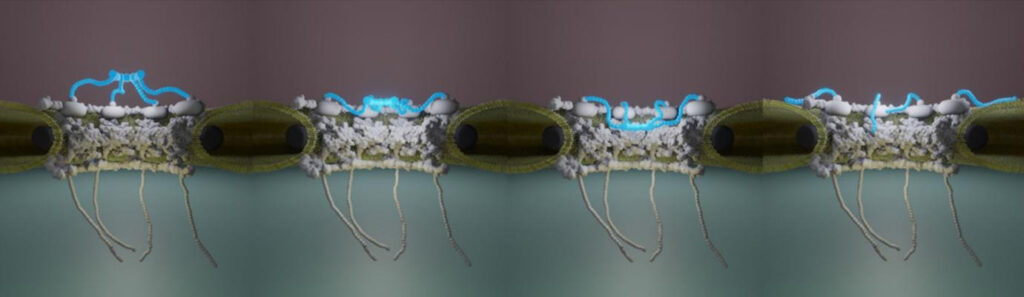
Nuclear pore complexes (NPCs) are the only gateways between the nucleus and cytoplasm in eukaryotic cells, facilitating the transport of selected cargoes of size from a few up to hundred nanometers. This versatility implies an important pore plasticity. Here, by combining atomic force microscopy (AFM) and single molecule localization microscopy (SMLM), a group led by France-BioImaging R&D team members Christine Doucet and Pierre Emmanuel Milhiet revealed that the NPC basket is very soft and explores a large conformational landscape: apart from its canonical basket shape, it dives into the central pore channel or opens, highlighting  how this structure can adapt, and let morphologically diverse cargoes shuttle through NPCs.

Vial et al., Nanoscale, 15, 5756-5770 (2023) 

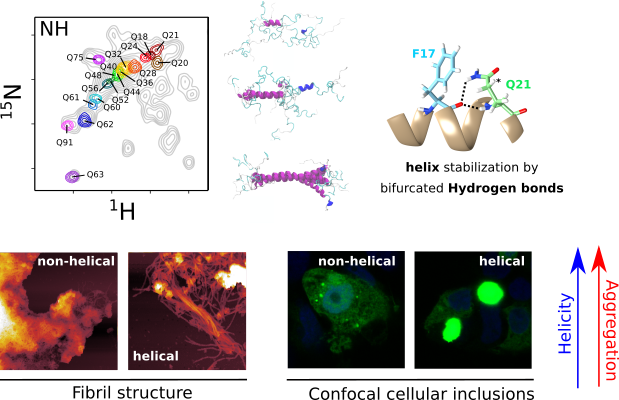
  • The structure of pathogenic huntingtin exon 1 defines the bases of its aggregation propensity

Structural Biology meets Correlative Imaging

Huntington’s disease is a neurodegenerative disorder caused by an extended polyglutamine (poly-Q) tract in huntingtin. Here, using NMR, the team of Pau Bernado (CBS Montpellier) demonstrated that this poly-Q tract adopts long α-helical conformations. By adding correlative Atomic Force Microscopy and Fluorescence Microscopy data obtained in the FranceBioImaging facility PIBBS in Montpellier, they could demonstrate that the stability of this α-helix is a stronger signature than the number of glutamines, in defining the aggregation kinetics and the structure of the resulting fibrils, potentially linked to their pathogenicity.

Elena Real et al., Nature Structural & Molecular Biology, 30, 309–320 (2023)

How to use Atomic Force Microscopy at France-BioImaging?

Atomic Force Microscopy is open to collaborations under Proof-of-concept studies via Euro-BioImaging webportal (www.eurobioimaging.eu/service)! At the Montpellier node of France-BioImaging, you will be in contact with Dr Luca Costa (costa@cbs.cnrs.fr) with whom you will talk about the feasibility and the inherent experimental constraints linked to the technique. The collaboration procedure is discussed on a case-by-case basis, depending on the duration and technicity of the required experiments. Feel free to submit your project!

Thanks to Christine Doucet and Emmanuel Margeat for providing helpful information!

1. Dahmane, S., Doucet, C., Le Gall, A., Chamontin, C., Dosset, P., Murcy, F., Fernandez, L., Salas, D., Rubinstein, E., Mougel, M., et al. (2019). Nanoscale organization of tetraspanins during HIV-1 budding by correlative dSTORM/AFM. Nanoscale 11, 6036–6044.

2. Vial, A., Costa, L., Dosset, P., Rosso, P., Boutières, G., Faklaris, O., Haschke, H., Milhiet, P.-E., and Doucet, C.M. (2023). Structure and mechanics of the human nuclear pore complex basket using correlative AFM-fluorescence superresolution microscopy. Nanoscale 15, 5756–5770.

Deadline: April 21th, 2023

The three national infrastructures ProFi, France-BioImaging and FRISBI along with the GIS IBiSA are pleased to announce a call for a funded access to IBiSA-labelled facilities.

Our aim is to promote IBiSA facilities networking through interdisciplinary research projects.

Applications should request access to at least two different IBiSA facilities from two disciplines (structural biology, Biological imaging and proteomics). The call is open to any academic laboratory.

The amount of the financial support will be up to 5000 € per application to cover facility costs.

Applications should be submitted to: Call-IBISA-FBI-FRISBI-PROFI@i2bc.paris-saclay.fr
using the template document https://mycore.core-cloud.net/index.php/s/VNLU2jgfcP4o0NN


The deadline for this inter-infrastructure access call is April 21, 2023.

All submitted proposals will be peer-reviewed by independent experts and the final funding will be approved by a committee comprising 2 representatives from each infrastructure as well as representatives
from GIS IBISA. We advise the project PI to contact the chosen facilities in order to set-up the optimal experimental design.

Call description

Correlative X-ray imaging and electron microscopy (CXEM) is the combination of X-ray imaging and electron microscopy. It is a correlative approach that makes it possible to characterise a sample of interest and locate a structure of interest in a non-destructive way. Nicolas BROUILLY is in charge of the Electron Microscopy Unit of the PICsL imaging facility on the Marseille node of France BioImaging, where CXEM is used for developmental biology studies. As part of Euro-BioImaging’s Proof-of-Concept study, his facility is now accepting applications from external users for CXEM projects. Learn more about how this approach works and what it can be used for in the interview below. 

We are today talking about CXEM imaging. Please provide a short summary of this type of imaging and tell us some applications:

Nicolas Brouilly: It is often very useful to combine 2 imaging modalities to take advantage of each while trying to lower their respective drawbacks. For example, by combining Light Microscopy and Electron Microscopy, we obtain the popular CLEM (for Correlative Light and Electron Microscopy). Visible light can then be used in combination with EM either:

  • To target a precise region of interest ;
  • To localize molecules within the ultrastructural information obtained by EM.

Using the same acronym building, CXEM corresponds to Correlative X-ray and Electron Microscopy. X-rays are photons of shorter wavelength than those from visible light, and can again be used to characterize a sample in 2 different ways:

  • To use their ability to easily go through tissues in order to record the 3D morphology of a sample: either by computed x-ray micro-tomography (or micro-CT) for micrometric resolution of big samples (mm to cm range) or by Soft X ray tomography for nanometric resolution of small samples (100’s of nm to um range);
  • To use a focused beam of high energy x-rays to analyse the localization of the elements of a sample: X-Ray Fluorescence microscopy (or XRF).

Both modalities can be used to complement the ultrastructural information obtained by electron microscopy. At the Marseille node of France BioImaging, in the Electron Microscopy Unit, we routinely use Correlative Micro-CT and Electron Microscopy to answer developmental biology questions.

What are some advantages of this technique that make it suited to addressing this type of question?

Nicolas Brouilly: The main advantage of Micro-CT (or Computed X-ray Tomography) is its ability to “see through” a sample and to reveal its overall organization in 3D without any labelling. The second advantage of Micro-CT is the fact that it is non-destructive. Thirdly, the contrast we usually give to samples for electron microscopy is compatible and even beneficial for X-ray imaging.

Altogether, this means that we can use X-ray tomography to map the microscale morphology of a sample in order to target a specific region of interest without having to go through the time-consuming and destructive collection of semi-thin sections.

We routinely use the micro-CT tool, not only to target a given organ or a given group of cells, but also to pre-orient the sample in order to cut it under a specific orientation. It is a timesaving tool within the frame of a 2D electron microscopy project, but it really is key within the frame of a 3D electron microscopy project given that Serial BlockFace and Focused Ion Beam techniques are destructive.

Tell us a bit more about a specific project that was done in your facility using this technology? What scientific questions were you addressing?

Nicolas Brouilly: Imagine that, first, you have a ball of yarn, second, you cannot untangle it, and third, you want to cut small bits of the thread at 24 cm from the end (not 22, not 26… 24 !). CXEM enabled us to do this on Drosophila gut. The micro-CT gave us the 3D map of the sample within the resin block. We could then use this map to find the best itinerary within the sample to make transverse sections of the portion of interest that was precisely indicated by the user on the micro-CT dataset. At the end of the day, the user was able to look at perfect transverse ultrathin TEM sections, at a precise position of this ball of yarn that Drosophila gut is. He could finally get precise metrics from this precise part of the gut in several samples. None of this could have been achieved without CXEM.

Like a ball of yarn… Above is an example of how CXEM can be used to find the best itinerary within a sample to make transverse sections of the portion of interest. On the left, the micro-CT provided a 3D map of the sample within the resin block. On the right, a transverse ultrathin TEM section of the drosphilia gut. Image courtesy of Nuno Luis (Schnorrer lab, IBDM) & Nicolas Brouilly (Electron Microscopy Facility, IBDM AMU/CNRS, France BioImaging).

For another example, you can have a look at the following paper where we used CXEM to map platelet aggregates within arteries in order to explore them by Serial BlockFace SEM, another example of “Find a needle in a haystack”. Have a look at movie S1, it is a wonder that we could not obtain without CXEM:

Estelle Carminita, Lydie Crescence, Nicolas Brouilly, Alexandre Altié, Laurence Panicot-Dubois, Christophe Dubois, DNAse-dependent, NET-independent pathway of thrombus formation in vivo, Proc Natl Acad Sci U S A. 2021 Jul 13;118(28)

Finally, below is the seminal paper from the Schwab lab at EMBL, which revealed the power of micro-CT to the Electron Microscopy community : 

Matthia A. Karreman, Luc Mercier, Nicole L. Schieber, Gergely Solecki, Guillaume Allio, Frank Winkler, Bernhard Ruthensteiner, Jacky G. Goetz, Yannick Schwab, Fast and precise targeting of single tumor cells in vivo by multimodal correlative microscopy, J Cell Sci (2016) 129 (2): 444–456.

Contact: nicolas.brouilly@univ-amu.fr

How to apply:

CXEM is part of the Euro-BioImagingProof-of-Concept study. The Proof-of-Concept study makes it possible to introduce exciting, new imaging technologies to our portfolio that were previously unavailable via our network. We are currently accepting applications to use these technologies at participating Nodes as part of the Proof-of-Concept study. Be part of this study – and contribute to community-wide continuous technological innovation!

All scientists, regardless of their affiliation, area of expertise or field of activity can benefit from Euro-BioImaging’s pan-European open access services. Potential users of these new technologies are encouraged to submit project proposals via our website. To do so, you can Login to access our application platform, choose the technology you want to use and the facility you wish to visit, then submit your proposal. All applications will be processed by the Euro-BioImaging Hub. As usual, users will benefit from advice and guidance by technical experts working at the Nodes, training opportunities, and data management services.

For more information: info@eurobioimaging.eu

From left to right: Nicolas BROUILLY (Head of facility), Aïcha AOUANE (Engineer Assistant), Fabrice RICHARD (Engineer)

Thanks to Nicolas Brouilly for the article!
Original article on: www.eurobioimaging.eu/news/cxem-finding-a-needle-in-a-haystack

Alors que les volumes et la complexité des données augmentent de manière sans précédent, leur suivi et partage devient de plus en plus un défi pour la communauté de la recherche en science de la vie (biologie, biomédicale…). Ce manque de traçabilité a un impact négatif reconnu sur la réutilisation des données publiées.
C’est pourquoi il est important de rendre les données « FAIR » – faciles à trouver, accessibles, interopérables, réutilisables – dès la conception des projets de recherche.

OMERO est la solution intégrée de référence pour gérer les données d’images pendant toute leur durée de vie, de l’acquisition à la publication.

Le RT-mfm du CNRS organise cette ANF avec la collaboration de France BioImaging et le EMBRC. Cette ANF est orientée vers les ingénieurs en plateforme d’imagerie et similaires. Elle a pour objectifs de former les stagiaires à la gestion FAIR de données au travers la base de données OMERO comme outil principal de gestion de données pour la microscopie et pendant tout le cycle de vie de la donnée : de l’acquisition à la publication. 

A l’issue de la formation les stagiaires seront capables de :
    • Comprendre les principes FAIR et leur application dans la pratique
    • Comprendre les enjeux et objectifs d’un Plan de Gestion de Données.
    • Connaître les schémas de mise en place d’une infrastructure OMERO avec les différents scenarios de déploiement
    • Savoir administrer les politiques d’accès selon les différentes configurations de groupes
    • Importer des images dans OMERO
    • Être capable de décrire les images avec des métadonnées riches
    • Analyser les données sur OMERO et sauver les résultats sur OMERO en association avec les données source
    • Annoter les images en masse à partir de tableaux
    • Chercher des images en fonction des différentes métadonnées
    • Être capable de créer des figures avec OMERO-figure

    • Apprendre les options d’interfaçage avec différents logiciels (ImageJ, Napari, CellProfiler, QPath,…)
    • Publier les données

Two projects recently received funding from the Chan-Zuckerberg Initiative (CZI), in which France-BioImaging members take part actively: COMULIS and NEUBIAS. Two community building activities breaking up frontiers to gather scientists around one goal: developing biological imaging.

COMULIS

COMULIS received a 2-year funding from CZI to expand their network both globally and sustainably. Being designed to harness the power of multimodal imaging (MMI) across scales, from basic to clinical diagnostics, this European initiative aims at facilitating access and training a new generation of scientists for whom multimodal imaging will be the new norm. Thanks to this grant, the project will be consolidated and it will help extend the collaborative and innovative network to establish a global multimodal imaging association (COMULISglobe) and ensure long term sustainability.

MMI integrates the best features of combined techniques and overcomes limitations faced when applying single modalities independently. MMI relies on the joint expertise from biologists, physicists, chemists, clinicians, and computer scientists, and depends on coordinated activities and knowledge transfer between technology developers and users. To achieve this inherently interdisciplinary goal, the ultimate goal is toestablish a network of scientists across continents and disciplines, from academia to industry, including transnational research facilities (e.g. synchrotrons, Euro-BioImaging ERIC), to foster and market MMI as a versatile tool in biomedical research and diagnostics.

COMULISglobe will help bridge the gap between biological and clinical imaging, identify, fund, and showcase novel multimodal pipelines, and develop, evaluate, and publish correlation software through dedicated networking activities, including conferences, training schools, open databases, and fellowships for lab exchanges, access to research infrastructures, and conference attendance. And, of course, all outputs of the project will be open access!

Please do not hesitate to join the community and help organize activities or publications – and please share the news, mobility and access grants available at: https://www.comulis.eu/comulisglobe-czi

Thanks to Perrine Paul-Gilloteaux, Bretagne-Loire node of France-BioImaging, for taking part of this amazing project!

NEUBIAS

The international Network of European BioImage Analysts (NEUBIAS), hosted by German BioImaging has also received a 2-year funding from the Chan Zuckerberg Initiative (CZI) as part of their Advancing Imaging through Collaborative Projects program. This grant will secure the sustainability of NEUBIAS, establish strong connections to similar initiatives, and share knowledge about state-of-the-art bioimage analysis tools and methods globally.

Spreading the profession of bioimage analysts and bioimage analysis knowledge internationally are the major aims of NEUBIAS. Modern life-sciences are unthinkable without advanced microscopy imaging techniques and quantitative bioimage analysis. This grant will help ensure novices and experts can access cutting edge techniques, reduce duplications of effort, and support everyone who is working to making new discoveries possible.

NEUBIAS had a tremendous impact on the community by training a powerful generation of bioimage analysts across Europe and beyond. The next step of this project will expand the network internationally and connect to related imaging and image analysis societies around the globe. With that in mind, the project includes travel grant opportunities for early-career bioimage analysts who seek to join NEUBIAS activities, explicitly including scientists outside central Europe. Besides, a dedicated team will work on collecting bioimage analysis teaching materials and make them accessible to the global imaging and life science community.

Great news for both projects that – we hope – will continue to write the great story of bioimaging!

Thanks to Florian Levet, Bordeaux node of France-BioImaging, for being a member of this fantastic project!