Correlative X-ray imaging and electron microscopy (CXEM) is the combination of X-ray imaging and electron microscopy. It is a correlative approach that makes it possible to characterise a sample of interest and locate a structure of interest in a non-destructive way. Nicolas BROUILLY is in charge of the Electron Microscopy Unit of the PICsL imaging facility on the Marseille node of France BioImaging, where CXEM is used for developmental biology studies. As part of Euro-BioImaging’s Proof-of-Concept study, his facility is now accepting applications from external users for CXEM projects. Learn more about how this approach works and what it can be used for in the interview below.
We are today talking about CXEM imaging. Please provide a short summary of this type of imaging and tell us some applications:
Nicolas Brouilly: It is often very useful to combine 2 imaging modalities to take advantage of each while trying to lower their respective drawbacks. For example, by combining Light Microscopy and Electron Microscopy, we obtain the popular CLEM (for Correlative Light and Electron Microscopy). Visible light can then be used in combination with EM either:
- To target a precise region of interest ;
- To localize molecules within the ultrastructural information obtained by EM.
Using the same acronym building, CXEM corresponds to Correlative X-ray and Electron Microscopy. X-rays are photons of shorter wavelength than those from visible light, and can again be used to characterize a sample in 2 different ways:
- To use their ability to easily go through tissues in order to record the 3D morphology of a sample: either by computed x-ray micro-tomography (or micro-CT) for micrometric resolution of big samples (mm to cm range) or by Soft X ray tomography for nanometric resolution of small samples (100’s of nm to um range);
- To use a focused beam of high energy x-rays to analyse the localization of the elements of a sample: X-Ray Fluorescence microscopy (or XRF).
Both modalities can be used to complement the ultrastructural information obtained by electron microscopy. At the Marseille node of France BioImaging, in the Electron Microscopy Unit, we routinely use Correlative Micro-CT and Electron Microscopy to answer developmental biology questions.
What are some advantages of this technique that make it suited to addressing this type of question?
Nicolas Brouilly: The main advantage of Micro-CT (or Computed X-ray Tomography) is its ability to “see through” a sample and to reveal its overall organization in 3D without any labelling. The second advantage of Micro-CT is the fact that it is non-destructive. Thirdly, the contrast we usually give to samples for electron microscopy is compatible and even beneficial for X-ray imaging.
Altogether, this means that we can use X-ray tomography to map the microscale morphology of a sample in order to target a specific region of interest without having to go through the time-consuming and destructive collection of semi-thin sections.
We routinely use the micro-CT tool, not only to target a given organ or a given group of cells, but also to pre-orient the sample in order to cut it under a specific orientation. It is a timesaving tool within the frame of a 2D electron microscopy project, but it really is key within the frame of a 3D electron microscopy project given that Serial BlockFace and Focused Ion Beam techniques are destructive.
Tell us a bit more about a specific project that was done in your facility using this technology? What scientific questions were you addressing?
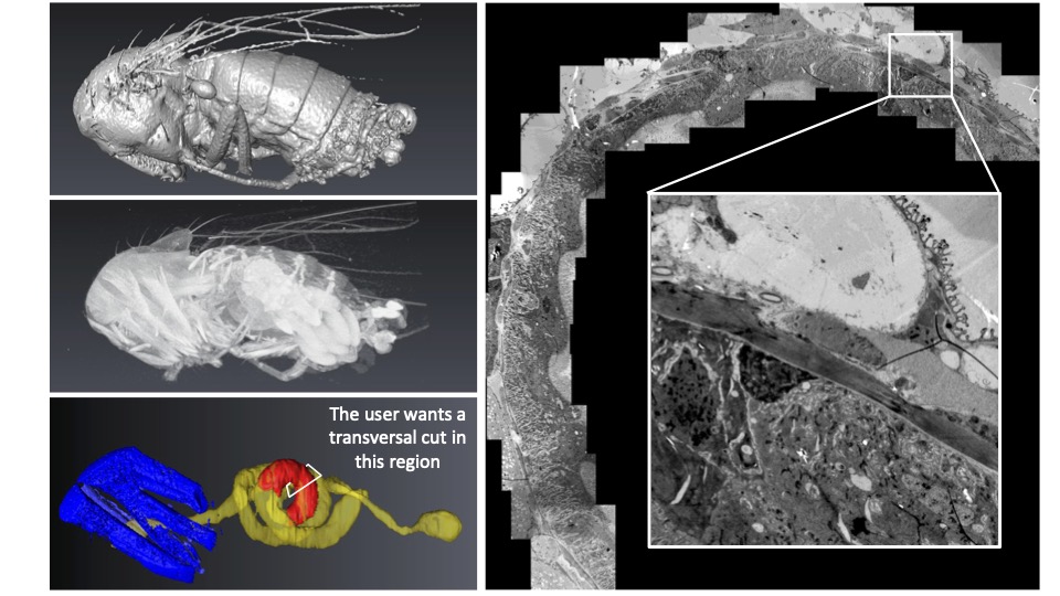
Nicolas Brouilly: Imagine that, first, you have a ball of yarn, second, you cannot untangle it, and third, you want to cut small bits of the thread at 24 cm from the end (not 22, not 26… 24 !). CXEM enabled us to do this on Drosophila gut. The micro-CT gave us the 3D map of the sample within the resin block. We could then use this map to find the best itinerary within the sample to make transverse sections of the portion of interest that was precisely indicated by the user on the micro-CT dataset. At the end of the day, the user was able to look at perfect transverse ultrathin TEM sections, at a precise position of this ball of yarn that Drosophila gut is. He could finally get precise metrics from this precise part of the gut in several samples. None of this could have been achieved without CXEM.
For another example, you can have a look at the following paper where we used CXEM to map platelet aggregates within arteries in order to explore them by Serial BlockFace SEM, another example of “Find a needle in a haystack”. Have a look at movie S1, it is a wonder that we could not obtain without CXEM:
Estelle Carminita, Lydie Crescence, Nicolas Brouilly, Alexandre Altié, Laurence Panicot-Dubois, Christophe Dubois, DNAse-dependent, NET-independent pathway of thrombus formation in vivo, Proc Natl Acad Sci U S A. 2021 Jul 13;118(28)
Finally, below is the seminal paper from the Schwab lab at EMBL, which revealed the power of micro-CT to the Electron Microscopy community :
Matthia A. Karreman, Luc Mercier, Nicole L. Schieber, Gergely Solecki, Guillaume Allio, Frank Winkler, Bernhard Ruthensteiner, Jacky G. Goetz, Yannick Schwab, Fast and precise targeting of single tumor cells in vivo by multimodal correlative microscopy, J Cell Sci (2016) 129 (2): 444–456.
Contact: nicolas.brouilly@univ-amu.fr
How to apply:
CXEM is part of the Euro-BioImagingProof-of-Concept study. The Proof-of-Concept study makes it possible to introduce exciting, new imaging technologies to our portfolio that were previously unavailable via our network. We are currently accepting applications to use these technologies at participating Nodes as part of the Proof-of-Concept study. Be part of this study – and contribute to community-wide continuous technological innovation!
All scientists, regardless of their affiliation, area of expertise or field of activity can benefit from Euro-BioImaging’s pan-European open access services. Potential users of these new technologies are encouraged to submit project proposals via our website. To do so, you can Login to access our application platform, choose the technology you want to use and the facility you wish to visit, then submit your proposal. All applications will be processed by the Euro-BioImaging Hub. As usual, users will benefit from advice and guidance by technical experts working at the Nodes, training opportunities, and data management services.
For more information: info@eurobioimaging.eu

Thanks to Nicolas Brouilly for the article!
Original article on: www.eurobioimaging.eu/news/cxem-finding-a-needle-in-a-haystack


