Interdisciplinary access call to Structural biology, Biological imaging and Proteomics facilities
▽ Scroll down
Tag: Microscopy
Deadline: April 15th, 2022
The three national infrastructures ProFi, France-BioImaging and FRISBI along with the GIS IBiSA are pleased to announce a call for a funded access to IBiSA-labelled facilities.
Our aim is to promote IBiSA facilities networking through interdisciplinary research projects.
Applications should request access to at least two different IBiSA facilities from two disciplines (structural biology, Biological imaging and proteomics). The call is open to any academic laboratory.
The amount of the financial support will be up to 5000 € per application to cover facility costs.
The deadline for this inter-infrastructure access call is 15 April 2022.
All submitted proposals will be peer-reviewed by independent experts and the final funding will be approved by a committee comprising 2 representatives from each infrastructure as well as representatives from GIS IBISA. We advise the project PI to contact the chosen facilities in order to set-up the optimal experimental design.
The France-BioImaging LSFM workgroup is pleased to announce an INSERM workshop on Light-Sheet Fluorescence Microscopy imaging technics.
This workshop will be divided in two parts:
A first theoretical part in Bordeaux from the 16th to the 18th of May 2022 which will cover basic principles, applications and challenges of LSFM imaging through seminars,
A second practical part along June 2022 where you will have the possibility to choose the set-up on which you want to be trained amongst many systems available within the France-BioImaging community.
If you want to discover, learn and/or deepen your knowledge about this vast and powerful family of 3D imaging technics, this training is for you!
You will find all the information (program, registration details, venue and accommodations, …) about this event on the following poster and website (Atelier 268): https://evenium-site.com/site/atelier-de-l-inserm-268
The IGDR (Institut de Génétique et de Développement de Rennes) and MRic facilities in Rennes organize an EMBO practical course “Sensing biophysical properties in living samples using light microscopy” from July 4th to July 9th 2022.
France BioImaging and all the French community aims to develop and promote innovative imaging technologies and methods. But microscopy images can also take an artistic, creative look and make the invisible world beautiful, allowing people to see the visual appeal of the life sciences.
We enjoyed the diversity of the images submitted with many different microscopy techniques, models and applications represented. A big thank you to all the participants!
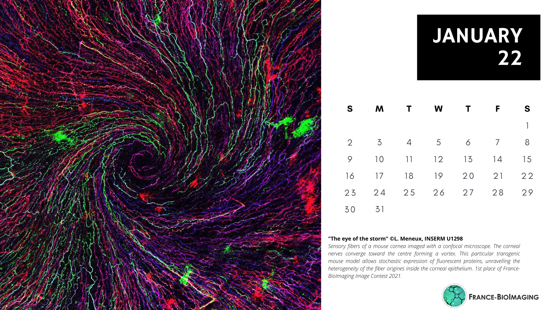
The National Coordination Team and the Executive Board are proud to announce the winners of the FBI Image Contest 2021:
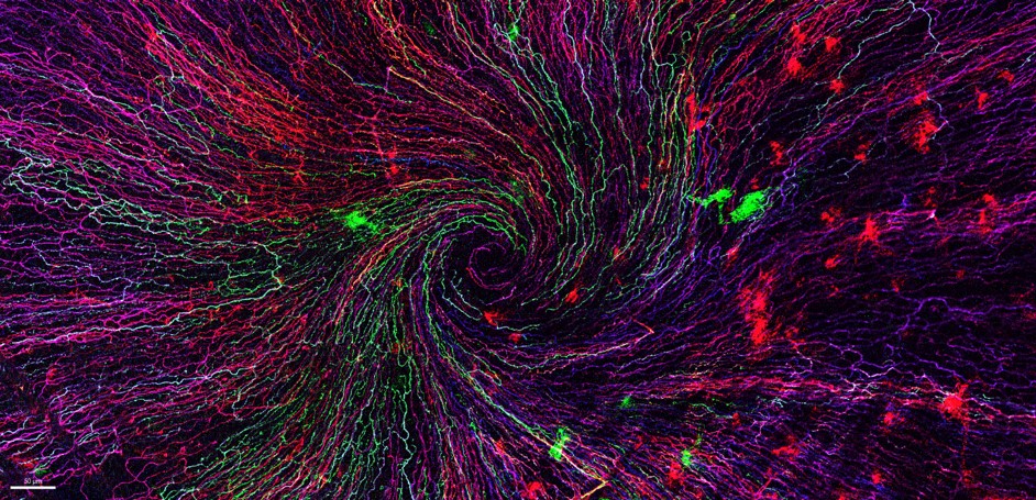
1st Place: Léna Meneux, Eye Team, Institut des Neurosciences de Montpellier
“The eye of the storm”
Sensory fibers of a mouse cornea imaged with a confocal microscope. The corneal nervesconverge toward the centre forming a vortex. This particular transgenic mouse model allows stochastic expression of fluorescent proteins, unravelling the heterogeneity of the fiber origines inside the corneal epithelium. Acknowledgements to Karine Loulier for the mouse model and Laetitia Hudececk for her help during the acquisition.
Confocal microscopy
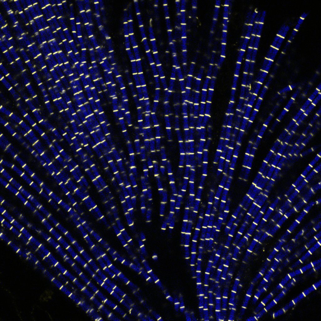
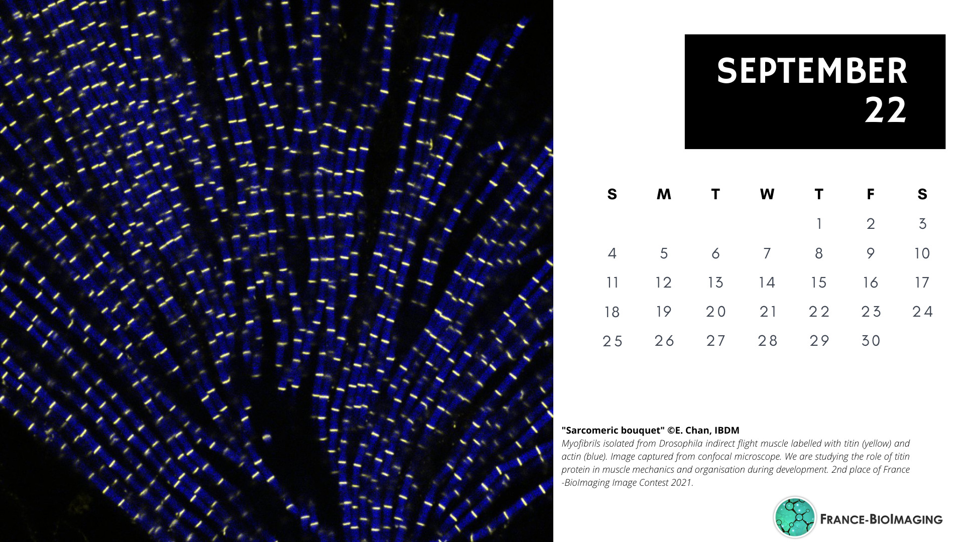
2nd Place:Eunice HoYee Chan, Muscle Dynamics Team, Developmental Biology Institute of Marseille (IBDM)
“Sarcomeric bouquet”
Myofibrils isolated from Drosophila indirect flight muscle labelled with titin (yellow) and actin (blue). Image captured from confocal microscope. We are studying the role of titin protein in muscle mechanics and organisation during development.
Confocal LSM880
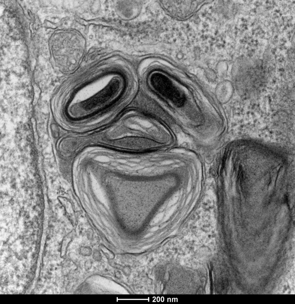
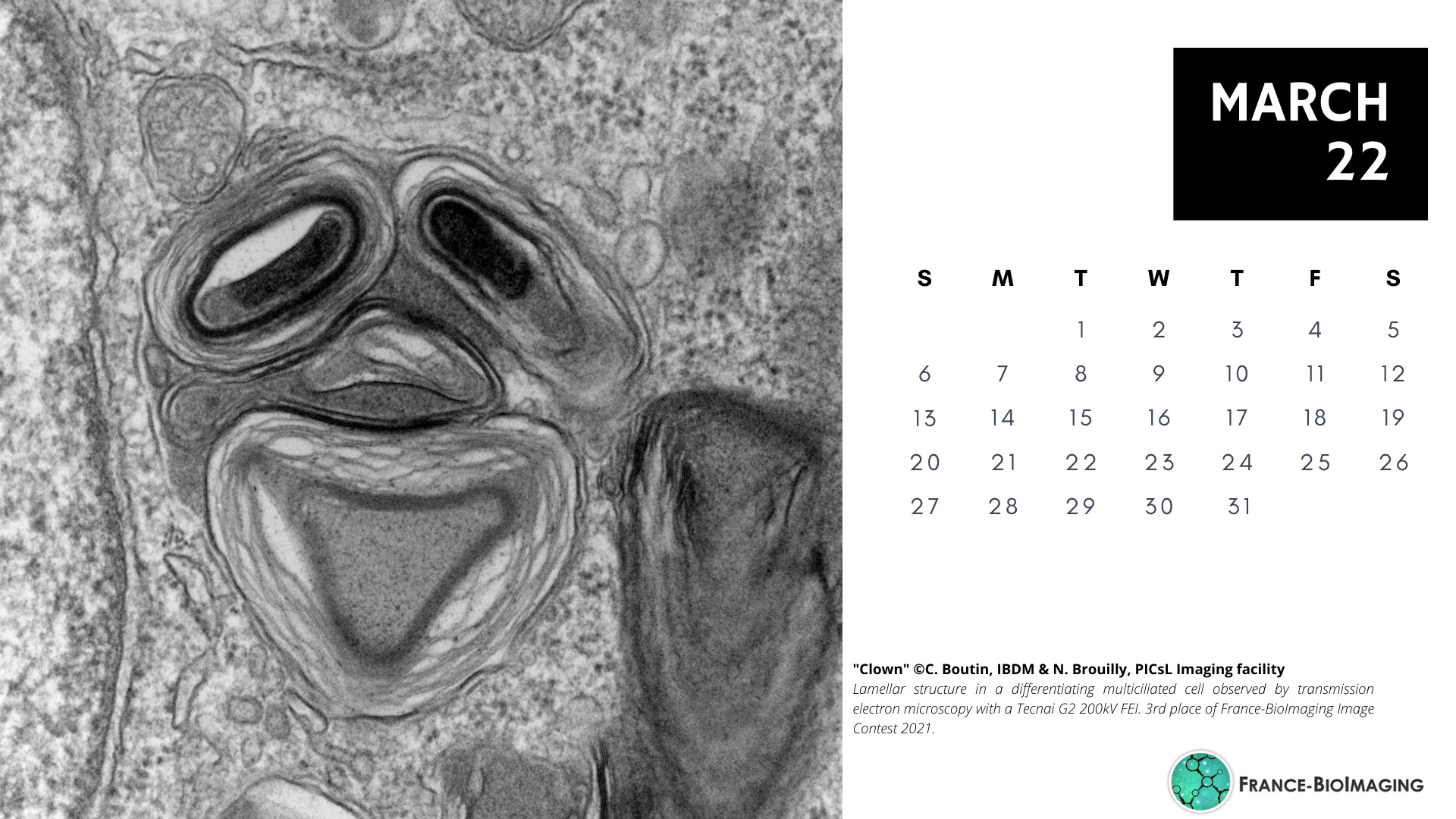
3rd Place:Camille Boutin, Biology of multiciliated cells Team, Developmental Biology Institute of Marseille (IBDM) &Nicolas Brouilly, PICsL Imaging facility, Electron Microscopy department
“Clown”
Lamellar structure in a differentiating multiciliated cell observed by transmission electron microscopy with a Tecnai G2 200kV FEI.
Transmission Electron Microscopy, Tecnai G2 200kV FEI
Congratulations to the winners!
Explore all the images submitted here:
As stated in the Terms & Conditions of the contest, foreign participants non-affiliated to a French institution are featured in the gallery, but were not evaluated as part of the contest.
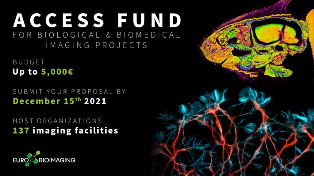
Euro-BioImaging first open call for user projects is open! If you have an idea for a biological or biomedical imaging project that you, your student or your close colleague could carry out in one of Euro-BioImaging Nodes, including France-BioImaging, now is the time to make this project come true with financial support from the Euro-BioImaging Access Fund.
How it works:
Submit your project proposal through the Euro-BioImaging web portal between October 20 and December 15, 2021, and indicate that you want to apply for the Euro-BioImaging Access Fund in order to be considered for a grant of up to 5.000 Euros to access the imaging services at a Euro-BioImaging Node. Projects will be evaluated by a committee of independent reviewers. Successful applicants will be notified by late January 2022 and successful projects should be started before July 2022.
What the funding covers:
The Euro-BioImaging Access Fund covers the user’s travel and accommodation costs as well as access and consumable costs at the imaging facilities that are part of Euro-BioImaging Nodes. For remote access projects shipment costs are also covered. Each successful applicant is eligible for up to 5.000 Euros of support.
Who is eligible:
All academic scientists, regardless of gender, nationality, home institution, career phase, or field of interest, are eligible to apply. We strongly encourage early career researchers to apply for this grant.
Projects should include transnational access to a Euro-BioImaging Node, i.e. the applicant’s home institution is in a different country than the Node where the project is to be performed. Due to the current sanitary situation, projects with non transnational access are elligible but transnational access will have priority.
All the external users/collaborators of France BioImaging facilities/labs are eligible:
any users from outside the institutional perimeter of France BioImaging nodes (i.e. from outside the following institutions: Aix-Marseille Université, Université de Montpellier, Université de Bordeaux, Université de Nantes, Université de Rennes 1, Université Paris-Saclay, Université de Paris, Université PSL -Paris Sciences & Lettres-, Généthon, Ecole Polytechnique, Institut Pasteur) who would like to use imaging technologies in one of FBI nodes: Paris Centre, Paris Ile-de-France-Sud, Marseille, Montpellier, Bordeaux, Bretagne-Loire. They can be French or international users – EU and non-EU
or users from a France BioImaging regional Node who want to access an equipment available in another FBI regional node.
Evaluation:
All applications will be evaluated for scientific excellence by a committee of independent reviewers. Selected projects will be assessed for technical feasibility and if needed receive technical advice from the Node providing the service.
How to apply:
Applicants are invited to visit our website to discover the range of technologies provided by Euro-BioImaging Nodes. Applicants will then follow the user access process described here: https://www.eurobioimaging.eu/about-us/how-to-access and indicate that they wish to apply for the Euro-BioImaging Access Fund in the application form.
SPAOM is a joint effort from Red Española de Microscopía Óptica Avanzada (REMOA) and the Portuguese Platform of Biomedical Imaging (PPBI), to organize an annual congress covering new applications in optical microscopy and image analysis. This year’s programme offers 5 plenary sessions + 5 community workshops + numerous workshops and tech bites from commercial partners.
REGISTRATION AND ATTENDANCE IS FREE FOR ALL PARTICIPANTS
There will be also flash-talks, and a prize for the best communication from the submitted abstracts! The full list of speakers and scientific programme can be found here: https://igc.events.idloom.com/spaom2021
(the event will be online, through the Hopin platform, transmitted from Oeiras @IGCiencia)
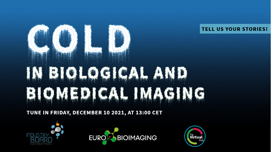
Euro-BioImaging is pleased to announce that the next special edition of the Virtual Pub, organized by Euro-BioImaging and the Euro-BioImaging Industry Board, will take place on December 10, 2021. In line with the season, this edition will focus on the theme of “COLD” in imaging.
They are inviting abstracts for 5 minute Flash talks about “COLD” in imaging. Be creative! Possible interpretations of “COLD” include:
Biology of the cold – from cold receptors and arctic organisms to brain reactions to cold and infections with common cold viruses
Cold-related metabolism including fat metabolism
Cold technologies – from cooling and freezing samples to low-temperature and cryo-imaging techniques
Keeping your sample or equipment cool – from reducing laser output and keeping high-power magnetic coils cool to incubating your sample at controlled low temperatures
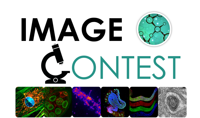
The France BioImaging Image Contest is back for its 3rd edition!
This image contest is open to all within the imaging community: core facility staff and users, R&D labs teams and co-workers, students… Submit your best microscopy images for a chance to showcase your skills, research and creativity to the French bioimaging community and beyond, allowing people to see the visual appeal of the life sciences. Images from the contest will be featured on France BioImaging communication tools, online and in print.
France BioImaging and all the French community aims to develop and promote innovative imaging technologies and methods. But microscopy images can also take an artistic, creative look and make the invisible world beautiful.
We are all eager to see your work !
Prizes
1 to 3 images will be awarded depending on the quantity and quality of the entries submitted. France BioImaging will cover the registration fees for one 2022 microscopy related event of the winners’ choice (FOM, ELMI, EMC, COMULIS conference, etc.).
Important: Only French or foreign participants affiliated to a French institution can enter the contest. Foreign participants non-affiliated to a French institution can submit images and will be featured in the gallery, but will not be evaluated as part of the contest.
Submission deadline: Friday, October 15th, 2021, 23h59 UTC+2.
The first practical workshop « Imaging Organoids: from the bench to the microscope » will take place in Bordeaux, from Mon 27th Sept to Fri 1st Oct 2021.
The aim of this workshop encompasses most of the workflow steps from the 3D-sample preparation (organoids, spheroids, encapsulated 3D cultures), how to process them (histology, staining), how to mount them (for upright, inverted etc…), and finally how to image them with various microscopy techniques (from super resolution microscopies -liveSR or STED- to microscopies dedicated to thick samples (two-photon, ultramicroscope), from fast optical scanning spinning-disk to High Content Screening -HiTech and LowTech-).
Bench work is possible thanks to the TBM Core Facility, and the microscopes are provided by the BIC facility and the LP2N (home-made setup).
This workshop is open to all (PhD students, Postdocs, Engineers, Technicians, Researchers and Teaching Assistants) who would like to learn how to prepare their 3D samples for photon microscopy.
The number of attendees will depend on the rules that will prevail at this time. Seats will be limited.
Each seminar will be preceded by a talk from our industry partners: Corning, StemCell, TreeFrog Therapeutics and Idylle. They will be accessible freely on a dedicated streaming platform, upon ad-hoc registration which will be set soon.
France BioImaging primary mission is to develop, promote, disseminate and provide access to innovative instruments and imaging technologies in the field of bioimaging to scientists. Fostering the technological transfers is at the heart of this mission, and for this France BioImaging relies on a strong association of leading R&D research teams with core facilities.
However, several bottlenecks exist and often hamper or prevent successful technology transfer:
A lack of human resource leads to difficulties in transferring and stabilizing the technology which is not enough user-friendly
A technology that is too specific, with not enough user base
A difficulty to contract with industry through institutional offices for industrial valuation
In the context of image analysis: the instability of open software economical model, inter-operability, large data handling and algorithm complexity
As a way to tackle these bottlenecks, France BioImaging launched in January 2021 its first “FBI Internal Call 2021: Technology transfer from the R&D teams to the core facilities” to promote the transfer of new technologies (instrumentation, probes, staining methods, software, data analysis or data visualization) from the R&D teams to the facilities of France BioImaging, for access and service to end-users. The outcome of the transfer project had to ensure for the prototype to be usable by the end-users until the interpretation of the data. The project had also to include a sustainability plan and a training plan to guide both facility staff and end-users toward autonomy.
The project selection was organized by the National Coordination of France-BioImaging and applications were assessed according to the following evaluation criteria:
Innovation and originality of the proposal
Scientific quality, implementation, timeline
Competitive positioning
Adequacy of resources with the proposed project
Economic impact and tech transfer potential and perspectives
Estimation of the user market and potential for user adoption
Plan for training and sustainability.
For the first edition of the “FBI Internal Call 2021: Technology transfer from the R&D teams to the core facilities”, 5 projects were selected:
Icy@FBI: Jean-Christophe Olivo-Marin (IPDM Node): Broadening the scope of applications of Icy (http://icy.bioimageanalysis.org/) by implementing several key new bioimage analysis components
BIC-HCS-SMLM: Jean-Baptiste Sibarita (Bordeaux Node), Technological transfer of a Single-Molecule-based High Content Screening platform to the Bordeaux Imaging Center
CloudFISH: Marcello Nollmann (Montpellier Node), A tool for the analysis of single-molecule RNA and DNA FISH images
MorphoNet: Emmanuel Faure (Montpellier Node), An interactive online morphological browser to explore complex multi-scale data
BioImageIT (https://bioimageit.github.io/#/about): Jean Salamero, Sylvain Prigent (IPDM Node), An open source framework for integration of image data management with analysis
Each selected project was awarded with a 80k€ grant for salary and/or equipment, and several positions are currently available: https://france-bioimaging.org/jobs/
This call will be renewed in 2023.
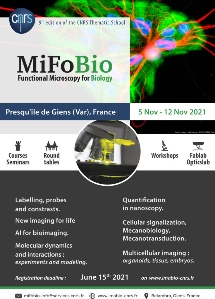
Don’t forget to register before June 15th for MiFoBio autumn school, 5-12th November 2021 (Presqu’île de Giens)!
We use cookies on our website to give you the most relevant experience by remembering your preferences and repeat visits. By clicking “Accept All”, you consent to the use of ALL the cookies. However, you may visit "Cookie Settings" to provide a controlled consent.
This website uses cookies to improve your experience while you navigate through the website. Out of these, the cookies that are categorized as necessary are stored on your browser as they are essential for the working of basic functionalities of the website. We also use third-party cookies that help us analyze and understand how you use this website. These cookies will be stored in your browser only with your consent. You also have the option to opt-out of these cookies. But opting out of some of these cookies may affect your browsing experience.
Necessary cookies are absolutely essential for the website to function properly. These cookies ensure basic functionalities and security features of the website, anonymously.
Cookie
Duration
Description
cookielawinfo-checkbox-analytics
11 months
This cookie is set by GDPR Cookie Consent plugin. The cookie is used to store the user consent for the cookies in the category "Analytics".
cookielawinfo-checkbox-functional
11 months
The cookie is set by GDPR cookie consent to record the user consent for the cookies in the category "Functional".
cookielawinfo-checkbox-necessary
11 months
This cookie is set by GDPR Cookie Consent plugin. The cookies is used to store the user consent for the cookies in the category "Necessary".
cookielawinfo-checkbox-others
11 months
This cookie is set by GDPR Cookie Consent plugin. The cookie is used to store the user consent for the cookies in the category "Other.
cookielawinfo-checkbox-performance
11 months
This cookie is set by GDPR Cookie Consent plugin. The cookie is used to store the user consent for the cookies in the category "Performance".
viewed_cookie_policy
11 months
The cookie is set by the GDPR Cookie Consent plugin and is used to store whether or not user has consented to the use of cookies. It does not store any personal data.
Functional cookies help to perform certain functionalities like sharing the content of the website on social media platforms, collect feedbacks, and other third-party features.
Performance cookies are used to understand and analyze the key performance indexes of the website which helps in delivering a better user experience for the visitors.
Analytical cookies are used to understand how visitors interact with the website. These cookies help provide information on metrics the number of visitors, bounce rate, traffic source, etc.
Advertisement cookies are used to provide visitors with relevant ads and marketing campaigns. These cookies track visitors across websites and collect information to provide customized ads.


