Developed by the Serpico Inria-CNRS-Institut Curie Joint Team, member of the IPDM-BioImage Informatics node of France-BioImaging (FBI), this open-source framework could be a huge step forward in bioimaging management and analysis.
Bioimaging has a broad range of applications, addressing a variety of biological models at diverse scales of life. Thus, descriptions of novel computational approaches are often focused on target case studies. To tackle any scenario in biological imaging is a major challenge, that needs the conception and the development of a unified solution.
With this in mind, the BioImageIT project aims at providing a middleware that integrates data management with analysis using existing softwares (Omero, BioFormats, Fiji, napari, Scipy, pytorch…). The mission of BioImageIT was to design a graphical user interface (GUI) that allows any scientist without coding skills to annotate and analyze datasets using various software. By being user-centered, open-source and cross-platform (Windows, MacOS, Linux), BioImageIT created a management tool that is definitely accessible and well documented.
Started in late 2019, the project, funded by France-BioImaging, is now being deployed in 10 FBI imaging facilities. As it is a first step, the BioImageIT project have the ambition to expand the dissemination of the middleware throughout France and even further, Europe.
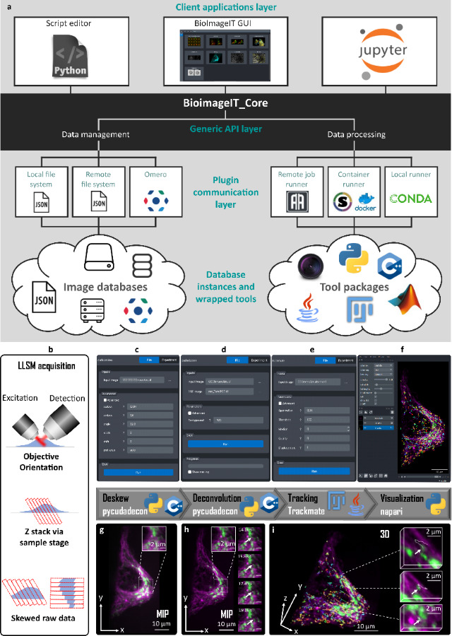
BioImageIT overview. a, Schematic view of BioImageIT architecture. The BioImageIT core is composed of data management and data processing functionalities. Users can access plugins by a script editor, Jupyter or the BioImageIT graphical interface (GUI). Data management functionalities exploit local files, remote files or databases such as OMERO. Data processing can perform computations in remote jobs, containers, or local runners. Image analysis is provided by plugins written in different languages. Developers can implement their own plugins in BioImageIT and design their own Graphical Interface. (b-i) LLSM processing workflow gathered in BioImageIT. Hela cell line expressing CD-M6PR-eGFP were stained with Tubulin TrackerTM Deep Red for Microtubules. b, Due to the geometry of LLS scanning, raw 3D images are skewed. c, g, First, realignment (deskew) of raw stacks is performed using Pycudadecon. d, h, Richardson Lucy deconvolution is performed using Pycudadecon. e, CD-M6PR-eGFP vesicles are tracked using Trackmate(FiJi). f, i, Deconvolved stacks and tracks are rendered using napari.
Prigent, S., Valades-Cruz, C.A., Leconte, L. et al. BioImageIT: Open-source framework for integration of image data management with analysis. Nat Methods (2022).
https://doi.org/10.1038/s41592-022-01642-9