Cannot see the nanorods for the blood cells
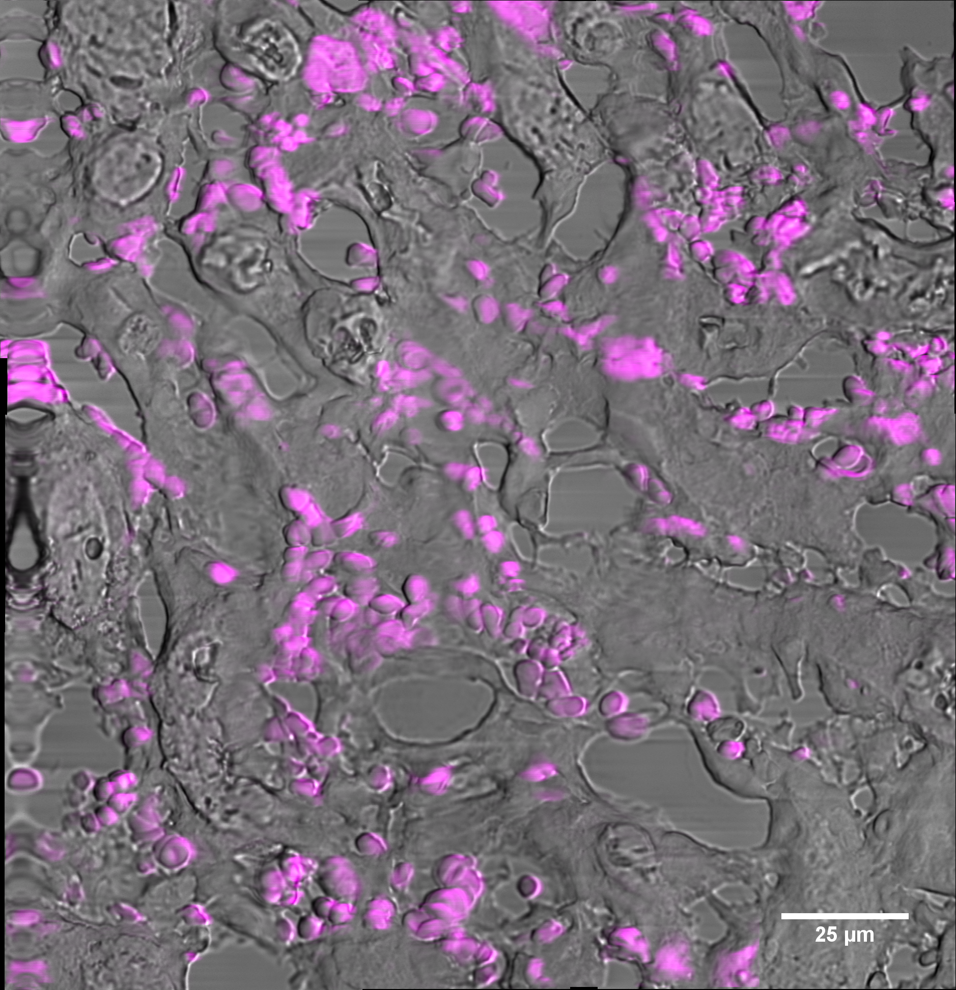
(Phothermal microscopy) Macrophages labelled with gold nanorods were injected into a mouse after kidney injury. Through photoaccoustic imaging, the cells were tracked over time and at the time of sacrifice, nanorod labelled cells were located in the kidney. The kidneys were prepared for photothermal imaging by slicing them to 7µm thick layers. Gold nanorods are generally easily detectable using photothermal microscopy and cells were expected to be located in the vasculature. However, the abundance of blood cells in the kidney resulted in us being unable to identify any nano rod labelled cells. The image shows an overlay of pseudo transmitted light and photothermal signal. The image was stitched from a grid of four by four individual images.