Meeting with Atitheb Chaiyasitdhi: Dive into hearing mechanism
We are pleased to introduce Atitheb Chaiyasitdhi, one of the winners of the “FBI Call for User Access Projects 2024.” Atitheb received a grant to access imaging services at one of the France-BioImaging facilities.
In this interview, discover his research project focused on auditory mechanotransduction and hearing loss in locusts.
To start, could you tell us a bit about yourself? What has been your academic journey so far, and what is your current role or area of work?
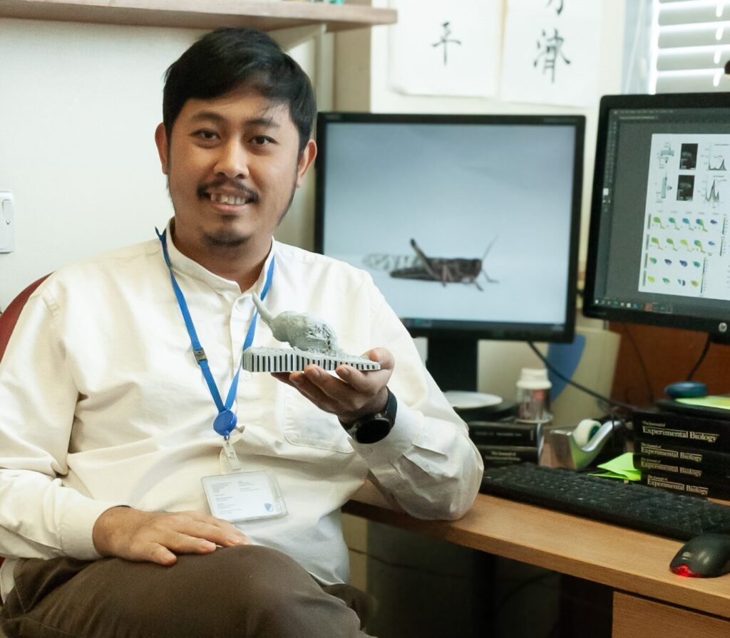
My name is Atitheb Chaiyasitdhi. I am a research fellow in Benjamin Warren’s lab at the University of Leicester in the United Kingdom. At the lab, we focus on auditory mechanotransduction and hearing loss in insects. Before joining the lab, I did my PhD at the Institut Curie in Paris, where I studied the biophysics of hearing in vertebrates.
What are you currently working on in your research? What is the main topic or challenge you’re exploring?
I have always been fascinated by how hearing works. Currently, I am investigating how insect ears operate. Unlike humans, insects possess ears in a wide variety of shapes and locations—sometimes on their legs, antennae, or abdomens. Despite this diversity, they all rely on a similar mechanosensitive structure known as a chordotonal organ. Interestingly, insects also use chordotonal organs to detect other mechanical cues, such as gravity or body position (proprioception), and these organs are located in different parts of the body as well.
This raises two key questions:
How does the chordotonal organ convert such a broad range of mechanical forces into electrical signals? And do these organs share a common underlying mechanism?
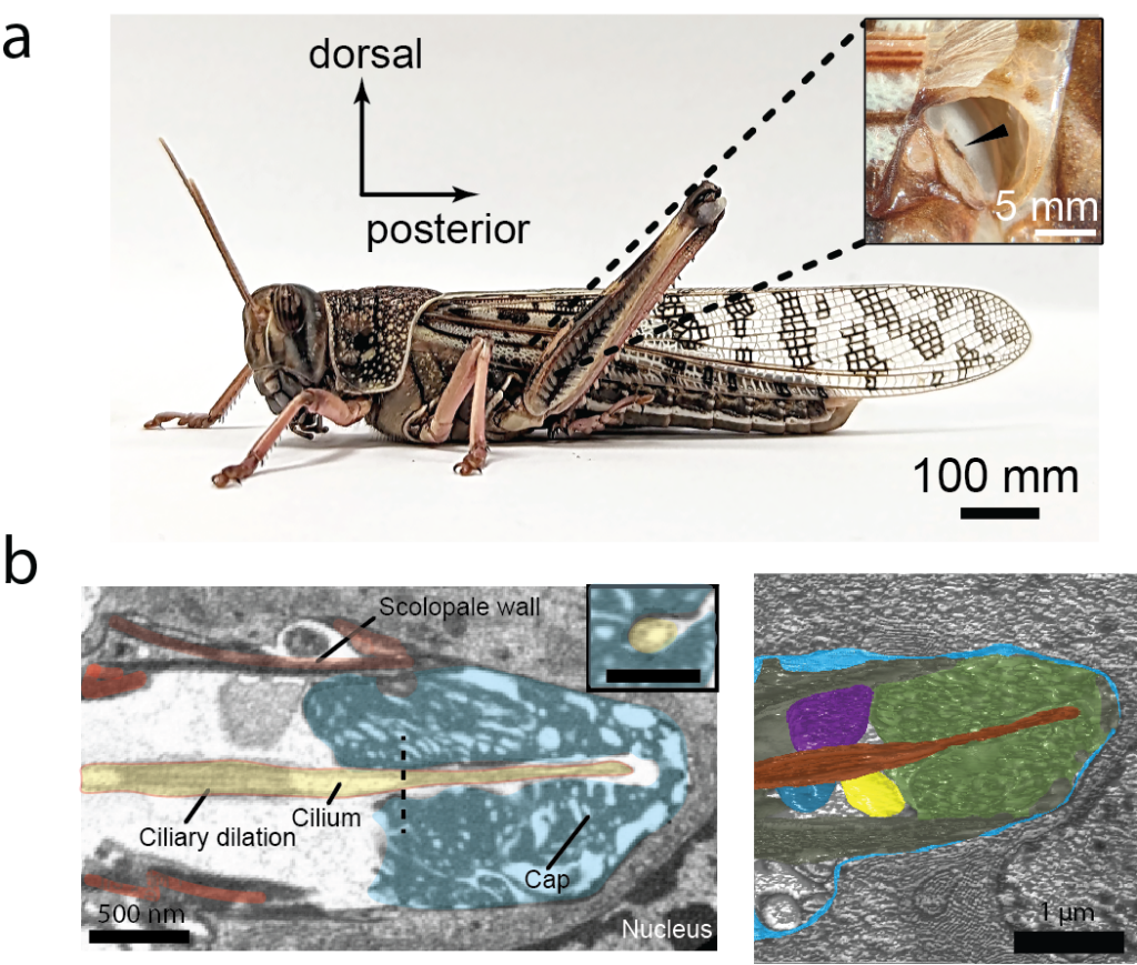
To explore these questions, I am focusing on the chordotonal organ in the locust ear. On one front, I am using a high-speed camera to capture sound-evoked motion of the organ and electrophysiology techniques to measure electrical current in the chordotonal neurons evoked by the sound.
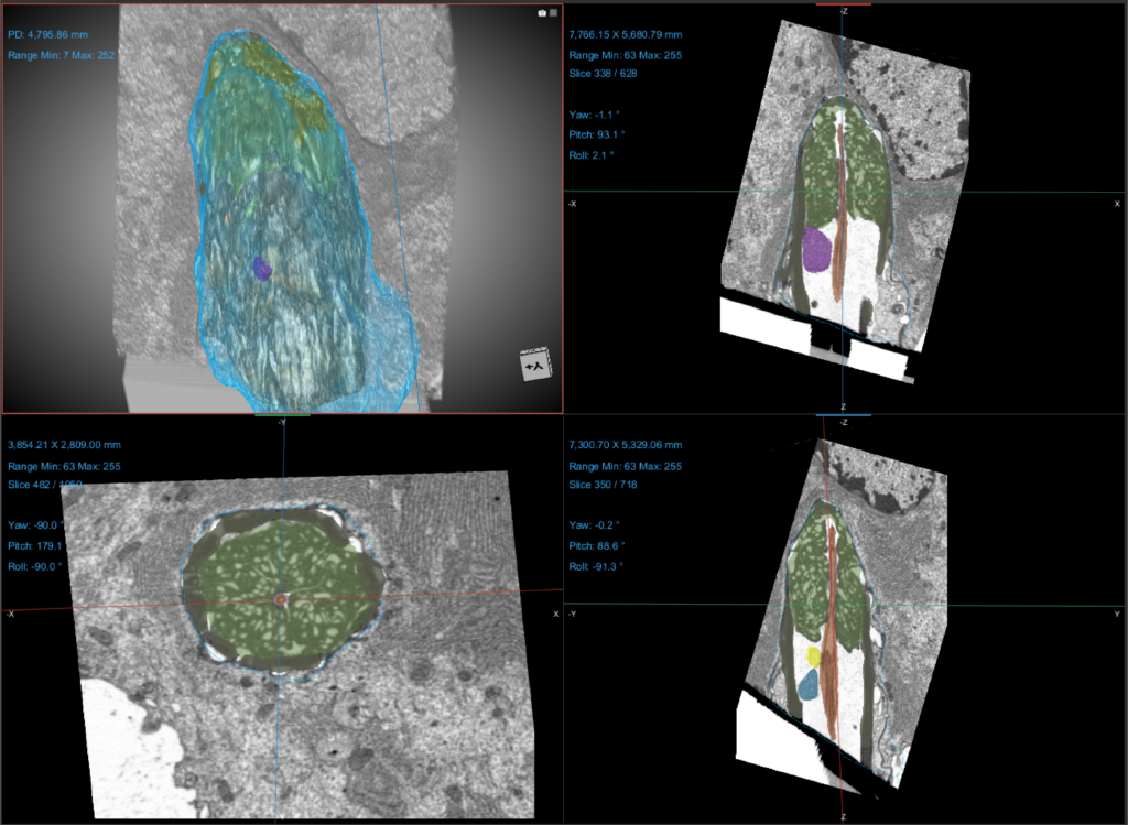
On another front, I am collaborating with Claire Boulogne at Plateforme Imagerie-Gif, using Focused Ion Beam Electron Microscopy (FIB-EM) to reconstruct the organ’s three-dimensional structure. These complementary approaches will provide insights into the mechanics of insect auditory transduction and bring us closer to solving the two questions I previously talked about.
At what point did you come across France-BioImaging, and what made you want to use its services or connect with the infrastructure?
I first learned about France-BioImaging during my PhD in France. At that time, I had the opportunity to collaborate with and use the excellent resources provided by one of the France-BioImaging Nodes—the Cell and Tissue Imaging Platform (PICT-IBiSA) at the Institut Curie. That experience left a strong impression on me, highlighting the expertise and support that France-BioImaging offers. Since then, I have been interested in collaborating with France-BioImaging platforms again.
Could you walk us through your experience accessing France-BioImaging? Which facility did you work with, how did the process go, and what stood out to you during your time there?
I worked with the Plateforme Imagerie-Gif in Gif-sur-Yvette, near Paris, using Focused Ion Beam Electron Microscopy (FIB-EM) with the support of Claire Boulogne, the lead engineer. Since I am based in the UK, and thanks to modern communication technology, our collaboration could start remotely. We discussed the experimental plan online, and I prepared the preliminary samples here before shipping them to the platform in France. Claire then carried out further sample preparation and image acquisition.
We worked closely, going back and forth to establish an optimal workflow, and soon began obtaining promising results afterwards. Despite the platform’s tight schedule, I always received prompt responses and consistent support from Claire.
What did microscopy bring to your project specifically? Were there insights or results you couldn’t have obtained otherwise?
The first ultrastructural image of a chordotonal organ was captured over 60 years ago, and it provided key insights into how the organ functions. In our lab, we have been able to resolve the mechanics of the chordotonal organ with unprecedented temporal resolution using high-speed cameras.
However, we still lack the spatial resolution to fully understand its structure. FIB-EM enables us to visualize the ultrastructure in three dimensions at a level of detail that has not been previously achieved. Seeing how each cellular component connects in 3D allows us to answer questions that arise from our high-speed recordings of sound-evoked motion in the organ.
At the same time, it opens up new questions and research opportunities. While there are other techniques, such as array tomography, they cannot match the spatial resolution of FIB-EM and would require hundreds of hours of manual effort.
Looking back, would you encourage other researchers to use France-BioImaging’s platforms and access program? What would you say to someone considering it?
Yes, I would. I would tell them that I have extensive experience working with France-BioImaging platforms, and the collaborations have always delivered reliable results. I trust their expertise and the quality of their support. In fact, I’ve already recommended France-BioImaging to colleagues here in the UK, as well as from the US and Germany. We are currently exploring the possibility of continuing our collaboration with Imagerie-Gif to investigate the diversity and evolution of chordotonal organs in various insect ears, even those involved in proprioception in the legs.
You can read Atitheb’s article preprint here: https://www.biorxiv.org/content/10.1101/2025.06.27.662008v1.full