Join the Imaris Workshop Day at Gustave Roussy Microscopy Facility!
Category: News from Nodes
Gustave Roussy Microscopy Facility, in collaboration with Imaris Software, is organizing the Imaris Workshop Day on Friday, February 14th.
This event includes a general presentation on Imaris, during which an Imaris expert will showcase various examples of its applications. Following the presentation, there will be an image analysis clinic where you can discuss the analysis of your own data*.
Workshop program:
11:00-12:00: Imaris presentation
13:00-16:00: Image analysis clinic
Location: Salle 2, Espace Maurice Tubiana, 20 Rue du Dr Pinel, 94805, Villejuif.
Registration is free of charge but mandatory. You can register here or click on the file below.
*If your data isn’t ready by then, we’ll find a similar dataset to discuss.
L’institut CIML (Centre d’Immunologie de Marseille Luminy), membre du nœud marseillais de France-BioImaging, organise en mars 2025 une formation dédié à la microscopie confocale spectrale.
L’objectif de cette formation, ouverte aux ingénieurs et chercheurs utilisant la microscopie confocale, est d’acquérir en mode spectral un panel 10 couleurs sur coupe de tissu et analyser les images réalisées.
Vous avez jusqu’au 27 janvier 2025 pour vous inscrire et retourner le questionnaire complété!
Contact:
Hélène Pastor: Chargée de formation et du développement des ressources humaines – Inserm demat-form.dr-marseille@inserm.fr
Inscription (aucune demande ne sera prise en compte sans le questionnaire):
Personnels Inserm ou non Inserm dans une structure mixte Inserm : inscription via www.sirene.inserm.fr + envoi du questionnaire à : demat-form.dr-marseille@inserm.fr (Région : Paca – Domaine : TS3 – Imagerie)
Autres personnels : formulaire d’inscription + questionnaire à transmettre à demat-form.dr-marseille@inserm.fr
From February 6 to 7, 2025, the University of Rouen Normandie will host the 8th edition of the France Cerebellum Club Days. This year’s event will include a session dedicated to cerebellum bioimaging, highlighted by the Primacen imaging platform, a member of the Normandie Node of France-BioImaging.
The France Cerebellum Club is an organization aimed at promoting exchanges between scientists involved in the study of the cerebellum in all its modalities, using a variety of analysis methods.
This new edition will bring together researchers and industrials to discuss the latest advances in the study of the cerebellum. On the program: plenary lectures, thematic sessions and workshops highlighting recent work on the development, functions and pathologies associated with this cerebral region. This year’s topics include the development and evolution of the cerebellum, innovations in applied bioimaging, organoid models and studies of connections between the cerebellum and other brain regions.
Two keynotes will surround these scientific days. Mari Sepp (Heidelberg, Germany) will present her work on the development and evolution of the cerebellum using single-cell genomics, while Christian Hansel (Chicago, USA) will discuss cerebellar instructive signals and their role in neocortical plasticity.
For more information and registration details, click here.
Le 3 décembre dernier, le Nœud Paris-Centre, membre de l’infrastructure France-BioImaging a organisé une journée intitulée placée sous le signe de la valorisation scientifique et du transfert de technologie. Accessible en mode hybride, elle a réuni une dizaine d’intervenants et une quarantaine d’auditeurs.
L’objectif de cette journée était de rendre accessible la valorisation de l’activité de recherche au plus grand nombre de chercheurs, étudiants, et acteurs du transfert technologique. À cette fin, les participants ont pu bénéficié d’une série de témoignages de chercheurs, de directeurs de startups (ABBELIGHT AVATAR MEDICAL, TWINCKLE FACTORY, INSCOPER) de responsables d’entreprises, et d’acteurs des services de valorisation qui ont converti un résultat scientifique dans le domaine de l’imagerie biologique en un produit ou un service commercialisé.
Ces témoignages ont permis aux intervenants de faire part de leur motivation et leur parcours de transfert technologique avec ses difficultés et ses satisfactions. Les directeurs de startups et responsables d’entreprises ont également présenté aux participants les temps clés de la valorisation d’une découverte scientifique, tels que les échanges entre industriels-chercheurs et la mise en place de la valorisation économique du travail scientifique initial.
Pour clore cette journée, une la table ronde finale a permis aux acteurs des services de valorisation de décrire la fonction, les moyens, ainsi que les modalités d’interaction avec les chercheurs et d’accompagnement du transfert technologique.
Restez connectés, les certaines sessions seront bientôt disponibles en replay!
On September 6, the Cellular Imaging Master, created in 2004 by Delphine Burel and Ludovic Galas at the University of Rouen Normandie, celebrated its 20th anniversary in partnership with the IBiSA PRIMACEN imaging facility.
Over 50 alumni attended the event, along with the Master’s coordinators and several teaching staff members. The day began with a review of the program’s 20 years and a presentation on international collaborations with the Universities of Turku and Abo Academy in Finland, followed by a vote for the new Master’s logo!
In the afternoon, participants enjoyed fun activities such as karting, bowling, and karaoke at Espace Loisirs Rouen, fostering a convivial atmosphere among students and teachers. Everyone agreed to meet again in five years for future celebrations.
This successful anniversary was made possible thanks to the efforts of the M2 IMAC 2024 class and the support of their sponsors!
Find out more about the IMAC Master’s program here.
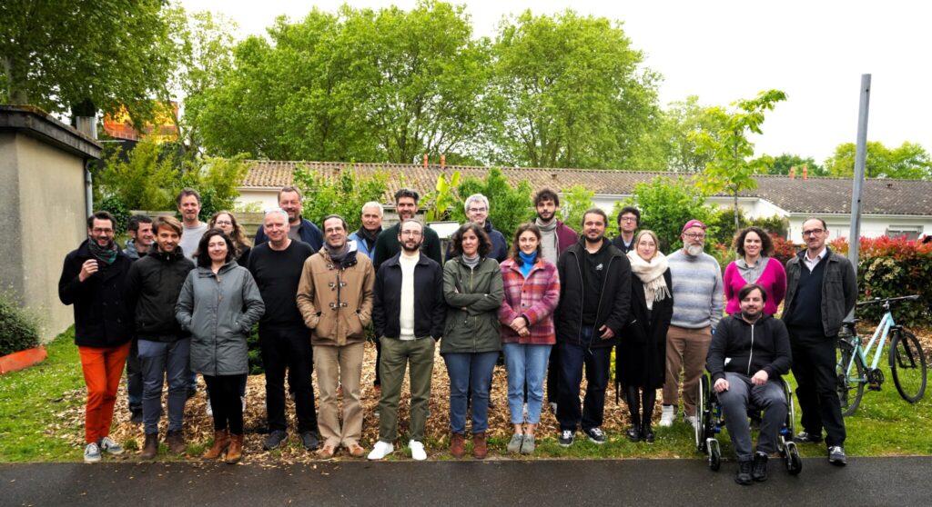
On September 27th, 2024, the Bordeaux Imaging Center (aka BIC) will be celebrating its 15th anniversary with a full-day conference at the Broca Auditorium, Centre Broca Nouvelle-Aquitaine, 146 rue Léo-Saignat, Bordeaux.
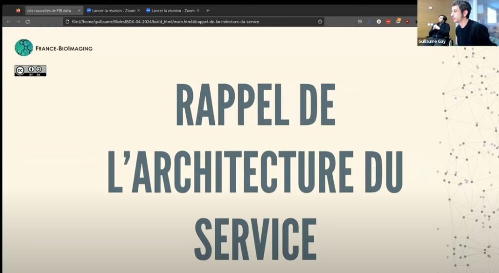
France-BioImaging’s roadmap for managing and analyzing data produced by the infrastructure spans several areas, supported by the transversal node Image Processing and Data Management (BioImage Informatics), as well as engineers and researchers distributed across various nodes.
Two geographically distributed teams are developing solutions: FBI.data for managing microscopy data, from metadata management to using data centers and regional computing centers, pooling efforts for the entire infrastructure; and F-BIAS to develop image analysis as a national service offering within the infrastructure. These distributed groups meet frequently via video conferencing and twice a year in person.
The FBI.Data and FB-IAS teams at the FBI Data Sprint Spring 2024 Edition
The Bordeaux Spring 2024 Edition allowed progress on the test deployment of the FBI.data solution, welcoming the latest F-BIAS recruits, and offering a live open desk. It also involved joint sessions between the two teams to address the challenge of making powerful but complex infrastructure accessible to our users, as well as discussing upcoming and ongoing challenges like the Lightmycells – Grand Challenge at grand-challenge.org.
The event also featured a public progress update via videoconference, with recordings available here:
As the FBI Correlative Light-Electron Microscopy workshop is currently happening at the Bordeaux Imaging Center (FBI Bordeaux node), what a better occasion to highlight a correlative microscopy technique: The Array Tomography.
Correlative Light and Electron microscopy (CLEM) increases our capacity of biological investigation. By combining light microscopy and electron microscopy, this complementary approach takes advantages of both techniques. In fact, light imaging provides valuable functional information thanks to its labeling power, whereas Electron microscopy excels at high resolution.
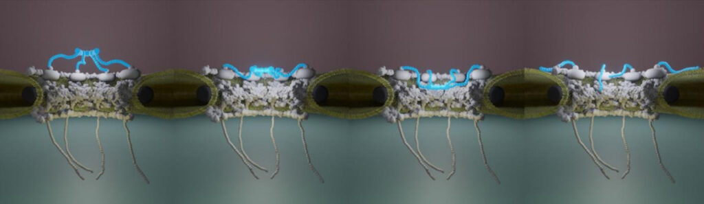
Where array tomography (AT) is special is that this technique is based on serial ultramicrotomy (cutting many sections less than a micrometer in thickness) of the sample, section collection onto support, and serial scanning EM (SEM) imaging.
An array of microscopy modes
Array tomography is a versatile microscopy method that offers opportunities to explore cell and tissues in three dimensions. This technique is well suited to image large tissue volumes of your sample with fine structural and molecular details.
Different modes are available, each having its own specificity and benefits:
The fluorescence microscopy AT mode (FM-AT) delivers volumetric resolution and molecular marker multiplexing highly superior to traditional fluorescence microscopies.
The electron microscopy AT mode (EM-AT) captures three-dimensional ultrastructure at size scales that would require prohibitive effort using traditional serial-section EM methods.
And of course, you can combine both modes in a unique one FM/EM-AT with three-dimensional light and electron images acquired in perfect volumetric data.
Why you should consider this technique next time?
The use of FM-AT should be considered for volumetric fluorescence imaging of fixed tissue specimens whenever there is need for very high resolution, high-order molecular multiplexing and/or rigorously depth-independent quantification of fluorescence signal intensities. Use of EM-AT offers perhaps the most convenient approach to volumetric electron microscopy available. Moreover, even though fields of applications are numerous, these attributes establish AT as an ideal choice for the most demanding analyses of diverse cellular architectures within mature and developing tissues such as brain tissue (neuroscientists, this technique is for you!).
Finally, while originally developed for EM, physical cutting of ultrathin sections was found to improve the axial resolution and provide accessibility to the sample for molecular labeling, beneficial for both, EM and light microscopy. Need another argument? The same or adjacent sections can be imaged with different modalities in correlative or even conjugate microscopy!
Get access to one of our services!
You need Array Tomography or another imaging technology or expertise that France-BioImaging provides? To get open access, please login via Euro-BioImaging website! You just have to choose the technology you want to use, then submit your proposal. All applications will be processed by the Euro-BioImaging Hub in close relation with France-BioImaging. And of course, all scientists regardless of their affiliation, area of expertise or field of activity can benefit from open access services! Users whose projects will be validated by Euro-BioImaging will benefit from a waiver for the access cost on France-BioImaging core facilities (https://france-bioimaging.org/access/).
Launched earlier this year incoordination with the African BioImaging Consortium and Imaging Africa and within the framework of the Horizon Europe Programme, the Africa-France Joint Initiative for Biological Imaging aims at extending its partnership with colleagues in Africa that have interest in using advanced microscopy approaches for their own research programs and projects. With this in mind, we have previously designed two calls for funding: one for access to FBI’s bioimaging core facilities, the other as a twinning program.
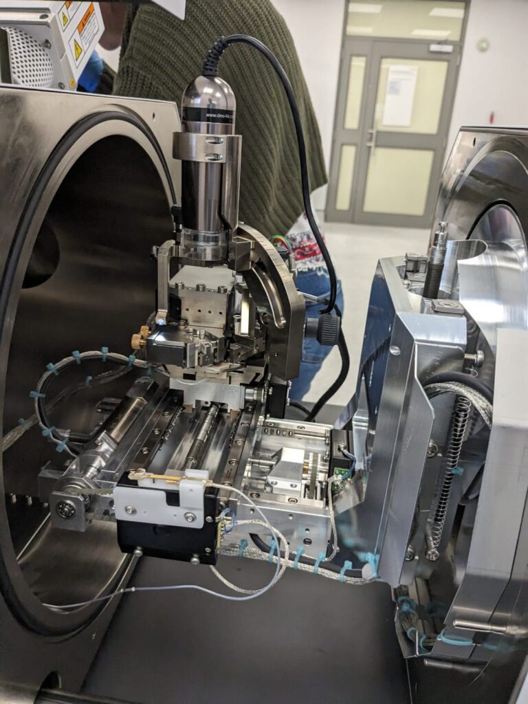
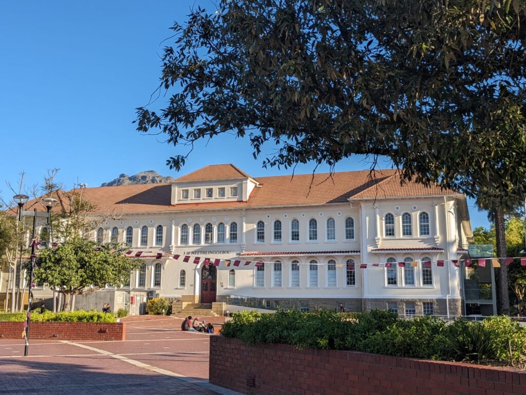
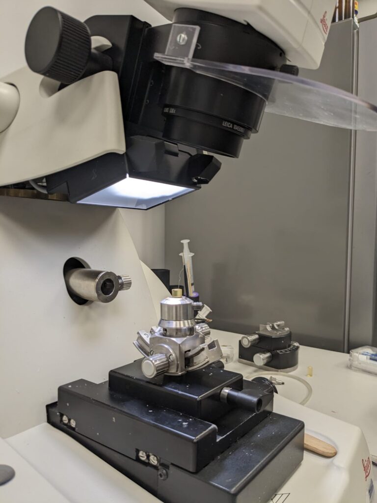
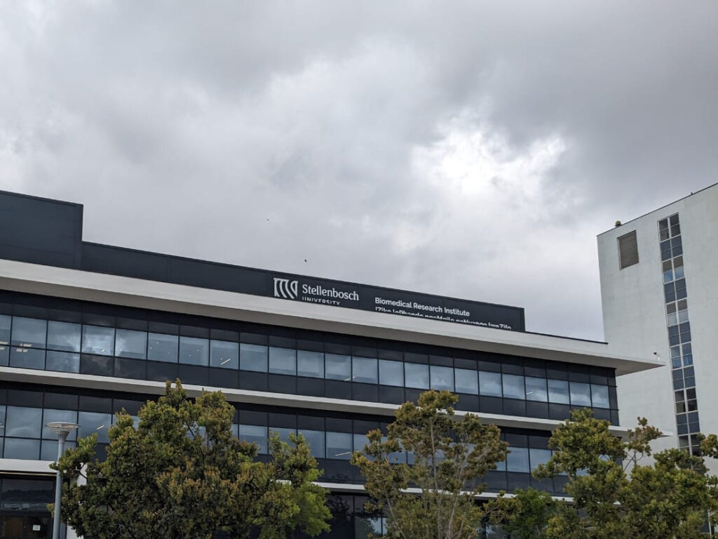
Good news! Our first project has started! Granted by our second call, the Twinning program has begun between Stellenbosch University and FBI-Paris Node. A fantastic experience based on sharing practices, knowledge transfer and many fruitful discussions on image analysis and correlative approaches between light sheet and serial block face microscopy techniques. For the South African partner, Madelaine Frazenburg (Stellenbosch University), it is the opportunity to see how other microscopy laboratories in France works but also to learn more about cryo-SEM and to study new kind of sample preparation methods. From the French side, Ludovic Leconte (Institut Curie, FBI Paris-Centre node) is indeed very interested in gaining new experience in electron microscopy mainly in Serial Block Face, another tissue section imaging that is not available on his site and for which the Stellenbosch imaging platform has the mastery.
Our warmest thanks to Lize Engelbrecht, Professor Ben Loos and Janica Conradie for making this event possible and for the warm welcome they extended. The second stage of this “Twinning” project will take place at Institut Curie next spring. We look forward to welcoming Madelaine!
Atomic Force Microscopy (AFM) is a scanning probe microscopy technique that relies on measuring the interaction forces between a sharp tip and the surface of a sample to generate high-resolution images of its surface features and mechanical properties. A very broad range of sample types can be imaged with this technique at a very high resolution – at sub-nanometer level for some of them! Discover the AFM at the Montpellier node of France-BioImaging with Christine Doucet from Integrative Biophysics of Membranes team of the Centre de Biochimie Structurale.
Quickly visualize dynamic biological processes with High-Speed AFM
AFM provides images in physiological conditions, in liquid, over a length-scale ranging from few nanometers (single biomolecules) to tens of micrometers (living cells). In fact, the resolution depends on the tip radius and sample properties. For some of them, you can routinely obtain a nanometer lateral resolution and Angstrom axial resolution!
You want a video-rate version of the biological samples you are imaging? The High-Speed AFM, permits the acquisition of movies at approximately 10 images per second, enabling the visualization at nanoscale of dynamic biological processes involving biomolecular interactions, diffusion or conformational changes. It delivers nanometric resolved images typically at the same speed as conventional fluorescence microscopes!
Unravel the chemical information of your sample by combining AFM with…
AFM in ambient conditions and in liquids has a key limitation in that it does not directly provide chemical information about the sample being imaged. However, this limitation can be overcome by combining AFM with other techniques to obtain additional information about the sample’s composition.
One commonly used technique in correlation with AFM is fluorescence microscopy. This combined approach of fluorescence labeling and AFM provides valuable insights into the chemical and biological properties of the sample. It was recently used on the Montpellier custom-made correlative AFM / fluorescence setup to observe the sublocalization of proteins in HIV-1 budding sites 1. They also used it to unambiguously attribute some unexpected configurations of the nucleoplasmic sides of Nuclear Pore Complexes 2. In these two cases, fluorescently-labeled proteins were imaged by dSTORM (direct STochastic Optical Reconstruction Microscopy). Of note, the lateral resolution of dSTORM and AFM are both in the 20 nm range with such samples, which makes their combination ideal!
In addition to fluorescence microscopy, AFM can also be correlated with other complementary techniques to obtain chemical information about the sample, such as Raman spectroscopy, Infrared Spectroscopy, X-Ray spectroscopy, microscopy and scattering.
Learn more about AFM applications
Here are 2 studies where Atomic Force Microscopy were essential:
Structure and mechanics of the human nuclear pore complex basket using correlative AFM-fluorescence superresolution microscopy
Combining mechanical and superresolution measurements to reveal the plasticity of the Nuclear Pore Complexes
Nuclear pore complexes (NPCs) are the only gateways between the nucleus and cytoplasm in eukaryotic cells, facilitating the transport of selected cargoes of size from a few up to hundred nanometers. This versatility implies an important pore plasticity. Here, by combining atomic force microscopy (AFM) and single molecule localization microscopy (SMLM), a group led by France-BioImaging R&D team members Christine Doucet and Pierre Emmanuel Milhiet revealed that the NPC basket is very soft and explores a large conformational landscape: apart from its canonical basket shape, it dives into the central pore channel or opens, highlighting how this structure can adapt, and let morphologically diverse cargoes shuttle through NPCs.
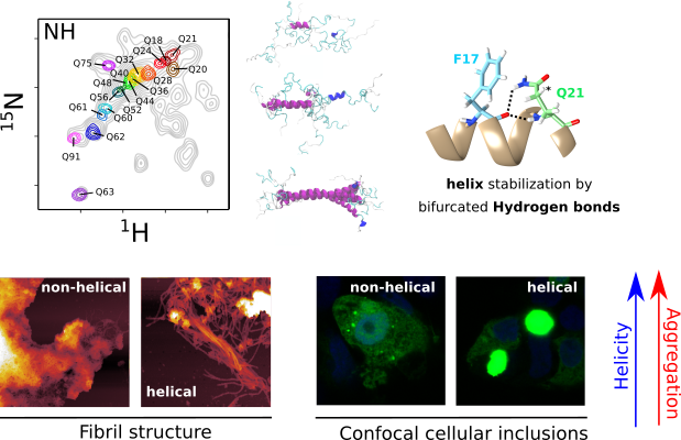
The structure of pathogenic huntingtin exon 1 defines the bases of its aggregation propensity
Structural Biology meets Correlative Imaging
Huntington’s disease is a neurodegenerative disorder caused by an extended polyglutamine (poly-Q) tract in huntingtin. Here, using NMR, the team of Pau Bernado (CBS Montpellier) demonstrated that this poly-Q tract adopts long α-helical conformations. By adding correlative Atomic Force Microscopy and Fluorescence Microscopy data obtained in the FranceBioImaging facility PIBBS in Montpellier, they could demonstrate that the stability of this α-helix is a stronger signature than the number of glutamines, in defining the aggregation kinetics and the structure of the resulting fibrils, potentially linked to their pathogenicity.
How to use Atomic Force Microscopy at France-BioImaging?
Atomic Force Microscopy is open to collaborations under Proof-of-concept studies via Euro-BioImaging webportal (www.eurobioimaging.eu/service)! At the Montpellier node of France-BioImaging, you will be in contact with Dr Luca Costa (costa@cbs.cnrs.fr) with whom you will talk about the feasibility and the inherent experimental constraints linked to the technique. The collaboration procedure is discussed on a case-by-case basis, depending on the duration and technicity of the required experiments. Feel free to submit your project!
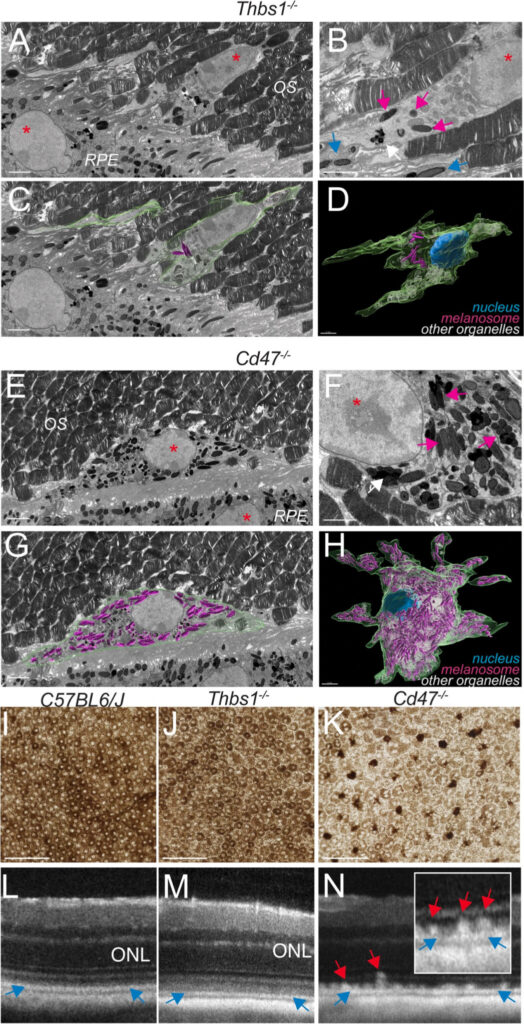
Age-related macular degeneration (AMD) affects more than 150 million people worldwide (early AMD) and 10 million of patients suffer from debilitating late stage AMD. Blurring central vision, this eye disease progresses over time, usually beginning when people are around their 50s or 60s by causing damage to the macula, in the retina. Researchers from the Institut de la Vision (Sorbonne Université, INSERM, CNRS, UMR_S 968) recently published about the AMD. Thanks to Serial Block-Face Scanning Electron Microscopy (SBF-SEM) experiments carried out at the ImagoSeine core facility (Institut Jacques Monod / FBI Paris-Centre node), they describe in this new study melanophages as a disease-progression marker.
Early or intermediate AMD is characterized by pigmentary changes and lipoproteinaceous debris accumulation between the photoreceptors and the melanosome-rich retinal pigment epithelium (RPE) or below the RPE. Later, AMD can be complicated by central choroidal neovascularization or by an expanding lesion of the photoreceptors. Even though patients with early or intermediate AMD can progress and develop late AMD, a large part of patients stay stable for years, underlining the potential usefulness of progress.
AMD is associated with the appearance of hyperreflective foci, with reflectivity comparable to melanocyte-containing RPE cells. Thbs1 and CD47 are both important for the elimination of these cells. In the absence of either of them, melanocyte-containing RPE cells would then accumulate. The goal was to determine the origin of these cells in the retina, and the main question was: are these cells RPE migrating to the wrong place, or melanosome phagocytes cells having ingested melanosomes?
SBF-SEM: the key to answer this question
The Serial Block-Face Scanning Electron Microscopy (SBF-SEM) is a 3D electron microscopy imaging technique, where an ultramicrotome is placed inside a SEM. Biological samples are beforehand stained with heavy metals and embedded in a plastic resin block. Inside the microscope, a thin-section is cut at the surface of the block and discarded. Then, an image of the surface of the block – therefore inside the sample – is made, using back-scattered electrons. The process of cutting and imaging is repeated automatically as many times as necessary to produce a 3D stack of images inside the sample, as it is progressively imaged and destroyed.
This technique allows 3D imaging of large samples for Electron Microscopy standards (up to several hundred microns in each of the X,Y,Z direction) at high resolution. This technique is often used to image whole cells, or even small pieces of tissues in 3D. The two major domains of application are to:
find a rare structure within a cell or tissue. The sample is imaged until the structure of interest is found.
understand the 3D spatial organization of organelles within cells, or of cells between them.
The benefits of bioimaging in this study
In the study, SBF-SEM was essential. As previously mentioned, AMD is associated with the appearance of hyperreflective foci, with reflectivity comparable to melanocyte-containing RPE cells. In the images produced by SBF-SEM, the retinal pigment epithelium (RPE) surrounding the melanophages in mice, where CD47 was inhibited, were markedly less pigmented and deformed compared to those where Thbs1 was blocked. This suggests that melanosomes have been transferred by phagocytosis from the RPE to nearby melanophages because they lack CD47. Finally, authors have shown that CD47 acts as a “don’t eat me” signal. The SBF-SEM was a great addition to this study where understanding the 3D spatial organization of the structure of interest was key.
Thanks to Jean-Marc Verbavatz for providing very helpful insights of the study!
Augustin, S., Lam, M., Lavalette, S. et al. Melanophages give rise to hyperreflective foci in AMD, a disease-progression marker. J Neuroinflammation 20, 28 (2023). https://doi.org/10.1186/s12974-023-02699-9
Get access to one of our services!
You need SBF-SEM or another imaging technology or expertise that France-BioImaging provides? To get open access, please login via Euro-BioImaging website! You just have to choose the technology you want to use, then submit your proposal. All applications will be processed by the Euro-BioImaging Hub in close relation with France-BioImaging. And of course, all scientists regardless of their affiliation, area of expertise or field of activity can benefit from open access services! Users whose projects will be validated by Euro-BioImaging will benefit from a waiver for the access cost on France-BioImaging core facilities (https://france-bioimaging.org/access/)
Massive intracellular accumulation of RPE-derived melanosomes in subretinal MPs of CD47−/−-mice causes subretinal melanophage formation and their clinical appearance as hyperreflective foci.
Correlative X-ray imaging and electron microscopy (CXEM) is the combination of X-ray imaging and electron microscopy. It is a correlative approach that makes it possible to characterise a sample of interest and locate a structure of interest in a non-destructive way. Nicolas BROUILLY is in charge of the Electron Microscopy Unit of the PICsL imaging facility on the Marseille node of France BioImaging, where CXEM is used for developmental biology studies. As part of Euro-BioImaging’s Proof-of-Concept study, his facility is now accepting applications from external users for CXEM projects. Learn more about how this approach works and what it can be used for in the interview below.
We are today talking about CXEM imaging. Please provide a short summary of this type of imaging and tell us some applications:
Nicolas Brouilly: It is often very useful to combine 2 imaging modalities to take advantage of each while trying to lower their respective drawbacks. For example, by combining Light Microscopy and Electron Microscopy, we obtain the popular CLEM (for Correlative Light and Electron Microscopy). Visible light can then be used in combination with EM either:
To target a precise region of interest ;
To localize molecules within the ultrastructural information obtained by EM.
Using the same acronym building, CXEM corresponds to Correlative X-ray and Electron Microscopy. X-rays are photons of shorter wavelength than those from visible light, and can again be used to characterize a sample in 2 different ways:
To use their ability to easily go through tissues in order to record the 3D morphology of a sample: either by computed x-ray micro-tomography (or micro-CT) for micrometric resolution of big samples (mm to cm range) or by Soft X ray tomography for nanometric resolution of small samples (100’s of nm to um range);
To use a focused beam of high energy x-rays to analyse the localization of the elements of a sample: X-Ray Fluorescence microscopy (or XRF).
Both modalities can be used to complement the ultrastructural information obtained by electron microscopy. At the Marseille node of France BioImaging, in the Electron Microscopy Unit, we routinely use Correlative Micro-CT and Electron Microscopy to answer developmental biology questions.
What are some advantages of this technique that make it suited to addressing this type of question?
Nicolas Brouilly: The main advantage of Micro-CT (or Computed X-ray Tomography) is its ability to “see through” a sample and to reveal its overall organization in 3D without any labelling. The second advantage of Micro-CT is the fact that it is non-destructive. Thirdly, the contrast we usually give to samples for electron microscopy is compatible and even beneficial for X-ray imaging.
Altogether, this means that we can use X-ray tomography to map the microscale morphology of a sample in order to target a specific region of interest without having to go through the time-consuming and destructive collection of semi-thin sections.
We routinely use the micro-CT tool, not only to target a given organ or a given group of cells, but also to pre-orient the sample in order to cut it under a specific orientation. It is a timesaving tool within the frame of a 2D electron microscopy project, but it really is key within the frame of a 3D electron microscopy project given that Serial BlockFace and Focused Ion Beam techniques are destructive.
Tell us a bit more about a specific project that was done in your facility using this technology? What scientific questions were you addressing?
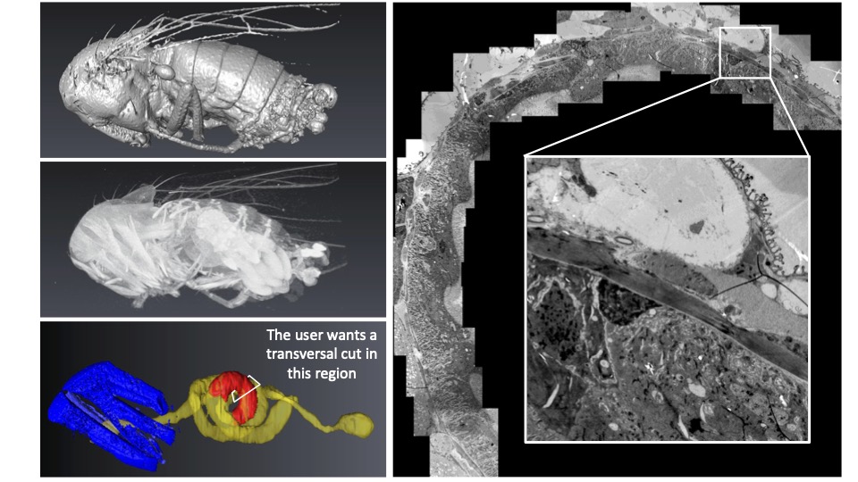
Nicolas Brouilly: Imagine that, first, you have a ball of yarn, second, you cannot untangle it, and third, you want to cut small bits of the thread at 24 cm from the end (not 22, not 26… 24 !). CXEM enabled us to do this on Drosophila gut. The micro-CT gave us the 3D map of the sample within the resin block. We could then use this map to find the best itinerary within the sample to make transverse sections of the portion of interest that was precisely indicated by the user on the micro-CT dataset. At the end of the day, the user was able to look at perfect transverse ultrathin TEM sections, at a precise position of this ball of yarn that Drosophila gut is. He could finally get precise metrics from this precise part of the gut in several samples. None of this could have been achieved without CXEM.
Like a ball of yarn… Above is an example of how CXEM can be used to find the best itinerary within a sample to make transverse sections of the portion of interest. On the left, the micro-CT provided a 3D map of the sample within the resin block. On the right, a transverse ultrathin TEM section of the drosphilia gut.Image courtesy of Nuno Luis (Schnorrer lab, IBDM) & Nicolas Brouilly (Electron Microscopy Facility, IBDM AMU/CNRS, France BioImaging).
For another example, you can have a look at the following paper where we used CXEM to map platelet aggregates within arteries in order to explore them by Serial BlockFace SEM, another example of “Find a needle in a haystack”. Have a look at movie S1, it is a wonder that we could not obtain without CXEM:
CXEM is part of the Euro-BioImagingProof-of-Concept study. The Proof-of-Concept study makes it possible to introduce exciting, new imaging technologies to our portfolio that were previously unavailable via our network. We are currently accepting applications to use these technologies at participating Nodes as part of the Proof-of-Concept study. Be part of this study – and contribute to community-wide continuous technological innovation!
All scientists, regardless of their affiliation, area of expertise or field of activity can benefit from Euro-BioImaging’s pan-European open access services. Potential users of these new technologies are encouraged to submit project proposals via our website. To do so, you can Login to access our application platform, choose the technology you want to use and the facility you wish to visit, then submit your proposal. All applications will be processed by the Euro-BioImaging Hub. As usual, users will benefit from advice and guidance by technical experts working at the Nodes, training opportunities, and data management services.
We use cookies on our website to give you the most relevant experience by remembering your preferences and repeat visits. By clicking “Accept All”, you consent to the use of ALL the cookies. However, you may visit "Cookie Settings" to provide a controlled consent.
This website uses cookies to improve your experience while you navigate through the website. Out of these, the cookies that are categorized as necessary are stored on your browser as they are essential for the working of basic functionalities of the website. We also use third-party cookies that help us analyze and understand how you use this website. These cookies will be stored in your browser only with your consent. You also have the option to opt-out of these cookies. But opting out of some of these cookies may affect your browsing experience.
Necessary cookies are absolutely essential for the website to function properly. These cookies ensure basic functionalities and security features of the website, anonymously.
Cookie
Duration
Description
cookielawinfo-checkbox-analytics
11 months
This cookie is set by GDPR Cookie Consent plugin. The cookie is used to store the user consent for the cookies in the category "Analytics".
cookielawinfo-checkbox-functional
11 months
The cookie is set by GDPR cookie consent to record the user consent for the cookies in the category "Functional".
cookielawinfo-checkbox-necessary
11 months
This cookie is set by GDPR Cookie Consent plugin. The cookies is used to store the user consent for the cookies in the category "Necessary".
cookielawinfo-checkbox-others
11 months
This cookie is set by GDPR Cookie Consent plugin. The cookie is used to store the user consent for the cookies in the category "Other.
cookielawinfo-checkbox-performance
11 months
This cookie is set by GDPR Cookie Consent plugin. The cookie is used to store the user consent for the cookies in the category "Performance".
viewed_cookie_policy
11 months
The cookie is set by the GDPR Cookie Consent plugin and is used to store whether or not user has consented to the use of cookies. It does not store any personal data.
Functional cookies help to perform certain functionalities like sharing the content of the website on social media platforms, collect feedbacks, and other third-party features.
Performance cookies are used to understand and analyze the key performance indexes of the website which helps in delivering a better user experience for the visitors.
Analytical cookies are used to understand how visitors interact with the website. These cookies help provide information on metrics the number of visitors, bounce rate, traffic source, etc.
Advertisement cookies are used to provide visitors with relevant ads and marketing campaigns. These cookies track visitors across websites and collect information to provide customized ads.