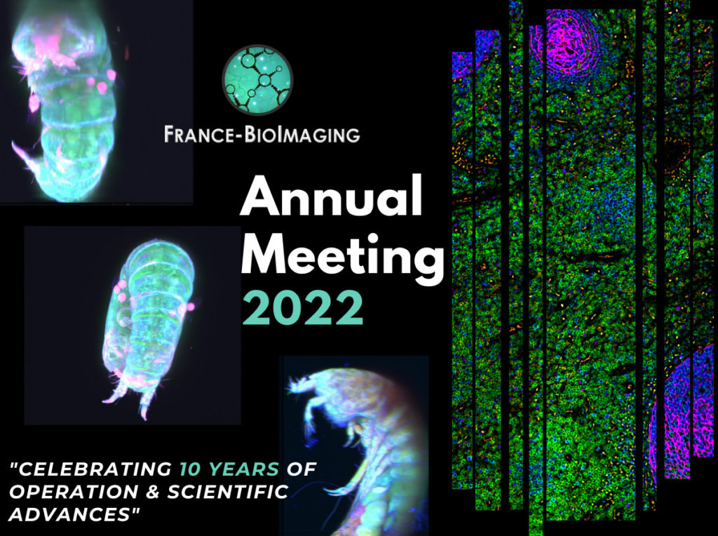
Ranging in size from 20 microns to 1 milimetre, Meiofauna is a crucial link between micro- and macro- marine ecosystems. Valentin Foulon, a research engineer in marine biology, is part of the Blue Revolution program that aims to develop a taxonomic identification protocol for these tiny creatures. He approached the Bretagne-Loire node of France-BioImaging, in Nantes, where a Single Plane Illumination Microscopy (SPIM) light-sheet available in open access helped him to further reach his goal.

Valentin Foulon loves imaging and microscopy – and he loves meiofauna. He works on the Blue Revolution program, whose goal is to accelerate the taxonomic identification of meiofauna using artificial intelligence algorithms. But while the confocal-microscopy-based protocol worked well for small plankton, the researchers quickly realized that it had to be adapted to work with meiofauna. 

Choosing the right microscopy approach

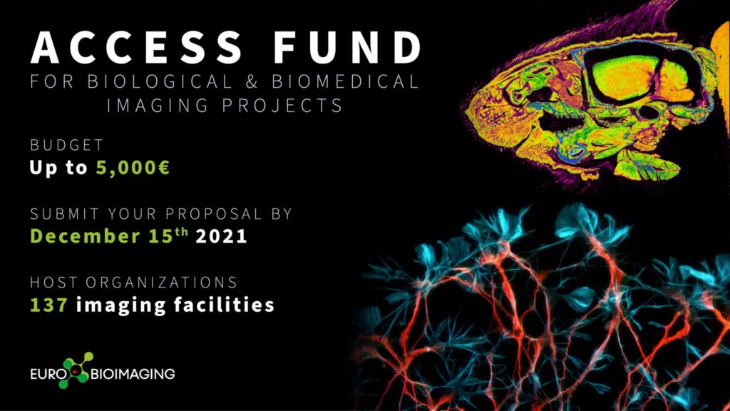
Identification of Meiofauna is usually operated manually on a classical microscope, which is “incredibly challenging” says Valentin. “And we only have one focal plane. To see important details, we need a 3D view of organisms.” explains Valentin. “That’s why we got curious about light-sheet imaging, because we would be able to see the organisms in 3D.” As France-BioImaging provides this type of microscopy in open-access in Nantes, “we clearly saw an opportunity there,” explains Valentin. His project proposal, which was submitted to the Euro-BioImaging pilot User Access fund, was fully funded by France-BioImaging, in a generous initiative introduced by France-BioImaging to fund all user projects to its facility received by Euro-BioImaging for the pilot User Access fund. 

“I had never tried light sheet microscopy before,” says Valentin, “And in Nantes, the lab didn’t have experience with marine organisms. Together, we found a compromise between different imaging parameters such as the resolution or the acquisition time.” Once the set-up was fine-tuned, he sent his samples, and the engineer at the lab did the imaging work. “It was a very nice experience,” expresses Valentin.  “I discovered a new technique, which is always enriching. I also think the lab was happy to work with a new sample type. And finally, we validated our proof-of-concept, proving that it is possible to image these organisms in 3D.

Image data management

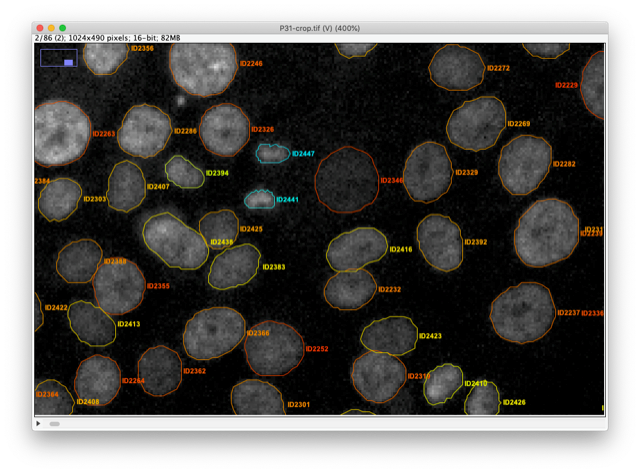
But the project doesn’t end there. “The next step is data management. We imaged between 200-300 different organisms. It’s not enough for automatic classification, but nevertheless, we have 6-7 TB of data. Now we must process the data for this classification with machine learning. It’s an essential part of the project, to make the link between sample imaging, and data processing. We are working to close that gap.” 

“The outcome of this project is to validate a proof-of-concept, from the sample collection to the image classification. With this new taxonomic identification protocol, we will be one step forward in the comprehension of meiofauna, these mysterious marine organisms, who may hold the key to understanding the impact of human activity on marine ecosystems,” concludes Valentin. 

Thank you Marianna Childress, communication officer of Euro-BioImaging, for the original article.

As the 2022 edition of the France-BioImaging Image Contest admissions is coming to an end, we wanted to highlight our previous winners and their projects. Here is a quick throwback to our 2021 winners.

Before getting to the heart of the matter, we want to remind you that you still have time (before November 11th) to submit your best images and try to win your registration fees for one 2023 microscopy-related event! Please make sure you upload your images on the following link:

FBI Image Contest 2022 – Last days to submit your images!

Last year, we enjoyed the winning images submitted for their artistic take and their quality. Thanks to Léna Meneux, Eunice HoYee Chan, Camille Boutin et Nicolas Brouilly for their beautiful images!

1st place: Léna Meneux, Eye Team, Institut des Neurosciences de Montpellier

"The eye of the storm"

Sensory fibers of a mouse cornea imaged with a confocal microscope. The corneal nerves converge toward the centre forming a vortex. This particular transgenic mouse model allows stochastic expression of fluorescent proteins, unravelling the heterogeneity of the fiber origines inside the corneal epithelium.
Acknowledgements to Karine Loulier for the mouse model and Laetitia Hudececk for her help during the acquisition.

In the Institut des Neurosciences de Montpellier since 2020, Léna is a PhD student working in the team Eye lead by Dr. Frédéric Michon. This team is investigating the mechanisms related to the preservation and the integrity of the anterior part of the eye, including the lacrimal gland, the tears and the cornea. Léna’s project focuses on the cellular and molecular effects of the corneal innervation on the corneal homeostasis. The project goes further as they aim at highlighting new targets able to prevent and/or repair corneal damage. 

The image she submitted for the 2021 France-BioImaging Image Contest (The eye of the storm) represents the sensory fibers of a mouse cornea. This innervation follows a typical pattern where all the nerves converge toward the centre forming a vortex. This particular transgenic mouse model allows random expression of fluorescent proteins, unravelling the heterogeneity of the fibers’ origin inside the corneal epithelium. As cornea is the most innervated tissue in the whole body, this model shows the differences between fibers. In pathological context, for example wound injury, it is thus possible to follow a specific fiber during the healing process.

France-Bioimaging sponsored her participation to the FOM (Focus on Microscopy) 2022 congress where she presented her project through a poster. Even though the congress was online, it gave her the opportunity to share her results with experts and as a consequence, to gather advice on her ongoing experiments.

2nd place: Eunice HoYee Chan, Muscle Dynamics Team, Developmental Biology Institute of Marseille (IBDM)

Myofibrils isolated from Drosophila indirect flight muscle labelled with titin (yellow) and actin (blue). Image captured from confocal microscope. We are studying the role of titin protein in muscle mechanics and organisation during development

"Sarcomeric bouquet"

Myofibrils isolated from Drosophila indirect flight muscle labelled with titin (yellow) and actin (blue). Image captured from confocal microscope. We are studying the role of titin protein in muscle mechanics and organisation during development.

Research engineer in Frank Schnorrer's team at Institut de Biologie du Développement de Marseille (IBDM), Eunice focuses her research on Drosophila muscle dynamic and organisation during development using advanced biophysical and imaging techniques.

The image she submitted named “Sarcomeric bouquet" was from one of her very first muscle myofibrils isolation experiment. She dissected flight muscles from flies and labelled the individualised myofibrils with Llama nanobodies conjugated with different epitopes. Those labelled myofibrils were then subjected to various imaging methods including standard confocal microscopy, super resolution microscopy and cryo electron-tomogram. Using these novel labelling tools and imaging techniques, her team could study the dynamic and organisation of muscles during development in details.

France-BioImaging sponsored her registration to the 49th European Muscle Conference in Prague (22-26 September 2022). As she is new to the muscle field, this conference offered a great opportunity to have a broad view on different kind of state-of-the-art imaging techniques. Besides, she gave a presentation during the conference, highlighting her work and initiating discussion.

3rd Place: Camille Boutin, Biology of multiciliated cells Team, Developmental Biology Institute of Marseille (IBDM) & Nicolas Brouilly, PICsL Imaging facility, Electron Microscopy department

Lamellar structure in a differentiating multiciliated cell observed by transmission electron microscopy with a Tecnai G2 200kV FEI.

"Clown"

Lamellar structure in a differentiating multiciliated cell observed by transmission electron microscopy with a Tecnai G2 200kV FEI.

Camille is a researcher in Laurent Kodjabachian’s group at the Institut de Biologie du Développement de Marseille (IBDM). She develops projects as a principal investigator on the compartmentalization and sizing of multiciliated cells. With this in mind, she routinely uses confocal and super-resolution microscopy but also scanning and transmission electron microscopy and tomography.

Nicolas is in charge of the Electron Microscopy Unit of the Plateforme d’imagerie commune du site de Luminy (PICsL). In addition to the routine sample preparation and 2D TEM imaging, this imaging facility offers, to internal and external users, advanced sample preparation (cryo-methods, immunolabelling...) and advanced imaging (tomography, CLEM, serial blockface…).

To understand the production of multiple centrioles in multiciliate cells, they focused on the deuterosome, a membrane-less organelle that has been described 50 years ago but whose composition, organisation and function remain unknown to this day. In this context they have developed an inducible multiciliated cells line. This image was taken during the initial characterisation of this cell line by transmission electron microscopy.

Thanks to the France-Bioimaging Image Contest, Nicolas participated to the COST COMULIS Conference that was held by the Cyprus Institute in Nikosia. It was a great opportunity to exchange with the people at the cutting edge of the multi-modal imaging field. The program covered subjects such as the sample preparation for multi-modal imaging, image analysis and integrated industrial partners.

Going to Rendez-Vous Carnot 2022? Drop by our booth and say hello! 12 & 13 October – Paris

In a few days, we will be travelling to Paris to attend the Rendez-Vous Carnot 2022! This is the fourth time that we will attend the forum as an exhibitor, in the Research Infrastructures Village. We are going to present France-BioImaging R&D ecosystem and the multiple advanced biological imaging technology developments taking place on FBI imaging platforms and R&D teams.

If you are in Paris between October 12 and October 13 attending the Rendez-Vous Carnot as well, be sure to drop by our booth and meet some of our colleagues at the venue:

  • Caroline Thiriet , France-BioImaging External Affairs Manager
  • Etienne Henry, France-BioImaging R&D and Tech-Transfer mission Officer
  • Jean Salamero, France-BioImaging Inter-Infrastructures Activities mission Officer

This year, we are pleased to share our booth with the French Infrastructure for Integrated Structural Biology (FRISBI). This infrastucture provides integrative structural biology approaches, from the molecular to the cellular level, integrating multi-resolution data from X-ray crystallography, small angle X-ray scattering, NMR, Cryo-EM and functional data including development for protein expression and crystallization.

The France-BioImaging Image Contest is back for its 4th edition!

This image contest is open to all within the imaging community: core facility staff and users, R&D labs teams and co-workers, students… Submit your best microscopy images for a chance to showcase your skills, research and creativity to the French bioimaging community and beyond, allowing people to see the visual appeal of the life sciences. Images from the contest will be featured on France-BioImaging communication tools, online and in print.

France-BioImaging and all the French community aims to develop and promote innovative imaging technologies and methods. But microscopy images can also take an artistic, creative look and make the invisible world beautiful.

We are all eager to see your work !

Prizes

1 to 3 images will be awarded depending on the quantity and quality of the entries submitted. France-BioImaging will cover the registration fees for one 2023 microscopy related event of the winners’ choice (FOM, ELMI, EMC, COMULIS conference, etc.).

Important: Only French or foreign participants affiliated to a French institution can enter the contest. Foreign participants non-affiliated to a French institution can submit images and will be featured in the gallery, but will not be evaluated as part of the contest.

Submission deadline: Friday, November 11th, 2022, 23h59 UTC+2. 

Click here to consult the terms and conditions of the contest. When you are ready, submit your entry by filling the form below. You can check out last editions’s entries for inspiration. One participant can submit several entries (up to 3).

(If you have any issues when submitting your image, please contact communication@france-bioimaging.org)

This form is currently closed for submissions.

Our three winners have been announced! Please discover all the submitted images on this following link: https://france-bioimaging.org/announcement/winning-images-fbi-image-contest-2022/

On December 13th and 14th 2022, we will have the pleasure to invite you to our Annual Meeting, to be hosted by FBI Bretagne-Loire Node at the Health Research Institute of the University of Nantes (Nantes, France).

2022 is an important landmark for France-BioImaging and its community, as the infrastructure is celebrating 10 years of operation and scientific advances. We will be happy to celebrate this milestone with all the members of the bioimaging community (within and outside the France-BioImaging community).

The Annual meeting will highlight France-BioImaging’s development as a research infrastructure and its node community accomplishments during these last 10 years, and the role they play in boosting innovation in bioimaging. Imaging scientists and users from the infrastructure’s nodes will present their key projects and demonstrate how they have profited from France-BioImaging and its community.

We look forward to meeting you there!

Gallery

Videos

Programme

Registration

Deadline to register: 04/12/2022 at 23:59

This form is currently closed for submissions.

Poster

INFOS PRATIQUES:

Adresse: Bâtiment IRS-UN 8 quai Moncousu 44000 Nantes

Plan d’accès https://sfrsante.univ-nantes.fr/medias/fichier/plan-acces-irs-un-1_1561112224023-pdf?ID_FICHE=1562669&INLINE=FALSE 

(arrivée directement à l’accueil, toutes les salles sont au rez de chaussé et seront fléchées)

Comment venir?

Arrêt de tram le plus proche: ligne 2 ou 3 du tramway Aimé Delrue

En train: 

Gare de Nantes à 20 minutes à Pied (préférez la sortie sud pour venir à pied). En bus ou tram compter 15 minutes.

Ligne de bus accessibles depuis arrêt sortie gare Sud C2, C3, 54 arrêt Hotel Dieu

Ligne de tram depuis la sortie Gare Nord : prendre la ligne 1 direction François Mitterand/Jamet et descendre à Commerce, continuer à pied (10 minutes de marche environ) 

En avion:

Navette aéroport  Navette aéroport Nantes Atlantique : horaires, tarif

Le départ et l’arrêt se font à cause de travaux de Hotel Dieu ( plus proche de l’IRS) et non pas Commerce. 

Taxis Nantais:

02 40 69 22 22 http://www.taxis-nantes.com/ 

02 40 85 40 85. https://heptaxis.com/ 

VTCs: 

LocalCab 09 80 66 62 82   TAXI / VTC – Réservez votre Chauffeur au Meilleur Prix ! (localcab.fr) 

Quelques Hôtels conseillés :

L’Hôtel  L’HÔTEL NANTES – Votre Boutique Hôtel idéalement placé centre-gare (nanteshotel.com) (135 à 195 euros la nuit avec petit déjeuner) 

Hôtel Amiral Hotel Nantes centre-ville – Hotel 3 étoiles – Hôtel Amiral | Nantes (hotel-nantes.fr) (128 à 150 euros avec petit déjeuner)

Hotel ibis Nantes Centre Gare Sud   ibis Nantes Centre Gare Sud – Hôtel 3 étoiles à Nantes – ALL (accor.com)  (110 à 130 euros)  ibis Styles Nantes Centre Place Royale 

Hôtel  ibis Styles Nantes Centre Place Royale Hôtel à Nantes – ibis Styles Nantes Centre Place Royale – ALL (accor.com) (85 à 95 euros) 

Prise en charge des missions: 

Se rapprocher de votre noeud FBI (fonds mission), sauf pour les intervenants qui seront directement contactés pour la prise en charge de leur missions. 

Save the date! The Electron Microscopy facility of Imagerie-Gif (I2BC, France-BioImaging), the Cryo-Electron Microscopy facility (I2BC, FRISBI) and the Cimex facility of Ecole Polytechnique are organizing a 5-day workshop from October 3rd to October 7th, 2022 on Transmission Electron Microscopy to explore the architecture of a virus in all its forms.

The aim of this workshop is to propagate knowledge about transmission electron microscopy applications and to outline the advantages of transversal studies combining structural biology and cell biology. Indeed, structural biology and cell biology approaches both use TEM but are rarely merged in the same studies.

The workshop will focus on the advantages of combining both approaches, which can be easily performed with the same equipment. The workshop will focus on a biological object whose study requires such multiscale approaches: a virus. The virus will be studied in vitro to resolve its high-resolution 3D structure, and will be observed in infected cells to determine the infection and replication mechanisms in situ.

The workshop targets students and young researchers. The training will focus on a given biological object, a virus, which will be studied by two complementary approaches:

  • Single particle analysis by cryo-electron microscopy, allowing high-resolution 3D reconstruction of particles purified in vitro. This part will be performed on a 200kV TEM on the Cimex facility.
  • Cellular tomography of infected cells with observation of the virus replication sites in situ and analysis of its interaction with cellular membranes. This workshop will cover the workflow from sample preparation and resin sections realisation, to acquisition and analysis of tomograms with a 120kV TEM.

Attendees will have a theoretical and practical overview of these two complementary techniques. The practical training will be particularly emphasised, to ensure that attendees will be able to apply the knowledge acquired in the workshop for their further research projects.


Susbscription here: https://www.azur-colloque.fr/DR04/inscription/preinscription/245

Preliminary programme

France BioImaging and all the French community aims to develop and promote innovative imaging technologies and methods. But microscopy images can also take an artistic, creative look and make the invisible world beautiful, allowing people to see the visual appeal of the life sciences. 

We enjoyed the diversity of the images submitted with many different microscopy techniques, models and applications represented. A big thank you to all the participants!

The National Coordination Team and the Executive Board are proud to announce the winners of the FBI Image Contest 2021:

  • 1st Place: Léna Meneux, Eye Team, Institut des Neurosciences de Montpellier

The eye of the storm

Sensory fibers of a mouse cornea imaged with a confocal microscope. The corneal nerves converge toward the centre forming a vortex. This particular transgenic mouse model allows stochastic expression of fluorescent proteins, unravelling the heterogeneity of the fiber origines inside the corneal epithelium.
Acknowledgements to Karine Loulier for the mouse model and Laetitia Hudececk for her help during the acquisition.

Confocal microscopy

  • 2nd Place: Eunice HoYee Chan, Muscle Dynamics Team, Developmental Biology Institute of Marseille (IBDM)
Myofibrils isolated from Drosophila indirect flight muscle labelled with titin (yellow) and actin (blue). Image captured from confocal microscope. We are studying the role of titin protein in muscle mechanics and organisation during development

“Sarcomeric bouquet”

Myofibrils isolated from Drosophila indirect flight muscle labelled with titin (yellow) and actin (blue). Image captured from confocal microscope. We are studying the role of titin protein in muscle mechanics and organisation during development.

Confocal LSM880
  • 3rd Place: Camille Boutin, Biology of multiciliated cells Team, Developmental Biology Institute of Marseille (IBDM) & Nicolas Brouilly, PICsL Imaging facility, Electron Microscopy department
Lamellar structure in a differentiating multiciliated cell observed by transmission electron microscopy with a Tecnai G2 200kV FEI.

“Clown”

Lamellar structure in a differentiating multiciliated cell observed by transmission electron microscopy with a Tecnai G2 200kV FEI.

Transmission Electron Microscopy, Tecnai G2 200kV FEI

Congratulations to the winners!


Explore all the images submitted here:

As stated in the Terms & Conditions of the contest, foreign participants non-affiliated to a French institution are featured in the gallery, but were not evaluated as part of the contest.

Euro-BioImaging first open call for user projects is open! If you have an idea for a biological or biomedical imaging project that you, your student or your close colleague could carry out in one of Euro-BioImaging Nodes, including France-BioImaging, now is the time to make this project come true with financial support from the Euro-BioImaging Access Fund.

How it works:

Submit your project proposal through the Euro-BioImaging web portal between October 20 and December 15, 2021, and indicate that you want to apply for the Euro-BioImaging Access Fund in order to be considered for a grant of up to 5.000 Euros to access the imaging services at a Euro-BioImaging Node. Projects will be evaluated by a committee of independent reviewers. Successful applicants will be notified by late January 2022 and successful projects should be started before July 2022.

What the funding covers:

The Euro-BioImaging Access Fund covers the user’s travel and accommodation costs as well as access and consumable costs at the imaging facilities that are part of Euro-BioImaging Nodes. For remote access projects shipment costs are also covered. Each successful applicant is eligible for up to 5.000 Euros of support.

Who is eligible:

All academic scientists, regardless of gender, nationality, home institution, career phase, or field of interest, are eligible to apply. We strongly encourage early career researchers to apply for this grant.

Projects should include transnational access to a Euro-BioImaging Node, i.e. the applicant’s home institution is in a different country than the Node where the project is to be performed. Due to the current sanitary situation, projects with non transnational access are elligible but transnational access will have priority.

All the external users/collaborators of France BioImaging facilities/labs are eligible:

  • any users from outside the institutional perimeter of France BioImaging nodes (i.e. from outside the following institutions: Aix-Marseille Université, Université de Montpellier, Université de Bordeaux, Université de Nantes, Université de Rennes 1, Université Paris-Saclay, Université de Paris, Université PSL -Paris Sciences & Lettres-, Généthon, Ecole Polytechnique, Institut Pasteur) who would like to use imaging technologies in one of FBI nodes: Paris Centre, Paris Ile-de-France-Sud, Marseille, Montpellier, Bordeaux, Bretagne-Loire. They can be French or international users – EU and non-EU
  • or users from a France BioImaging regional Node who want to access an equipment available in another FBI regional node.

Evaluation:

All applications will be evaluated for scientific excellence by a committee of independent reviewers. Selected projects will be assessed for technical feasibility and if needed receive technical advice from the Node providing the service.

How to apply:

Applicants are invited to visit our website to discover the range of technologies provided by Euro-BioImaging Nodes. Applicants will then follow the user access process described here: https://www.eurobioimaging.eu/about-us/how-to-access and indicate that they wish to apply for the Euro-BioImaging Access Fund in the application form.

The detailed procedure to apply to access one of France BioImaging facilities is available here: https://france-bioimaging.org/access/

Full details: https://www.eurobioimaging.eu/about-us/funding-user-access

The France BioImaging Image Contest is back for its 3rd edition!

This image contest is open to all within the imaging community: core facility staff and users, R&D labs teams and co-workers, students… Submit your best microscopy images for a chance to showcase your skills, research and creativity to the French bioimaging community and beyond, allowing people to see the visual appeal of the life sciences. Images from the contest will be featured on France BioImaging communication tools, online and in print.

France BioImaging and all the French community aims to develop and promote innovative imaging technologies and methods. But microscopy images can also take an artistic, creative look and make the invisible world beautiful.

We are all eager to see your work !

Prizes

1 to 3 images will be awarded depending on the quantity and quality of the entries submitted. France BioImaging will cover the registration fees for one 2022 microscopy related event of the winners’ choice (FOM, ELMI, EMC, COMULIS conference, etc.).

Important: Only French or foreign participants affiliated to a French institution can enter the contest. Foreign participants non-affiliated to a French institution can submit images and will be featured in the gallery, but will not be evaluated as part of the contest.

Submission deadline: Friday, October 15th, 2021, 23h59 UTC+2. 

Click here to consult the terms and conditions of the contest. When you are ready, submit your entry by filling the form below. You can check out last editions’s entries for inspiration. One participant can submit several entries (up to 3).


This form is currently closed for submissions.

A new version of TrackMate is available now, with major changes that improve its versatility. TrackMate now integrates state-of-the-art segmentation algorithms from machine-learning and deep-learning such as StarDist, Ilastik and Weka.

TrackMate[1] is a Fiji plugin that address cell or organelle tracking in Life-Science microscopy images. Its main goals are to be user-friendly, interoperable and to serve as a platform to accelerate the development of novel tracking algorithms and analysis pipelines.

With this new version we rewrote almost entirely TrackMate so that it can integrate state-of-the-art segmentation algorithms and benefit from their output. For instance, TrackMate can now store, display, save, load and exploit object contours in 2D.

We also made a new application programming interface that will facilitate and accelerate reusing TrackMate in other analysis pipelines and allow 3rd party contributors to add new segmentation algorithms in TrackMate in an easy way. We used this API ourselves to add 7 new segmentation algorithms to TrackMate:

For instance, the StarDist[2] algorithm is integrated as two different detectors. The first one uses the built-in deep-learning model that can segment cell nuclei in fluorescence image in a wide range of situation. The robustness of the StarDist algorithm in turn positively impacts the robustness of tracking and allows for better detection of cell divisions with TrackMate tracking algorithms. This will facilitate cell migration studies.

The TrackMate StarDist integration also allows for specifying and using a custom deep-learning model. For instance, we trained a specific model to detect T-cells imaged in bright-field microscopy and track them over time. Before the emergence of such detection algorithms, the tracking of label-free cells was difficult.

We also integrated the ilastik[3] segmentation software. A TrackMate user can input an ilastik classifier to detect objects then track them. We used them to study the bacterial growth of Neisseria meningitidis clones. The output of this analysis pipeline offers the lineage of each single cell along with its morphology and how it evolves across cell divisions.

The new capabilities of TrackMate can be used to address applications beyond tracking. For instance, it is now possible to use TrackMate to perform the segmentation of 3D objects using a slice-by-slice approach. This approach consists in segmenting objects in each 2D section of a 3D stack, then merging the segmentation results along Z in a subsequent step. This can be done in TrackMate, using the tracking step for merging. We implemented a novel tracking algorithm to foster this application, the overlap tracker. We could use this approach combining the cellpose[4] algorithm in 2D to segment 3D images of Arabidopsis thaliana floral meristem.

There are several other algorithms that are now offered to the TrackMate user, within a user-friendly software meant to interoperate with the key software of bioimage analysis. More importantly, TrackMate is an open-source academic software, and its new API will foster the development of new analysis pipeline with TrackMate and the integration of new algorithms by other developers, increasing the breadth of applications it can address for Life-Science researchers.


This new version of TrackMate is the product of a collaboration between the IAH facility (Institut Pasteur), part of the FBI Bioimage Informatics Node , the Jacquemet lab (Turku Bioscience Centre) , and the Dumenil lab (Institut Pasteur) .

Bringing TrackMate in the era of machine-learning and deep-learningDmitry Ershov, Minh-Son Phan, Joanna W. Pylvänäinen, Stéphane U. Rigaud, Laure Le Blanc, Arthur Charles-Orszag, James R. W. Conway, Romain F. Laine, Nathan H. Roy, Daria Bonazzi, Guillaume Duménil, Guillaume Jacquemet, Jean-Yves Tinevez bioRxiv 2021.09.03.458852; doi: https://doi.org/10.1101/2021.09.03.458852

Contact: Jean-Yves Tinevez


[1] https://imagej.net/plugins/trackmate/

[2] Alejandro F. Frangi, Julia A. Schnabel, Christos Davatzikos, Carlos Alberola-López, and Gabor Fichtinger Uwe Schmidt, Martin Weigert, Coleman Broaddus, and Gene Myers. Cell detection with star-convex polygons. In Alejandro F. Frangi, Julia A. Schnabel, Christos Davatzikos, Carlos Alberola-López, and Gabor Fichtinger, editors, Medical Image Computing and Computer Assisted Intervention – MICCAI 2018, pages 265–273, Cham, 2018. Springer International Publishing. doi:10.1007/978-3-030-00934-2_30.

[3] Stuart Berg, Dominik Kutra, Thorben Kroeger, Christoph N Straehle, Bernhard X Kausler, Carsten Haubold, Martin Schiegg, Janez Ales, Thorsten Beier, Markus Rudy, Kemal Eren, Jaime I Cervantes, Buote Xu, Fynn Beuttenmueller, Adrian Wolny, Chong Zhang, Ullrich Koethe, Fred A Hamprecht, and Anna Kreshuk. ilastik: interactive machine learning for (bio)image analysis. Nature Methods, 16(12):1226–1232, 2019. ISSN 1548-7105. doi:10.1038/s41592-019-0582-9.

[4] Carsen Stringer, Tim Wang, Michalis Michaelos, and Marius Pachitariu. Cellpose: a generalist algorithm for cellular segmentation. Nature Methods, 18(1):100–106, jan 2021. doi:10.1038/s41592-020-01018-x.

France BioImaging primary mission is to develop, promote, disseminate and provide access to innovative instruments and imaging technologies in the field of bioimaging to scientists. Fostering the technological transfers is at the heart of this mission, and for this France BioImaging relies on a strong association of leading R&D research teams with core facilities.

However, several bottlenecks exist and often hamper or prevent successful technology transfer:

  • A lack of human resource leads to difficulties in transferring and stabilizing the technology which is not enough user-friendly
  • A technology that is too specific, with not enough user base
  • A difficulty to contract with industry through institutional offices for industrial valuation
  • In the context of image analysis: the instability of open software economical model, inter-operability, large data handling and algorithm complexity

As a way to tackle these bottlenecks, France BioImaging launched in January 2021 its first “FBI Internal Call 2021: Technology transfer from the R&D teams to the core facilities” to promote the transfer of new technologies (instrumentation, probes, staining methods, software, data analysis or data visualization) from the R&D teams to the facilities of France BioImaging, for access and service to end-users. The outcome of the transfer project had to ensure for the prototype to be usable by the end-users until the interpretation of the data. The project had also to include a sustainability plan and a training plan to guide both facility staff and end-users toward autonomy.

The project selection was organized by the National Coordination of France-BioImaging and applications were assessed according to the following evaluation criteria:

  • Innovation and originality of the proposal
  • Scientific quality, implementation, timeline
  • Competitive positioning
  • Adequacy of resources with the proposed project
  • Economic impact and tech transfer potential and perspectives
  • Estimation of the user market and potential for user adoption
  • Plan for training and sustainability.

For the first edition of the “FBI Internal Call 2021: Technology transfer from the R&D teams to the core facilities”, 5 projects were selected:

  • Icy@FBI: Jean-Christophe Olivo-Marin (IPDM Node): Broadening the scope of applications of Icy (http://icy.bioimageanalysis.org/) by implementing several key new bioimage analysis components
  • BIC-HCS-SMLM: Jean-Baptiste Sibarita (Bordeaux Node), Technological transfer of a Single-Molecule-based High Content Screening platform to the Bordeaux Imaging Center
  • CloudFISH: Marcello Nollmann (Montpellier Node), A tool for the analysis of single-molecule RNA and DNA FISH images
  • MorphoNet: Emmanuel Faure (Montpellier Node), An interactive online morphological browser to explore complex multi-scale data
  • BioImageIT (https://bioimageit.github.io/#/about): Jean Salamero, Sylvain Prigent (IPDM Node), An open source framework for integration of image data management with analysis

Each selected project was awarded with a 80k€ grant for salary and/or equipment, and several positions are currently available: https://france-bioimaging.org/jobs/

This call will be renewed in 2023.