Meeting with Mariia Nazarova: Understanding chromatin regulation key factors
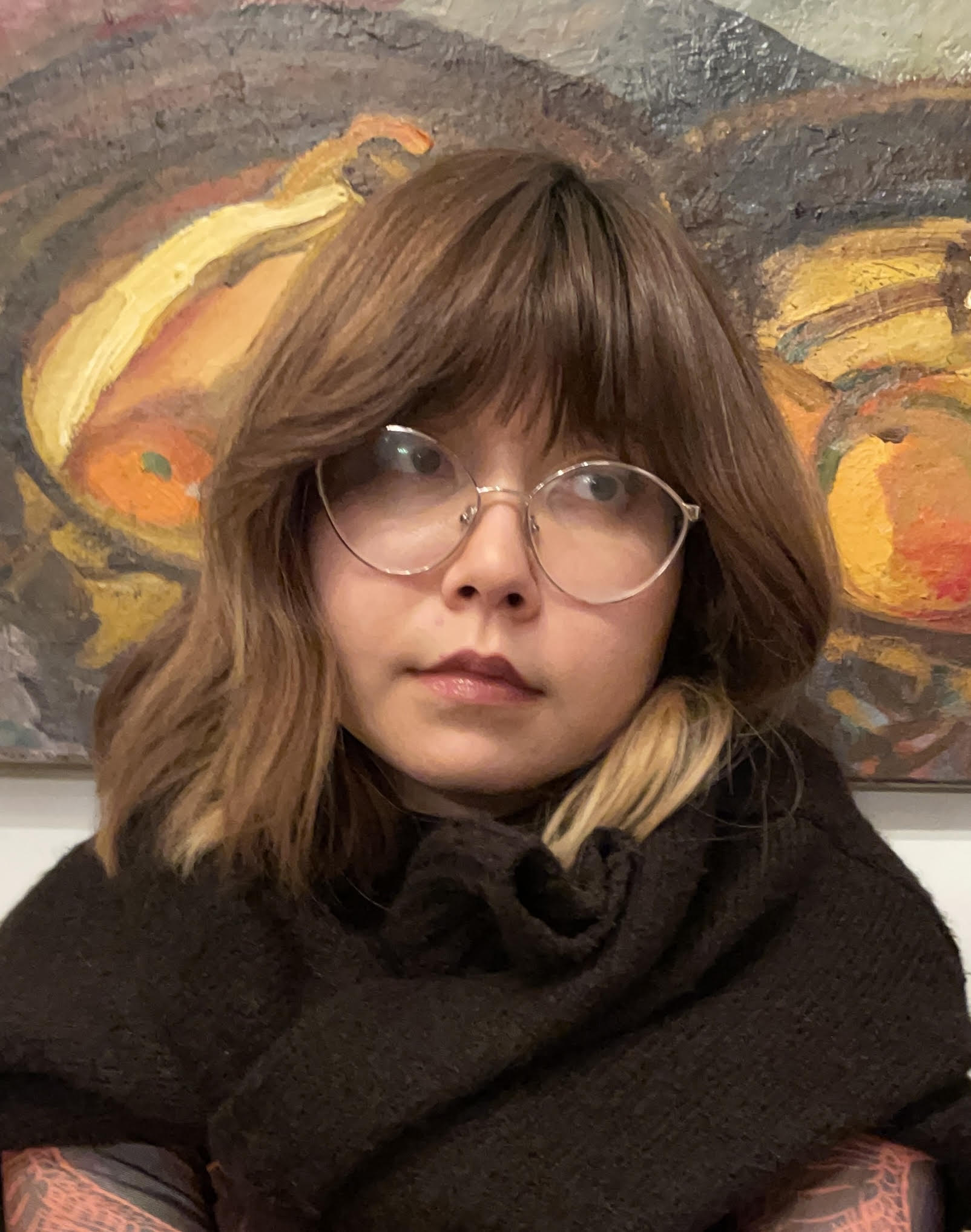
In this new user story, meet Maria Nazarova, a PhD student at IGBMC in Strasbourg.
For her research on chromatin regulation mechanisms, she benefited from the FBI Access Fund to access cutting-edge technology available at the MRI-CRBM platform in Montpellier.
But as is often the case in science, not everything went according to plan.
Read her story to find out more!
To start, could you tell us a bit about yourself? What has been your academic journey so far, and what is your current role or area of work?
I have always been interested in molecular biology, and I’ve been lucky to explore it from several angles starting with plant genetics, moving through cancer-related non-coding RNAs and immunoglobulin locus rearrangements, and finally arriving at my current favorite topic:
Chromatin dynamics and its role in crucial processes like transcription, replication, and repair
I’m now a 3rd-year PhD student in the Tom Sexton Lab at the IGBMC in Strasbourg, where I study how enhancers communicate with promoters using live-cell imaging.
What are you currently working on in your research? What is the main topic or challenge you’re exploring?
Even though enhancers are key chromatin regulatory elements in both normal cell function and disease, we still know surprisingly little about how they find, choose, and activate their target genes.
It seems these mechanisms aren’t universal, but instead depend on many variables such as gene type and function, genomic distance, nearby regulatory regions, and the local epigenetic environment.
My goal is to uncover some of those specific rules by modulating these factors in mouse stem cells and tracking how enhancer and promoter dynamics change during transcription under different conditions.
At what point did you come across France-BioImaging, and what made you want to use its services or connect with the infrastructure?
One of the main limitations of live-cell imaging is phototoxicity, which is especially important in my project because the cell line I use has three endogenous labels. Light Sheet technology seemed like the ideal solution for this.
To follow the dynamics of enhancer and promoter over long periods and across multiple transcriptional cycles without harming the cells, I need a system that can provide strong signal at low laser intensity.
Mounia Lagha, our collaborator from the Montpellier campus, shared the opportunity to apply for the France-BioImaging program, which I did and was fortunate to be selected.
Could you walk us through your experience accessing France-BioImaging? Which facility did you work with, how did the process go, and what stood out to you during your time there?
I spent two weeks at the Montpellier Ressources Imagerie (MRI) facility (MRI-CRBM, ed.), working with the Lattice Light Sheet microscope.
During the first week, I focused on learning the system — first with fixed samples provided by the team, and then moving to my own cells. Since my chromatin labels appear as single bright-ish spots on a background signal, it took some time to adjust, especially because I’m used to spinning disk microscopy.
The light sheet setup offers a very different perspective — both literally in terms of imaging geometry and in terms of how the data looks and behaves.
The second week was fully dedicated to data generation, though it came with some technical challenges.
The main issue was time resolution. Because I was working with three fluorophores, and the system had only one camera and needed to sequentially switch lasers, I could only get around 6 seconds per frame compared with the 1-1.5 seconds I’m used to with spinning disk.
That’s a big difference when you’re trying to follow the subtle, local dynamics of enhancer-promoter interactions in real time. So unfortunately, I wasn’t able to collect data I could use for quantitative analysis.
However, the experience itself was extremely valuable, and I’m very grateful for the opportunity. The MRI team was welcoming and supportive throughout my stay.
I am especially thankful to Virginie Georget, who guided me through the imaging process and was deeply committed to helping me get the most out of the system. Her knowledge, patience, and willingness to adapt the setup to my experiment made a big difference.
Even though the data didn’t turn out as hoped, I came back with a much deeper understanding of microscopy techniques in general and a lot of ideas for how to better design future imaging experiments.
What did microscopy bring to your project specifically? Were there insights or results you couldn’t have obtained otherwise?
Microscopy, especially live-cell imaging, is absolutely essential for my project. It allows me to directly follow the spatial and temporal behavior of enhancer-promoter pairs inside the nucleus, in real time.
Even though the data from the Lattice Light Sheet setup couldn’t be used in the end, it pushed me to think more deeply about the technical needs of my project, and it gave me first-hand experience with a powerful imaging technology that could still be extremely useful under different experimental conditions.
Looking back, would you encourage other researchers to use France-BioImaging’s platforms and access program? What would you say to someone considering it?
Yes, absolutely. The experience is not only useful for data generation, but also incredibly enriching from a learning and technical development perspective. It gives you access to cutting-edge technologies and expertise that you might not have in your home institution.
My advice would be to plan ahead as much as possible, communicate clearly with the hosting team about your needs and expectations, and if your project involves live imaging, try to negotiate at least three weeks, especially if you’re planning to use a new system. Two weeks pass very quickly, and having more time makes a huge difference.