News From FBI Marseille Node, March-April 2017
The FBI Marseille node is composed of the PICsL light and electron microscopy core facility and three associated research and development (R&D) labs. The FBI-PICsL facility is located on the Marseille Luminy campus and hosted by two institutes: the Centre d’Immunologie de Marseille Luminy (CIML) and the Institut de Biologie du Développement de Marseille (IBDM). The R&D labs comprise two labs of these institutes (Lenne lab at IBDM and Marguet lab at CIML) and the biophotonics lab of the Fresnel institute (Mosaic group led by H. Rigneault). The FBI-PICsL facility via its microscopy resources is dedicated to help its users deciphering cellular mechanisms in the fields of cell biology, immunology and developmental biology.

In 2017, the FBI-PICsL facility comprises 40 setups. Altogether, these microscopes cover a wide range of temporal and spatial scales from the second to milliseconds and from single molecules to entire organisms. This repertoire of microscopes is composed of cutting-edge light microscopes (FCS, FCCS, STED 3D, PALM/dSTORM, Fast polarization-resolved spinning disk, Light sheet microscopes) among which 8 are homemade and 3 electron microscopes (two TEMs and one SBF-SEM). Ten staff members manage the facility under the scientific supervision of Pierre-François Lenne (IBDM) and Didier Marguet (CIML). The FBI-PICsL facility staff is also involved in teaching microscopy techniques to the facility users and within courses open to external academic and industrial users.
New set-ups on the FBI-PICsL facility
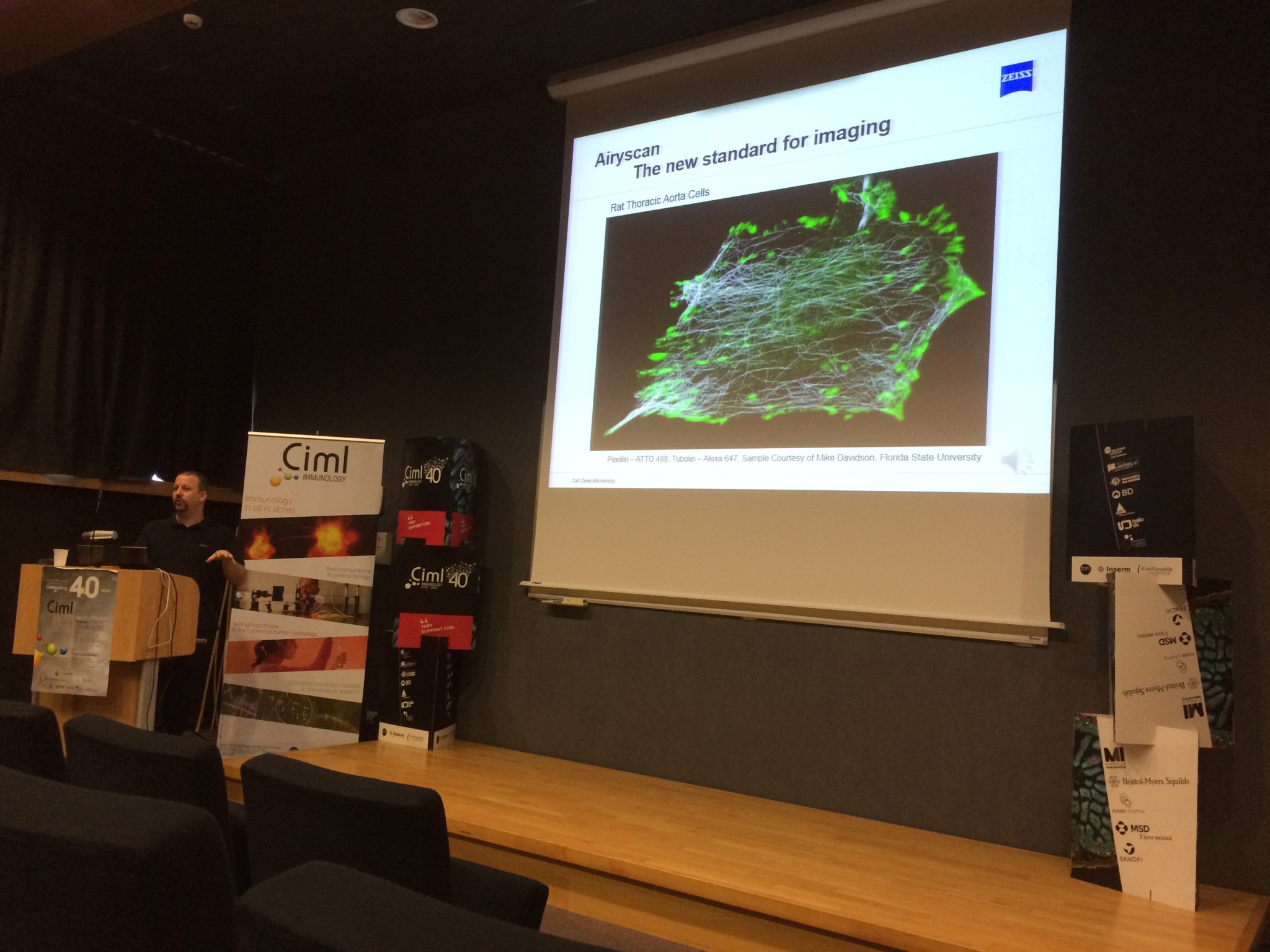
LSM 880 Fast-Airyscan

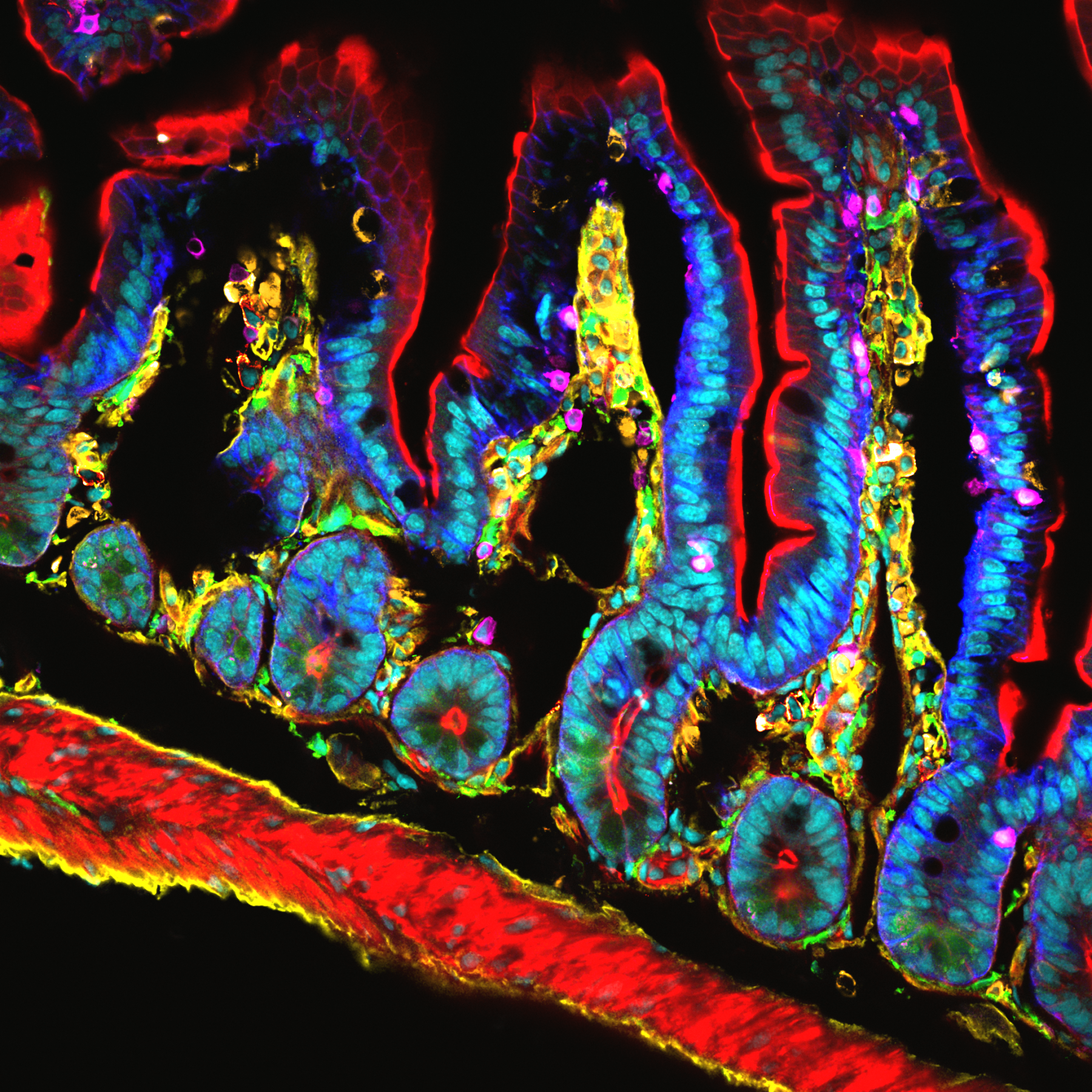
A new confocal microscope using the technology Airyscan (LSM 880 Fast-Airyscan, Zeiss) has been purchased through a co-funding FBI/Inserm/CNRS/Region PACA. The Airyscan improves spatial resolution (1.7 x) by imaging the Airy disk onto a concentrically arranged hexagonal detector array. Its detection area consists of 32 single detector elements, each of which acts like a very small pinhole. The confocal pinhole itself remains open and doesn’t block light – thus all photons of the whole Airy disk are collected. The signals from all detector elements are then reassigned to their correct position, producing an image with increased signal-to-noise ratio and resolution.
FEI Teneo VS
The FBI-PICsL facility also recently homed an electron microscope dedicated to automated 3D imaging.
Revealing the complex 3D architecture of cells and tissues is crucial for the structure-function correlation in biological systems. However, in most of the electron microscopy experiments, only a small portion of the sample is imaged. For instance, one 70nm-section of an average vertebrate cell only represents 0.4-1.0% of its volume. Beyond the fact that this tiny volume might not be representative to account for the entire cell ultrastructure, the sectioning also avoids a definite visualization of the cellular organization.
Until recently, the imaging of big volumes (tens to hundreds of cubic micrometres) by electron microscopy was tedious because it involved manual serial sectioning. Beyond the need of super-skilled people, manual serial sectioning has a limited z resolution of 70nm and is a time-consuming process at the sectioning, imaging and post-processing steps. Within the last 10 years, the Serial Block-Face Scanning Electron Microscopy (SBF-SEM) has emerged as the method of choice for the imaging of big volumes by electron microscopy.
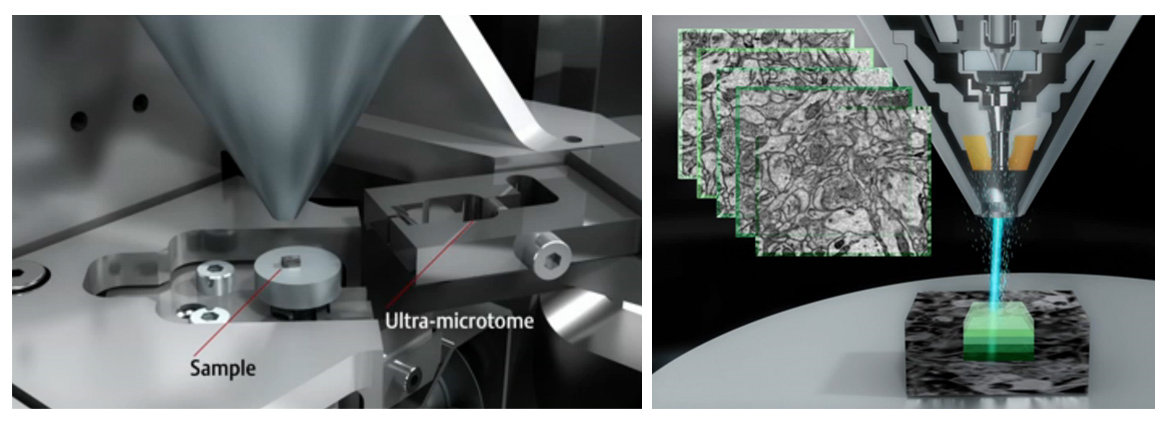
The concept behind SBF-SEM is to have a microtome within the chamber of a scanning electron microscope (see Figure). This set-up enables the iterative sectioning and imaging of the sample block-face for hundreds of microns in a fully automated way (Figure). When an electron beam hits a sample, it interacts with it and yields several signals that can be detected and assigned back to the scanned positions on the sample surface. Among these signals, the backscattered electrons can be collected to retrieve information on the sample electron density at a given point. Routinely, a working pixel size in SBF-SEM datasets is ±5nm and the slicing can be as thin as 20-30 nm.
In addition to be an excellent microscope in classical SEM mode, the Teneo VS microscope by FEI is currently the most advanced microscope to carry out SBF-SEM. It does not only allow you to cut and image the sample in a fully automated way but it also gives the possibility to avoid charging artefacts during imaging by putting the sample in low vacuum conditions. By limiting the charging, the microscope gives you the opportunity to retrieve more information from the sample. For instance, the sample can be scanned with different acceleration voltages in order to retrieve ultrastructural information at different depths (10nm – 20nm – 30nm – 40nm). When this multi-energy imaging is combined to a 40nm mechanical slicing, it gives rise to previously inconceivable datasets of hundreds of cubic microns of tissue with a voxel size of 5x5x10nm !
Since the delivery of the FEI Teneo VS microscope at the FBI-PICsL facility in December 2016, we tested the microscope on a broad range of biological samples in classical SEM mode (fly egg-laying apparatus, Xenope embryo, mouse face…) and in SBF-SEM mode (cell pellets, Drosophila muscle, mouse brain, marine sponges, Xenope embryo…). In particular, the group of Laurent Kodjabachian (IBDM), working on multiciliated cells, wanted to image the same cell type in both classical SEM mode and in SBF-SEM. We could 1. assess the penetrance of a mutant phenotype in classical SEM mode and 2. localize the basal bodies of these multi-ciliated cells in SBF-SEM (see Figure below).
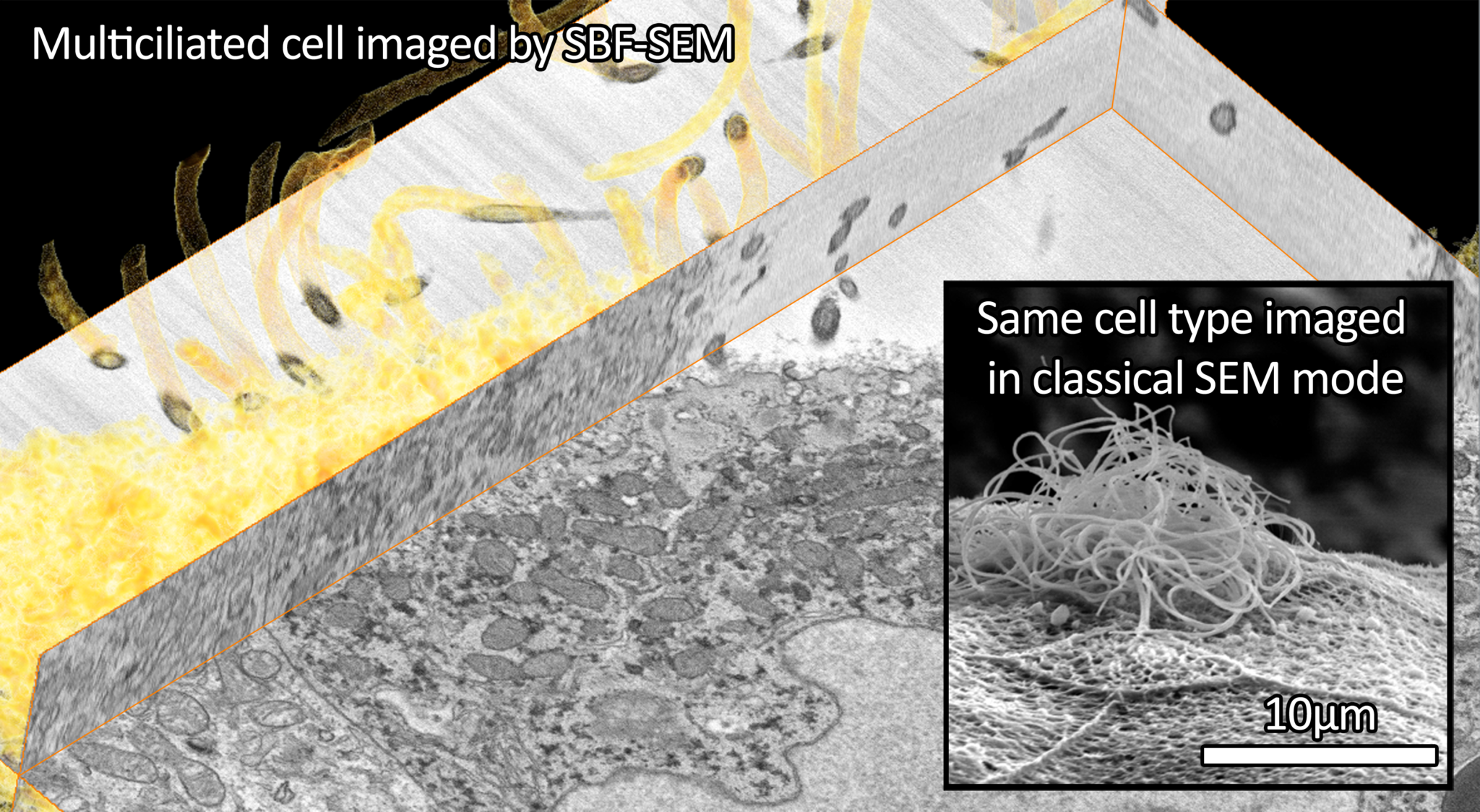
Photo credits: Virginie Thome and Nicolas Brouilly
Major implications of the FBI-PICsL facility
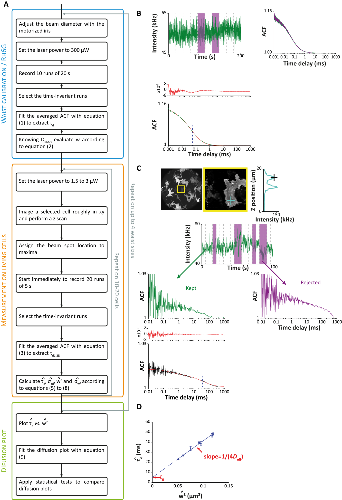
Spot variation fluorescence correlation spectroscopy (svFCS) (Hai-Tao He and Didier Marguet’s team)
In collaboration with Nicolas Bertaux (Institut Fresnel, AMU, Centrale Marseille), members of the Hai-Tao He and Didier Marguet team published a user guide for characterizing plasma membranes subdomains in living cells by sv-FCS2. This book chapter aims to serve as a guide for setting and applying the svFCS methodology to study the plasma membrane of both adherent and nonadherent cell types.
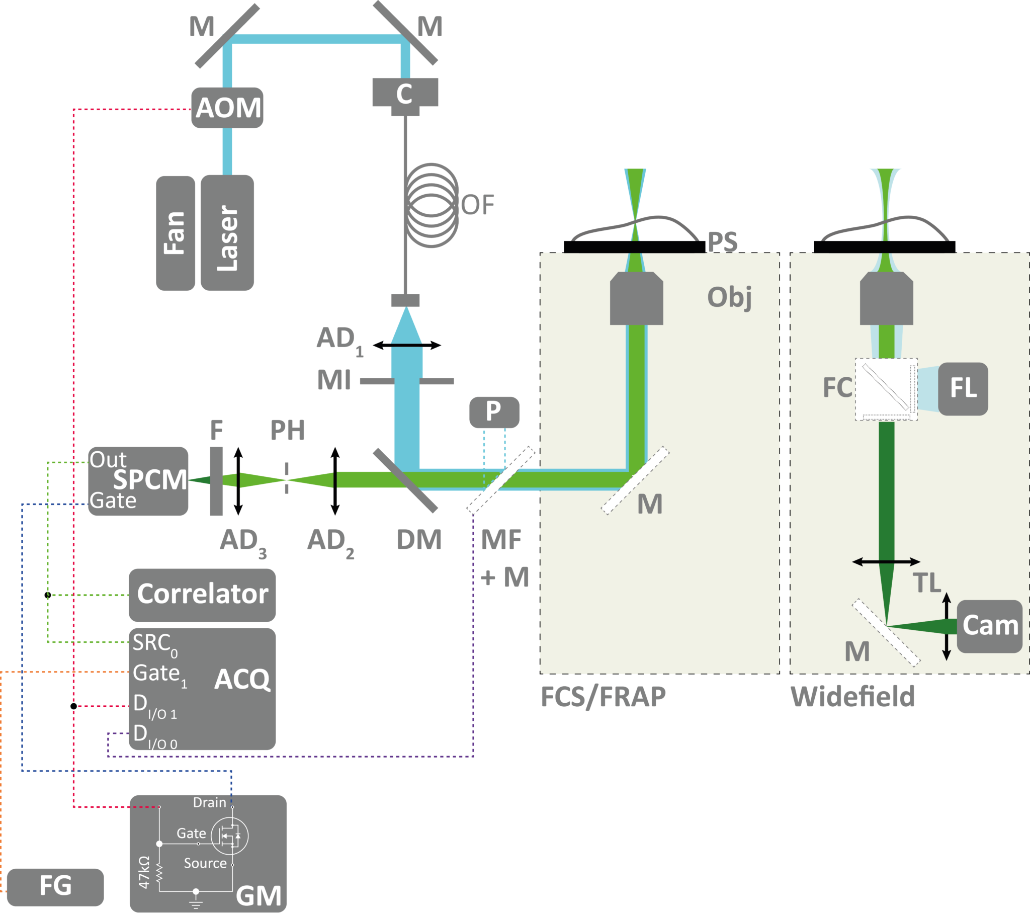
recovery after photobleaching (FRAP).
(Click to view the image in full size)
This technique has been used to understand a genetic disease by studying molecular diffusion at the plasma membrane. In collaboration with Christophe Lamaze (Institut Curie) and Céline Galès (Institut des Maladies Métaboliques et Cardiovasculaires de Toulouse), Hai-Tao He and Didier Marguet’s team worked on decoding the dysfunction of molecular mechanisms involving interferon-γ (IFN-γ), key protein for immune defense. This protein is not able to fulfil its natural function as a protective cytokine when its receptor is “blocked” in the wrong place on the cell membrane: a modification to the immune response causing severe infections sometimes even life-threatening for young children.
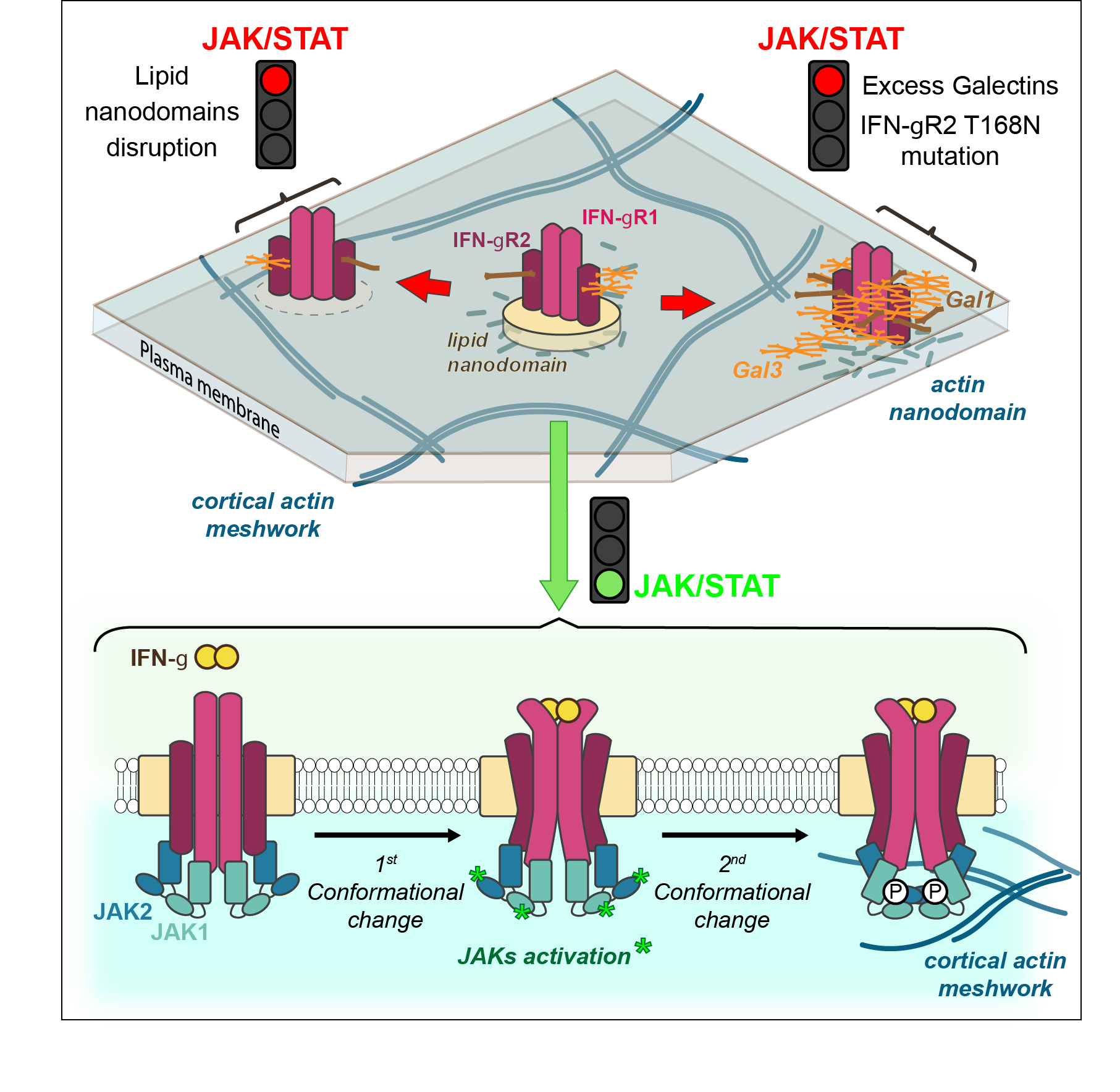
Lipid and Actin Nanodomains Is Critical for JAK
Activation
IFN-γ receptor is composed of two protein chains to which are generally associated six sugars. However, some patients diagnosed with Mendelian susceptibility to mycobacterial diseases syndrome (MSMD) have seven sugars. This simple gain of glycosylation affects greatly the immune response against mycobacteria.
Using their expertise in biophysics and more particularly in svFCS, this CIML team showed the existence of nanodomains and their fundamental role in signaling activity. “It was completely unexpected that only one additional sugar could modify the way the receptor is localized on the surface of the cells, affecting greatly the later signaling events”, explained Hai-Tao He. “IFN-γ receptor is then delocalized towards another membrane nanodomain populated by galectin proteins, that bind to this additional sugar” added Yannick Hamon2 (cf Figure)
As therapeutic target, galectins are an appealing perspective, particularly as turning off their expression canceled in vitro the pathologic effect linked to the glycosylation gain of the IFN-γ receptor. However, therapeutic programs towards patients requires the exploration and understanding of molecular mechanisms by uniting expertise of biologists and physicians but also mathematicians and physicists as it was the case in this discovery4.
News
In March 2017, a workshop on super-resolution is organized in Marseille by Sébastien Mailfert, Jean-Bernard Fiche and Orestis Faklaris on multicolor STORM an dSPT-pointillism data analysis, this workshop is dedicated to advanced researchers in this field. This event is supported by FBI.
The FBI-PICsL facility is also homing the 14th RTmfm convention from the 20th to the 22nd of March 2017. This convention is a great opportunity for imaging facilities staff to share their experiences and ideas on the past, present and future of their work (technical development, big data issues and solutions, link with industry).
Our facility also yearly organizes the following course: Scientific platform, instrumental sharing, how to build up and develop a service. The aim of the course is to acquire, by alternating courses, exercises and practical conditions, methods and constraints to the development of a provision of scientific equipment service. We insist on how to develop a contributory model for managing a scientific platform.
References
- Mailfert, S., Hamon, Y., Bertaux, N., He, H-T. & Marguet, D. Methods in Cell Biology, 139, 1-22 (2017)
- Blouin, C., Hamon, Y. et al. Cell, 166, 920-934 (2016)
- http://www.cell.com/cell/fulltext/S0092-8674(16)30909-6