A new intestinal cell junction has been discovered thanks to STED microscopy
Cellular junctions are essential to the integrity of epithelia, which cover most of our organs. In an article published in the journal PNAS, scientists, with among them members of our FBI Marseille node, reveal the existence of a new category of cell junctions. Using Stimulated emission depletion (STED) microscopy, they have put forward the need to reconsider the organization of intestinal cell junction described as such for more than 40 years.
Imaging intestine with STED
Stimulated emission depletion (STED) microscopy is a super-resolution technique that bypasses the diffraction limit of light microscopy to increase resolution. In our case, scientists were able to resolve the organization of complexes located at cell junctions with a resolution of a few tens of nanometers thanks to STED. Moreover, STED tripled the spatial resolution in the junctional plane and, using cryosections, they achieved imaging with a seven times greater spatial resolution compared to approaches that would use confocal microscopy and thus, without physical sectioning.
Although the resolution of STED is at least an order of magnitude lower than that of electron microscopy, the combination of STED with immunostaining reveals organization up to then unknown as multiple proteins can be efficiently labeled at the same time.
Three types of intestinal cell junctions
The intestine is covered with cells, most of which absorb the nutrients we ingest. These cells are joined together by three types of junctions which coexist and provide different functions, ranging from the selective filtration of certain ions to the mechanical maintenance of the epithelial layer. These junctions, the tight junction, the adherens junction, also called zonula adherens, and the desmosomes, were discovered in the 1960s and their constituent elements as well as their organization were proposed during the 1980s and 1990s.
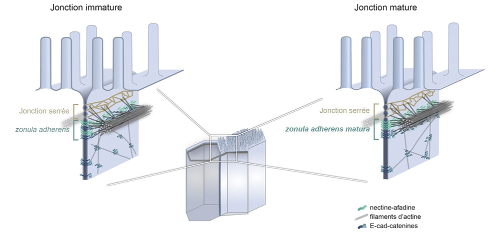
The adherens junction in particular is established as being organized into a belt of adhesion proteins anchored to the membrane, the cadherins, and supported by filaments, the actin filaments. This junction has an important mechanical role in the cell, for example by impacting the shape of the cell. The zonula adherens (ZA), a fundamental module of epithelial cell–cell adhesion initially observed in intestinal cells, is believed to comprise a single contractile actin belt linked via E-cadherin-catenin to the ones of neighboring cells.
How did microscopy help reevaluate our current knowledge?
By observing the adherens junction of epithelial cells obtained from human intestinal biopsies, or from human cells in culture using STED super-resolution microscopy, scientists have made a very surprising discovery. They show that the ZA consists of two distinct belts of adhesive complexes, a basal one with E-cad-catenin and an apical one with nectin–afadin. Contrary to the prevailing view, the major actin belt aligns with nectin and afadin, not E-cad-catenin.
The authors further demonstrate that this organization depends on the cell maturation state and that the classical ZA found in textbooks corresponds to a less mature state of the intestinal junction. Therefore, they decided to call the junction found in mature cells the zonula adherens matura. Genetic and physical perturbations show that afadin is essential for force transmission across cell junctions. This work redefines the intestinal ZA architecture and prompts a reevaluation of how forces propagate within an epithelial sheet.
Not only, these results are important to better understand the adhesion and mechanics of epithelial cells, but these two essential characteristics of the epithelia are particularly affected in cancers of epithelial origin, which represent 80% to 90% of current cancers. This discovery is, thus, a step forward to the comprehension of cancers and to their treatment.
Get access to one of our services!
You need FRAP, two photon FLIM-FRET, PALM/dSTORM at France-BioImaging? To get open access, please login via Euro-BioImaging website! You just have to choose the technology you want to use, then submit your proposal. All applications will be processed by the Euro-BioImaging Hub in close relation with France-BioImaging. And of course, all scientists regardless of their affiliation, area of expertise or field of activity can benefit from open access services! Users whose projects will be validated by Euro-BioImaging will benefit from a waiver for the access cost on France-BioImaging core facilities (https://france-bioimaging.org/access/).
Mangeol, P., Massey-Harroche, D., Sebbagh, M., Richard, F., Le Bivic, A., & Lenne, P. F. (2024). The zonula adherens matura redefines the apical junction of intestinal epithelia. Proceedings of the National Academy of Sciences, 121(9), e2316722121. https://doi.org/10.1073/pnas.2316722121
Sources : https://www.insb.cnrs.fr/fr/cnrsinfo/une-nouvelle-categorie-de-jonctions-cellulaires-dans-lintestin
https://www.ibdm.univ-amu.fr/redefining-epithelial-junctions-with-nanoscopy/