One of our external users got his work published in Nature Communications
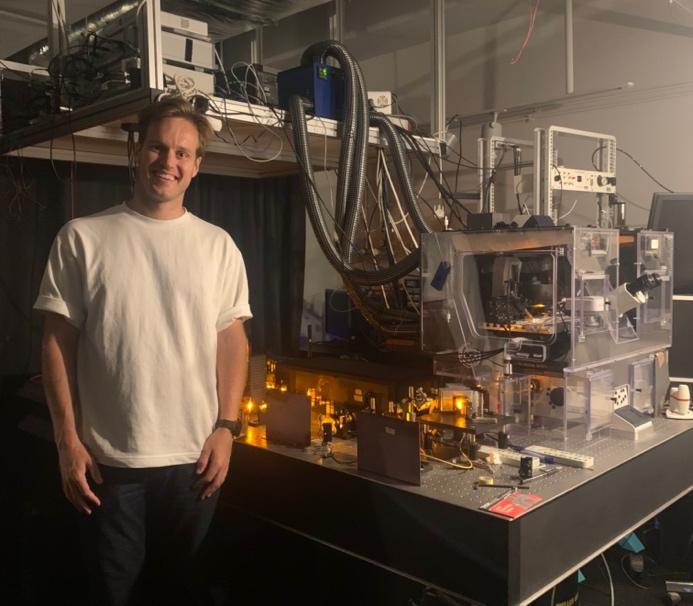
In 2022, in a project funded by the Euro-BioImaging pilot User Access Fund, Andrew Boyce, postdoctoral fellow at the University of Calgary, used state-of-the-art microscopy techniques at the Bordeaux Imaging Center, part of the Bordeaux node of France-BioImaging, for panoptical visualization of brain tissue in vivo. His work just got published in a Nature Communications article!
The advantages that shadow imaging has in visualizing brain tissue
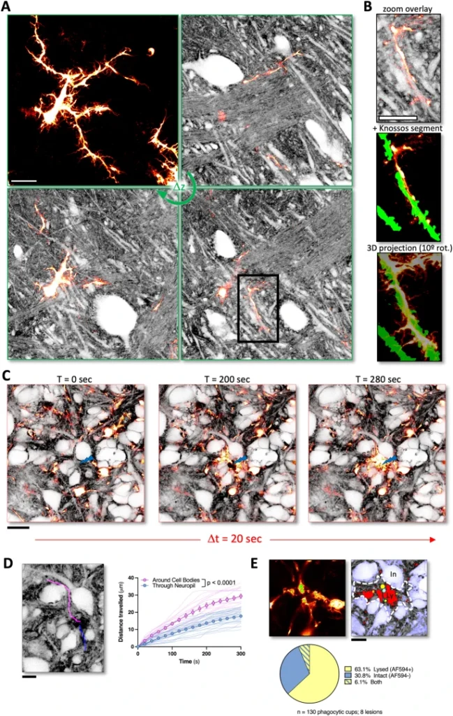
Progress in neuroscience research hinges on technical advances in visualizing living brain tissue with high fidelity and facility. Unfortunately, current neuroanatomical imaging approaches either require tissue fixation (electron microscopy), do not have cellular resolution (magnetic resonance imaging) or only give a fragmented view (fluorescence microscopy).
In this study, scientists have shown how regular light microscopy together with fluorescence labeling of the interstitial fluid in the extracellular space provide the information lacking in the previous techniques. Basically, it’s like looking at the negative of a photo! Called Shadow Imaging, they have demonstrated the power of this approach on revealing neurons, microglia, tumor cells and blood capillaries together with their complete anatomical tissue contexts.
The perfect combination between the right technology and the right expertise
How did Andrew Boyce contribute to this study? Interested in blebbing neuron dendrites during cell death caused by a stroke, he only had, at that time, results from how cells in culture reacted during this kind of events but no information about the neighboring cells.
Thanks to Euro-BioImaging pilot User Access Fund, he came to Bordeaux to use a combination of live cell 3D stimulated emission depletion (3D-STED) microscopy and super-resolution shadow imaging (SUSHI), and adapt shadow imaging approaches to conventional confocal microscopy (COSHI). Shadow imaging allows you to visualize fine details of cell-cell interactions and the extracellular space in an unbiased manner. Brain tissue is complex and shadow imaging is a technique that allows researchers to visualize all of the cells in this very complex architecture in an unbiased manner so it was the perfect match for Andrew’s research project!
A career-boosting experience
“This has really been an incredible experience. It’s amazing to be trained by the person and team who pioneered this technique. I’m very thankful for this opportunity and humbled to be over here,”reflects Andrew from Bordeaux.
When asked how his visit to Bordeaux might impact his career, Andrew explains that he can certainly imagine doing shadow imaging in the future. He would eventually like to run his own lab at an academic institution in Canada. Being able to bring an exciting and novel technique like this back to Canada would be really impactful for his career, and for science in general.
“Shadow imaging is a very exciting technique for studying brain tissue, but getting started on sample preparation and adapting this technique to your research question can be daunting. Coming to the Bordeaux Imaging Center to work with the person who developed this approach and get expert training is a dream come true and will make a big difference to my research. This is an amazing opportunity made possible by the Euro-BioImaging Pilot User Access fund with the goal of making shadow imaging more accessible across diverse platforms,” concludes Andrew.
at the Bordeaux Imaging Center (www.eurobioimaging.eu/news/using-super-resolution-live-cell-imaging-to-understand-cell-death-during-stroke)
Get access to one of our services!
You want to be the next user to get access to state-of-the-art technologies that France-BioImaging provides? Please login via Euro-BioImaging website! You just have to choose the technology you want to use, then submit your proposal. All applications will be processed by the Euro-BioImaging Hub in close relation with France-BioImaging. And of course, all scientists regardless of their affiliation, area of expertise or field of activity can benefit from open access services! Users whose projects will be validated by Euro-BioImaging will benefit from a waiver for the access cost on France-BioImaging core facilities (france-bioimaging.org/access)
Thanks to Marianna Childress and Andrew Boyce for the original article!
Dembitskaya, Y., Boyce, A.K.J., Idziak, A. et al. Shadow imaging for panoptical visualization of brain tissue in vivo. Nat Commun 14, 6411 (2023). https://doi.org/10.1038/s41467-023-42055-2