Chemical imaging methods : Infrared and Raman Microscopy in Cellular and Tissue bioimaging — June 20, 2018 — 9am-12pm
▽ Scroll down
Author: caroline.thiriet
This conference is organized by the “Réseau d’Imagerie Cellulaire Paris-Saclay”.
The two most common techniques of vibrational micro-spectroscopy are infrared (IR) and Raman. These two sophisticated tools enable to visualize the inherent vibrational spectra of biochemical constituents of a cell or a tissue. Therefore, IR and Raman microscopy provide the specific and distinct “fingerprint” spectrum of each cell and offer the powerful possibility of high contrast images without any external labeling.
Recently, significant developments of these approaches provided a better access of these two techniques by the biologist community. Currently, IR and Raman microscopy are used for tissues, biopsies, animal and plant analyses in order to visualize proteins (C-H3 bonds), lipids (C-H2 bonds), water (O-H bonds), membranes, myelin, chromophores such as flavins, etc …
Imaging has become a key technique in life science to achieve high-standards research and results. Among the three major components of imaging based research – laboratory experiment, microscopy and image analysis – nearly 60% of life science researchers acknowledged that “image analysis is the most difficult part”. Therefore, training researchers on image data analysis is crucial to ensure that they can get the most out of the data that they generate.
Several initiatives are active in developing training solutions in image data management and analysis Neubias, EuroBioimaging (EuBI), Global BioImaging (GBI) and ELIXIR. GBI/ELIXIR/EuBI/NEUBIAS are jointly organizing a hackaton on image analysis training material re-usability, that aims to bring together training providers from the imaging community and contributors to the ELIXIR Training Platform in order to create a curated collection of training materials on data management and analysis and facilitate their Findability, Accessibility, Interoperability and Re-usability.
Audience: Bioimage analysts, trainers and developers from NEUBIAS, EuroBioImaging and Global BioImaging, as well as ELIXIR’s Bioschemas and TeSS developers, and anyone willing to contribute, to: (i) foster new collaborations between ELIXIR and key initiatives from the image analysis community, (ii) create a curated collection of training materials on data management and analysis and (iii) facilitate their Findability, Accessibility, Interoperability and Re-usability.
Registration and more information can be found here.
Thank you for participating to this new edition MiFoBio 2018 by proposing a workshop. As announced in the spring, it will be necessary to explain the interest of your project and give a synopsis. It is therefore important to prepare your workshop project upstream. Evaluation of proposals will follow quickly in November. Depending on the returns, a second call will be made on a more restricted perimeter.
These workshops must fit into one of the themes below. The objective is to deal at least in part with one of the common questions so that we can then discuss at the end of the school the contributions of different technologies for the same subject.
Biological, physical, photochemical question on the themes identified below:
Mitochondria and metabolism
Regulation of gene expression, maintenance of information integrity, nucleus
Synapse, neuron, brain,
Interaction between cells or with the environment, mechanobiology.
Membrane and receivers, signaling,
biofilm
Development, regeneration, evolution
organoid
2. New technological, instrumental or probe and marker development
3. Implementation of at least two significantly different techniques for the same question
4. Instrumental engineering and systems engineering (characterization of instruments, peak methods, new practices)
5. Image Bar: Image analysis
6. FabLab MiFoBio: small practical realization: optics, electronics or analysis
7. Outside class (to be justified)
For the record, a workshop aims to share knowledge and know-how in the field of biological imaging and biophotonics through practice. It is worn by two academics at most and must allow the participants to practice. The workshops implement methodological and instrumental approaches and deal with a scientific issue that is often interdisciplinary.
Cordially
The MiFoBio organizing team
Dear colleagues, dear FBI community,
The National Coordination and Industry Board are proud to announce that the winners of the FBI Image Contest 2017 are:
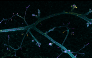
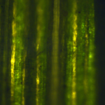
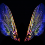
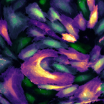
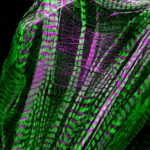
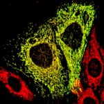
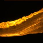
1. Marie Irondelle – PiCT Institut Curie with “Biology’s Little Venice”
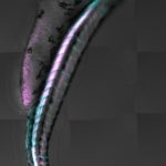
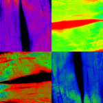
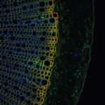
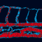
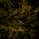
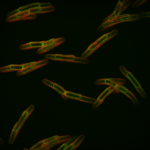
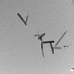
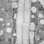
Application de la stratégie de double réaction chimique BLISS aux unités p-hydroxyphényle et guaïaicyle de la lignine sur coupe de racine de lin. Observation du double marquage (+ autofluorescence) par microscopie confocale et représentation par projection maximale d’intensité. Taille de l’image : 510 x 510 microns.
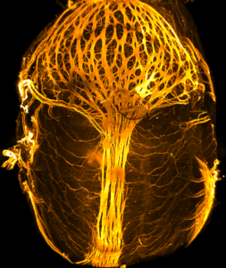
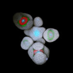
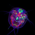
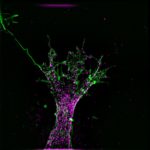
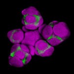
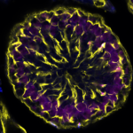
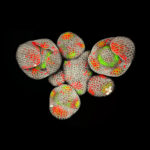
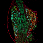
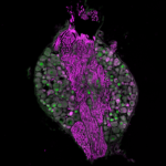
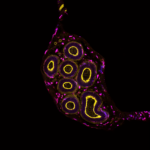
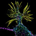
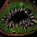
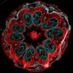
FBI Industry Committee Special Prize: Nathanaël Prunet – California Institute of Technology with “Arabidopsis Inflorescence”
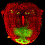
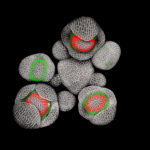
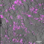
This is a live Arabidopsis inflorescence with young flower buds developing at the periphery. Cell walls have been stained with propidium iodide (grey). Fluorescent reporters were used to monitor the expression of the APETALA3 (AP3, green) and SUPERMAN (SUP, red) genes. AP3 is required for the development of stamens (the male organs), while SUP establishes the boundary between the male and female part of the flower. This picture was acquired using live confocal imaging, which allows us to describe the expression of several genes in both space and time, in the same live biological samples, with a precise cellular resolution. It finally allows us to understand a question that has been elusive for 25 years: how the male/female boundary is established during the formation of the flower. My research aims at understanding how flower buds are patterned as they form.
Jonathan Daniel, Institut des Sciences Moléculaires
Laurence Dubreil, APEX-UMR703 PAnTher INRA Oniris
Pierre-Olivier Strale, Interdisciplinary Institute for Neuroscience
Clémence Simon, Unité de Glycobiologie Structurale et Fonctionnelle, UMR8576
Jérémie Teillon, INSERM U1034
Morgane Rabineau, Inserm
Eve Gazave & Nicolas Rabet, Institut Jacques Monod-CNRS
Nathanaël Prunet, Caltech, Meyerowitz lab
Françoise Geffroy, CEA-DRF-NeuroSpin-UNIRS, Midas Team
Valeria Davi, ImagoSeine – Institut Jacques Monod – CNRS
Anna Smirnova, University of Strasbourg – GMGM
Debora Olivier, Institut Pasteur
Orestis Faklaris, Institut Jacques Monod
Xavier Baudin, Institut Jacques Monod
Mathieu P. Dailly, CMAS
Lucie Sengmanivong, UMR 144, Institut Curie, Paris
Marie Irondelle, Institut Curie
Thank you also to the core facilities staff and heads for having forwarded the contest to their users and for providing them state of the art bioimaging.
The National Coordination
Perrine Paul-Gilloteaux, bio-image analyst, CNRS research engineer and project manager of our Bio-image informatics node, received last month the 2017 CNRS Crystal Prize, awarding her contributions to French research.
A perfect occasion to highlight her career and her work with France BioImaging. What is eC-CLEM? How can our field deal with the massive amount of data produced? What future developments can we expect in the realm of bio-image informatics? Read the interview below to find out more.
Perrine Paul-Gilloteaux
Could you introduce yourself briefly?
My name is Perrine Paul-Gilloteaux, I’m a CNRS Research Engineer. I have a background in electrical engineering, signal and image processing, and did my PhD in augmented reality for neurosurgery through surgical microscope. I started working in bio-image analysis for microscopy in Ireland, and joined the Curie Institute on the PICT IBISA facility in 2010. I moved to Nantes in 2015, and now work in a biomedical research institute. I am also the project manager of the France BioImaging node Bio-Image Informatics IPDM (Image Processing & Data Management), and work closely with the national coordination on the aspect of data management.
I define myself as a bio-image analyst, meaning that I do my best to bridge the gap between microscopy, image analysis and biology. This means that I’m involved in data management, data processing and data analysis projects, that I provide as a service in facilities or work on as research topics.
How long have you been involved with FBI, and what main projects have you carried out with us?
I’ve been involved in FBI from its inception. I started working within the transversal IPDM working group, where we first defined the state of our management systems and worked on the interoperability of our data bases. I managed the setup of the Curie Image Database, supported by France Bio Imaging, based on the OpenImadis system. In 2015, I was nominated project manager of the IPDM node, led by Jean-Christophe Olivo-Marin and Charles Kervrann. One important part of my mission is to work with the national coordination on the data management aspect in FBI. For this, we started by making a survey of resources and management system on site. This question of data management is now central, and FBI collaborates with other infrastructures at the European level: EuroBioImaging and ELIXIR, but also at the national level with other national infrastructures in biology using microscopies, and with the French Institute of BioInformatics (ELIXIR French node).
You have developed a software called eC-CLEM. Could you explain what it consists of?
For this project, I’ve worked closely with Xavier Heiligenstein (Curie Institute, FBI working group on multimodal imaging). Ec-CLEM (for Easy-Cell-Correlative Light to Electron Microscopy) is a software designed to help correlative microscopies. The purpose is to help the fusion of information obtained by different modalities of microscopy on the same sample (for example electronic microscopy, photonic microscopy, atomic force microscopy, etc.). The software allows to register, i.e. align in the same system of coordinates, multidimensional images with big scale and resolution differences, either with a manual input of the user, either automatically when possible. In addition, it provides an estimation of the error of alignment, based on statistical methods, and detects the deformations that the sample may have undergone. I’ve developed a set of algorithms implemented as plugins for the ICY platform. [note: Perrine has published a paper about eC-CLEM in Nature Methods] During the development of this set of tools, I was greatly helped by the ICY coding parties (Hackatons) organized at Pasteur with the support of FBI, and I would encourage developers to attend such events, as there are always new things to be learnt.
Perrine presents her work during the Crystal Prize Ceremony at Nantes University in December 2017.
Bio-image informatics have taken the center stage lately, as more and more people realize how crucial image processing is for research. Could you expand a bit on that?
It is entirely true, and this is the reason why FBI has had a transversal node on that activity since its creation. I’ve cofounded a network of bio-image analysts (NEUBIAS) for that exact purpose also. The size and the number of data to be processed, the large amount of different questions to be answered from imaging and the interplaying between acquisition and processing to generate imaging data and analysis data, have led to a complexity of analysis which requires expert tools but also expert people. Bio-image informatics is a field of research by itself, and it is now recognized as such. It is bridging the gap between image processing research and biology research based on imaging. It can be seen also from the recent Nobel prizes in chemistry: in cryo-tomography or in super resolution light microscopy, both developments were relying on image processing as an essential part.
What are going to be, according to you, the next big steps and developments in the realm of image processing and data management?
The novelties in our field is two-sided: from one side we have data exploding in size and number, and on the other side, machine learning -and in particular deep learning- benefits from progress in hardware and opens the way to big progress in analysis and in particular in feature recognition (segmentation and tracking).
Regarding data management, the big issues to be solved need to involve the whole imaging community, but also to seek expertise from other fields with the same problems. Technical solutions, both from the software side (with management software such as OpenImadis, Omero, Bioemergences in France BioImaging nodes) or the hardware side (optimized hardware systems, optimized protocols of data transfer) are on their way, but will not be useful if the biologists do not put effort in data curation and data selection.
Up to this day, even with machine learning, tuning a software or a protocol to respond to a particular problem and a particular set of data requires a lot of effort, either to set up the learning network or train it in the case of machine learning, either to combine algorithm for a specific question in adapted workflows or to develop more performing algorithm. It means that we need well trained expert able to master both the image processing aspect and the biological questions behind.
On what will your FBI working group focus in 2018? What can we expect from you? (in terms of new developments, priorities, events etc.)
The priority is definitely to deal with the explosion of data we are facing. In addition to the directions exposed in the previous question (software and hardware solutions), one direction taken by IPDM on data management is the definition of quality data. For this multi-faceted topic, we have already started to set up tools to measure the quality of the data produced in term of resolution for example, based on the expertise in metrology of our facilities members, that we want to demonstrate in 2018. We will also concretize the collaborations between FBI and the other national infrastructures by running tests, for example on the speed of data transfers between node, in order to make sure that at the end of 2018, each user of the FBI nodes can easily access and process her/his data from anywhere. A technical catalogue of software and hardware resources is under construction, to allow FBI nodes and beyond to benefit wisely from the tools and networks created by FBI. In the first semester of 2018, we will be organizing an event to discuss and define the changes in bio-image informatics that deep learning could bring about (further information to come soon, please refer to the FBI site).
Paul-Gilloteaux, Perrine, Heiligenstein Xavier et al. “EC-CLEM: flexible multidimensional registration software for correlative microscopies.” Nature Methods, vol. 14, no. 2, 2017, pp. 102–103., doi:10.1038/nmeth.4170. https://www.nature.com/articles/nmeth.4170
AAP Support to Events
PEP 2018 (Photothermal effects in plasmonics. Special focus on biology) – by Guillaume Baffou, Institut Fresnel, Marseille. Funded by FBI Coordination.
AAP User Access
Identification of the local transcriptome in mature excitatory presynaptic boutons, by Anne-Sophie Hafner (Max Planck for Brain Research, Schuman Department), hosted by Bordeaux Node.
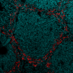
Effect of vitamin A on splenic dendritic cells subset localization, by Jennifer Gommerman (University of Toronto), hosted by Paris-Centre (Curie).
AAP Tech & Meth Transfer
Cryo-CLEM implementation at Bordeaux Imaging Center, Monica Fernandez Monreal (BIC), hosted by Paris-Centre Node (Pasteur – Ultrapole).
A message from the organizers:
Registration to the Bioimage Analysis Symposium of the 2nd NEUBIAS conference, in Szeged (Hungary), 31/01 to 02/02, 2018, is IMMINENTLY CLOSING !
We have extended the registration deadline to January 10th, 6pm (CET).
The Network of European Bioimage Analysts (NEUBIAS) is happy to confirm:
– 35+ talks with Keynotes Gaudenz Danuser, Gene Myers and Marleen DeBruijne, – 70+ posters – 22 Software packages in live demo at the Open source Software Lounge – 4 industry workshops – Call4help: A special BioImage Data Clinics session for participants to present and tackle their analysis problem with experts – 220+ participants
– Free shuttles set from Budapest airport for NEUBIAS participants. You can access the current program here:
The NEUBIAS conference is a forum to exchange the newest findings, applications, and cutting-edge developments in Bioimage Analysis, machine learning, data mining, and storage. European Bioimage Analysts organize this event bringing together an international, interdisciplinary community of about 250 leading scientists in the life and computer sciences. The 3-days Symposium reflects the need to foster the networking between image analysis Developers and their end-users. The interest and needs for image processing in life sciences are heavily growing, a reality that is reflected by the success of earlier events of NEUBIAS in Lisbon in February 2017.
The organizing committee is assembling a top-notch program featuring both academic and industry presenters which will include Image restoration, storage & management, 3D & 4D image analysis, tools for high content analysis, data mining and open source developments. Aside from plenary talks and abstract presentations, we will have dedicated “Call 4 Help” sessions to team up tool providers with life scientists facing roadblocks in their image analysis, and two satellite “training schools” for “early career scientists” and “advanced Bioimage Analysts”. NEUBIAS Symposium will also feature the latest updates on open source software tools, components, and packages, in plenary talks and during the “Open Source Software Lounge” session which has been a great success in Lisbon, and where participants can meet developers and power users of a wide range of Open tools for Bioimage analysis in Live demo format.
The 2nd Annual Conference of NEUBIAS, gathering the whole BioImage Analysis Community into a multi-faceted event, offers two Training schools, a Working Meeting (Taggathon) and a Large Symposium dedicated to Scientific Developments and Open Tools in BioImage Analysis.
Training Schools:
Training School #6: Bioimage Analysis for your research (for Early Career Investigators)
“Learn how to build Bioimage Analysis workflows across software tools & scripts”Digital image processing (DIP), Building workflows 2D, 3D & time-lapse, Extracting numerical data from bioimages, Toolboxes, Workflow automation (scripting).Sessions to solve your problem with an Analyst.
Registration to the Training Schools open until November 9th, 2017.
Symposium:
Topics Highlights of the Symposium
BioImage Analysis in Life Science, Developments, Machine Learning, Bioimage Data Mining, Storage, etc…
Open source Software Lounge, Call4help, Open Tools, Industry workshops, Panel Discussions, more…
Abstracts submission deadline: November 11th, 2017.
Les 15èmes Assises Nationales des Plateformes du réseau RT-mfm
se dérouleront à Rouen en 2018 (PRIMACEN, université de Rouen, Mont Saint Aignan)
du 19 Mars 13h au 21 Mars 13h.
Le formulaire d’inscription aux Assises se trouve à l’adresse : https://goo.gl/forms/oHBJkdjikkb5TrS83
la date limite d’inscription est le 20 décembre 2017 à 18h00.
Le programme est en cours d’élaboration, faites-nous savoir si vous souhaitez voir développer un thème ou proposer une présentation orale ou un intervenant.
Les propositions sont à envoyer à france.lam@upmc.fr et olivier.renaud@curie.fr.
Votre inscription définitive pour participer aux Assises vous sera confirmée par email fin janvier/début février 2018.
Cordialement,
France & Olivier
Pour le RT-mfm
Le Storage Day est une conférence organisée par l’INRA (Unité Ingenum) avec la participation d’autres Instituts et dispositifs de recherche (CIRAD, IGBMC, Biogenouest) autour des questions de la gestion de la donnée scientifique, et en particulier son stockage.
Cette journée propose des retours d’expérience de différentes communautés (Imagerie, NGS, SHS, etc.) autour de problématiques en lien avec la “vraie vie” sous la forme de présentations synthétiques (15 minutes). Ces retours d’expériences auront pour objet aussi bien la mise en valeur de “success stories” que la mise en évidence des écueils/difficultés auxquelles se trouvent confrontées les communautés.
The Electron Microscopy platform of the University and DIMNP are pleased to announce the volume EM conference which will take place at the new “Fac de Medecine” of Montpellier on December 14-15th.
Volume EM is a set of methods that allow to obtain high resolution 3D ultrastructural data, using scanning electron microscopy coupled to different automatic serial sectioning techniques. With these methods it is now possible to obtain high resolution 3D data within a few hours or days. Volume EM is of rapidly growing interest at the international level with applications in many fields of research.
During the conference, high level scientists and application developers will present the principles of these methods and selected examples of applications for research.
A “zebrafish challenge” has also been proposed to different microscope manufacturers, for which they had to image an imposed portion of zebrafish embryo using the equipment of their choice. The objective will be to analyse the results obtained in conditions close to “real research”. The data will be presented and commented at the end of the conference, highlighting advantages and limits of each type of method.
The conference will be entirely in English. Inscription is free.. For any question, please write to volumeem@umontpellier.fr
The dissemination of emerging technologies to end-users is a key objective of FranceBioImaging. It is indeed essential that developers can obtain feedback from the end-users on their technologies. It is equally important that end-users can feed the thoughts and work of the developers. France BioImaging has thus invested in the dissemination of recently developed technologies in the Paris Centre node in the form of short videos.
The first two videos focus respectively on a fast-developing correlative imaging method that combines fluorescence microscopy and electron microscopy, and on a powerful reversible fluorescent protein labeling technology. These two technologies (as well as others currently developed in the Paris Centre node of FranceBioImaging) led to the creation of two start-ups (CryoCapCell and Twinkle Bioscience) thus illustrating another side of the dissemination action engaged by the actors of FranceBioImaging.
We are proud to present these videos created in collaboration with Picta Productions and the Paris Centre Node. Xavier Heiligenstein (Curie Institute) and Arnaud Gautier (ENS) present their research and their work, supported by France BioImaging in their inception.
This research has led to the creation of CryoCapCell, which develops and manufactures new products for sample preparation in the field of electron microscopy, such as the CryoCapsule and the HPM Light µ machine.
Relevant Publications:
Paul-Gilloteaux, Perrine, Xavier Heiligenstein, Martin Belle, Marie-Charlotte Domart, Banafshe Larijani, Lucy Collinson, Graça Raposo, and Jean Salamero. “EC-CLEM: flexible multidimensional registration software for correlative microscopies.”Nature Methods 14, no. 2 (2017): 102-03. doi:10.1038/nmeth.4170. (http://rdcu.be/oVA9)
Heiligenstein, Xavier, Martin Belle, Frederic Eyraud, Graça Raposo, Jean Salamero, and Jerome Heiligenstein. “The HPM Live μ–From Live Cell Imaging to High Pressure Freezing in Less than 2 Seconds for Correlative Microscopy Approaches.”Microscopy and Microanalysis 23, no. S1 (2017): 1276-277. doi:10.1017/s1431927617007048.
Heiligenstein, Xavier, Ilse Hurbain, Cédric Delevoye, Jean Salamero, Claude Antony, and Graca Raposo. “Step by Step Manipulation of the CryoCapsule with HPM High Pressure Freezers.”Methods in Cell Biology Correlative Light and Electron Microscopy II, 2014, 259-74. doi:10.1016/b978-0-12-801075-4.00012-4.
Research and development of the FAST technology is now undertaken through the startup Twinkle Bioscience.FAST offers new perspectives for cellular imaging, notably for high content screening or genome editing.
Relevant Publications:
Pimenta, Frederico M., Giovanni Chiappetta, Thomas Le Saux, Joëlle Vinh, Ludovic Jullien, and Arnaud Gautier. “Chromophore Renewal and Fluorogen-Binding Tags: A Match Made to Last.”Scientific Reports 7, no. 1 (2017). doi:10.1038/s41598-017-12400-9.
Li, Chenge, Alison Tebo, and Arnaud Gautier. “Fluorogenic Labeling Strategies for Biological Imaging.”International Journal of Molecular Sciences 18, no. 7 (2017): 1473. doi:10.3390/ijms18071473.
Jullien, Ludovic, and Arnaud Gautier. “Des sondes fluorescentes hybrides pour l’imagerie « à la demande » des protéines cellulaires.”Médecine/sciences 33, no. 6–7 (2017): 576-78. doi:10.1051/medsci/20173306006.
Li, Chenge, Marie-Aude Plamont, Hanna L. Sladitschek, Vanessa Rodrigues, Isabelle Aujard, Pierre Neveu, Thomas Le Saux, Ludovic Jullien, and Arnaud Gautier. “Dynamic multicolor protein labeling in living cells.”Chem. Sci. 8, no. 8 (2017): 5598-605. doi:10.1039/c7sc01364g.
Plamont, Marie-Aude, Emmanuelle Billon-Denis, Sylvie Maurin, Carole Gauron, Frederico M. Pimenta, Christian G. Specht, Jian Shi, Jérôme Quérard, Buyan Pan, Julien Rossignol, Karine Moncoq, Nelly Morellet, Michel Volovitch, Ewen Lescop, Yong Chen, Antoine Triller, Sophie Vriz, Thomas Le Saux, Ludovic Jullien, and Arnaud Gautier. “Small fluorescence-activating and absorption-shifting tag for tunable protein imaging in vivo.”Proceedings of the National Academy of Sciences 113, no. 3 (2015): 497-502. doi:10.1073/pnas.1513094113.
Highlighted in ‘This week in PNAS’ in PNAS113 (3), 465-467 (2016).
Registration for the 2018 Conference on Quantitative BioImaging (QBI) is open and space is limited! Those who have submitted abstracts for the conference are being given the chance to register first before the registration availability is publicly advertised. Space is limited, so please, as soon as possible, register online at: https://www. quantitativebioimaging.com/apps/registration/
As in previous years, we have no registration fee for academic participants. Space is limited so please register as early as possible. The details of the program are being finalized by the organizers and will be made available on the conference website.
Information on hotels near the conference location is available on the conference website. The listed hotels are within walking distance of public bus stops. General details about the public bus routes to and from and the conference site are available on the website.
This will be the sixth conference in this series focusing on the quantitative analysis of bioimaging data in an interdisciplinary manner, and is to be held from January 4-6, 2018, at the Georg-August-Universität in Göttingen, Germany. It will bring together researchers from engineering, (bio)physics, biology, and chemistry who work on quantitative aspects of microscopy.
Our Keynote Speakers are:
Stefan Hell, Max Planck Institute for Biophysical Chemistry,
Göttingen, Germany.
Paul French, Imperial College London, London, UK.
Theo Lasser, École Polytechnique Fédérale de Lausanne, Lausanne,
Switzerland.
2018 Meeting Special Sessions:
In addition to contributed sessions we are planning a diverse range
of special sessions including:
Digital microscopy and image informatics
Software design for quantitative microscopy image analysis
We use cookies on our website to give you the most relevant experience by remembering your preferences and repeat visits. By clicking “Accept All”, you consent to the use of ALL the cookies. However, you may visit "Cookie Settings" to provide a controlled consent.
This website uses cookies to improve your experience while you navigate through the website. Out of these, the cookies that are categorized as necessary are stored on your browser as they are essential for the working of basic functionalities of the website. We also use third-party cookies that help us analyze and understand how you use this website. These cookies will be stored in your browser only with your consent. You also have the option to opt-out of these cookies. But opting out of some of these cookies may affect your browsing experience.
Necessary cookies are absolutely essential for the website to function properly. These cookies ensure basic functionalities and security features of the website, anonymously.
Cookie
Duration
Description
cookielawinfo-checkbox-analytics
11 months
This cookie is set by GDPR Cookie Consent plugin. The cookie is used to store the user consent for the cookies in the category "Analytics".
cookielawinfo-checkbox-functional
11 months
The cookie is set by GDPR cookie consent to record the user consent for the cookies in the category "Functional".
cookielawinfo-checkbox-necessary
11 months
This cookie is set by GDPR Cookie Consent plugin. The cookies is used to store the user consent for the cookies in the category "Necessary".
cookielawinfo-checkbox-others
11 months
This cookie is set by GDPR Cookie Consent plugin. The cookie is used to store the user consent for the cookies in the category "Other.
cookielawinfo-checkbox-performance
11 months
This cookie is set by GDPR Cookie Consent plugin. The cookie is used to store the user consent for the cookies in the category "Performance".
viewed_cookie_policy
11 months
The cookie is set by the GDPR Cookie Consent plugin and is used to store whether or not user has consented to the use of cookies. It does not store any personal data.
Functional cookies help to perform certain functionalities like sharing the content of the website on social media platforms, collect feedbacks, and other third-party features.
Performance cookies are used to understand and analyze the key performance indexes of the website which helps in delivering a better user experience for the visitors.
Analytical cookies are used to understand how visitors interact with the website. These cookies help provide information on metrics the number of visitors, bounce rate, traffic source, etc.
Advertisement cookies are used to provide visitors with relevant ads and marketing campaigns. These cookies track visitors across websites and collect information to provide customized ads.