The 8th Abercrombie meeting, celebrating the work of Michael Abercrombie, will be held from 11-14 September 2017 in Oxford and will address the key, exciting new findings and emerging approaches in the study of cell migration across a range of biological contexts, both in vitro and in vivo as well as providing an excellent platform for open and constructive discussions between researchers from world-leading labs.
Poster abstract submission and registration are open!
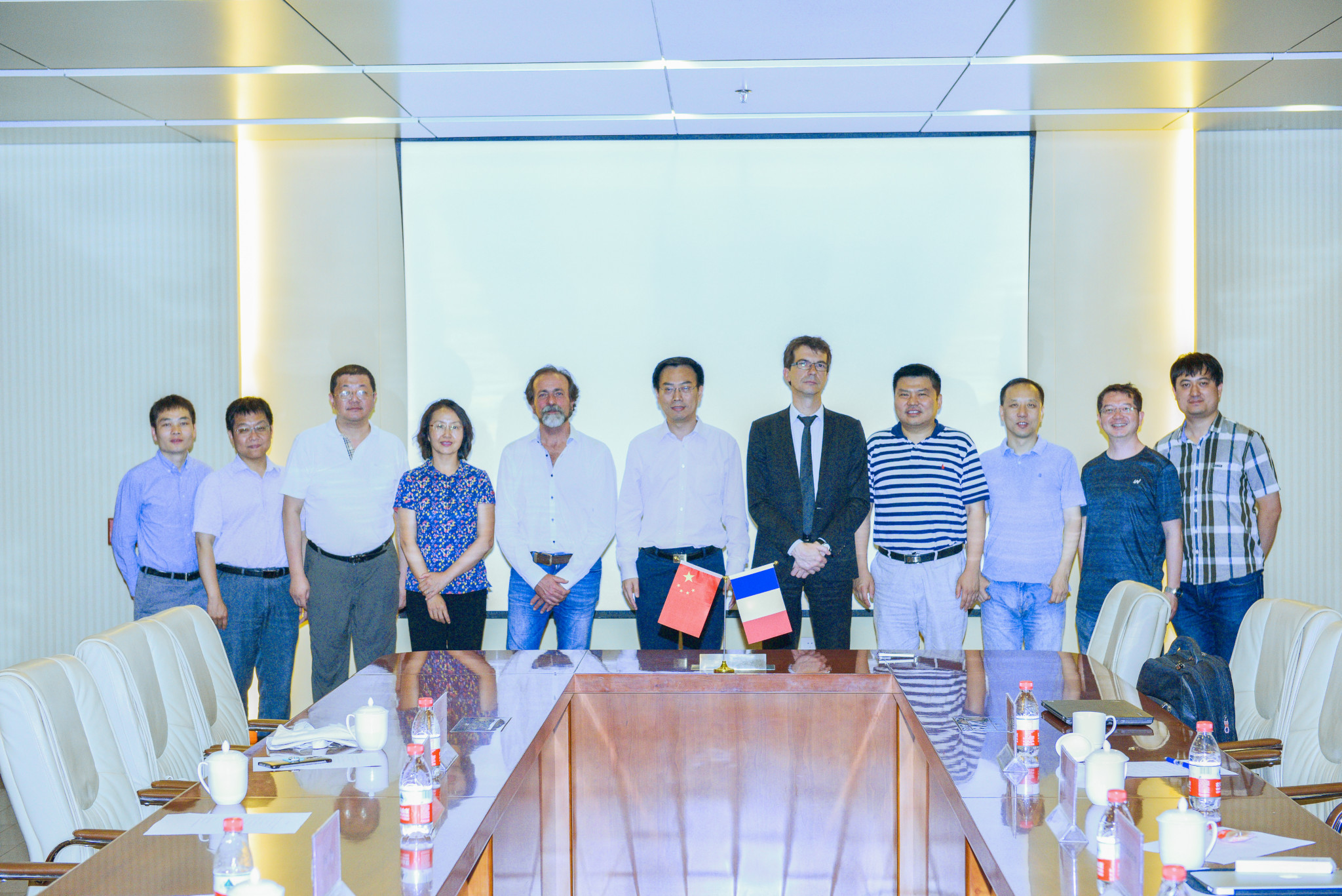
France BioImaging has initiated a partnership with the Institute of Biophysics of the Chinese Academy of Science and the Institute of Molecular Medicine of Peking University. The envisioned collaboration should be articulated around the following goals:
- To reinforce joint research activities and publications;
- To develop joint training activities for diverse categories of personnel, including imaging core facility staff;
- To exchange information and materials in those fields which are of interest to both parties;
- To organize joint conferences and academic programs;
- To develop grant proposals for joint research, infrastructure development (center and/or consortium);
- To foster technology transfer between each parties.
Several French institutions will take part in the partnership. We hope that the France BioImaging users and partners will be able to benefit from this partnership starting in 2018.
Electron microscopy PhD course
15th to 20th of October 2017, Core Facility for Integrated Microscopy, University of Copenhagen.
All details are available here: http://cfim.ku.dk/events/em-2017/
The course provides an essential grounding in the basic principles of electron microscopy, including electron optics, electromagnetic lenses, principles of transmission and scanning electron microscopy, electron sources, vacuum systems, specimen-electron interactions and diffraction. Biological specimen preparation will constitute a major part of the course, including methods of chemical fixation and cryo-preservation. Finally, advanced electron microscope techniques such as Cryo-techniques, immunogold labeling, electron tomography, and data analysis/visualization will be introduced towards the end of the course. The state-of-the-art facilities available at CFIM allow for a strong practical element, with time for each student to gain hands-on experience of both transmission and scanning electron microscopes. The course will be run by experienced microscopists in a relaxed atmosphere with the aim of promoting discussion and exchange of ideas between students and tutors.
The registration deadline is the 15th of September 2017.
We are pleased to announce the Coalition for Live Imaging Paris-Saclay (CLIPS) 2017 Symposium.
CLIPS 2017 is dedicated to highlighting innovative technologies in live imaging, spanning multiple scales from architectural and molecular microscopy to the modeling of biological processes. By probing and predicting the cellular state in a living organism, the aim is to unravel the processes underlying the building of a normal or a pathological state (adaptability, pathogenesis, therapeutic).
For its first edition, CLIPS 2017 also aims at gathering multidisciplinary scientists from the University Paris-Saclay and wider community to promote collaboration and innovation. Please join the round table, too (Tuesday September 12th, 5-6pm) just before the gala dinner!
Target audience: multidisciplinary scientists in the fields of live imaging
Are you a life-scientist needing to quantify microscopy images?
Do you want to learn how to co-localise, deconvolve, automate, track, handle massive data, and record work-flows?
If yes, this school is for you!
This one-week school provides a hands-on introduction to image processing and analysis, with an emphasis on biologically relevant examples. Participants will learn the fundamentals of image analysis, including macro programming in ImageJ/Fiji, MATLAB, as well as a range of focused topics What you will learn You will learn the fundamentals of image analysis, including basic macro programming in ImageJ/Fiji as well as other software solutions. In the first part of the week, it will also cover the process of image formation as it pertains to image analysis: Resolution, correct exposure, point-spread functions, detector noise, Shannon’s sampling theorem, and aliasing. All with a clear focus on application in the lab. In the second half of the week there will be a number of focused topics, building on what was learnt during the first days:
- Scripting in MATLAB
- Tracking particles and cells in time-lapse recordings
- De-convolution of microscopy images
- Stitching and registration of stacks of large image data
Deadline for application: August 15, 2017 at 12pm
The Executive Board of France-BioImaging has decided to support the following projects:
AAP Support to Events
- Workshop on Bioimage informatics, Rennes, June 27 2017 – by Thomas Walter (Curie Institute). Funded by FBI Coordination.
- Conférence « La microscopie à feuille de lumière », Villejuif, October 4, 2017 – by Laurent Combettes (Univ. Paris Sud). Funded by Ile-de-France Sud Node.
- Multiscale Live Imaging in Human, Animal and Plant Health Symposium, Gif-sur-Yvette, September 12-13, 2017 – by Sylvia Bruneau (INRA – Gif sur Yvette). Funded by Ile-de-France Sud Node.
AAP User Access
- Fusion, fission and maturation of early endosomes, by Xian Hu, University of Oslo. Hosted by Paris-Centre Node (Curie).
AAP Tech & Meth Transfer
- Claire Chardes (Université Aix Marseille – IBDM), “soSPIM experiments on celegans and drosophila embryos”. Hosted by Bordeaux Node.
- Ming Lei (Institute of Biophysics, Chinese Academy of Sciences), “Using live cell & superresolution imaging to study triptolide’s anti-tumor activity”. 2 months. Hosted by Paris Centre (PICT Curie).
The single-molecule localization microscopy symposium (SMLMS) 2017, is a 3 day conference being held at Guy’s Campus, King’s College London between August 30th and September 1st.
Registration is open until July 28th.
Abstract submission deadline: June 30th.
More information & registration: http://smlms.org/
This year, the Focus on Microscopy Conference took place in sunny Bordeaux, France, from April 9th to April 12th. France BioImaging participated in two ways: with a booth in the exposition hall, and through a symposium co-organized with Euro BioImaging, held on Wednesday 12th.
The France BioImaging Booth
Many curious attendees stopped by our booth to enquire about our activities. In particular, foreign visitors were curious to know how the infrastructure was organized, as they expressed the intention to set up similar services in their own countries. Industrials were equally intrigued by our organization, and expressed the desire to work with us towards the dissemination of their new available technologies.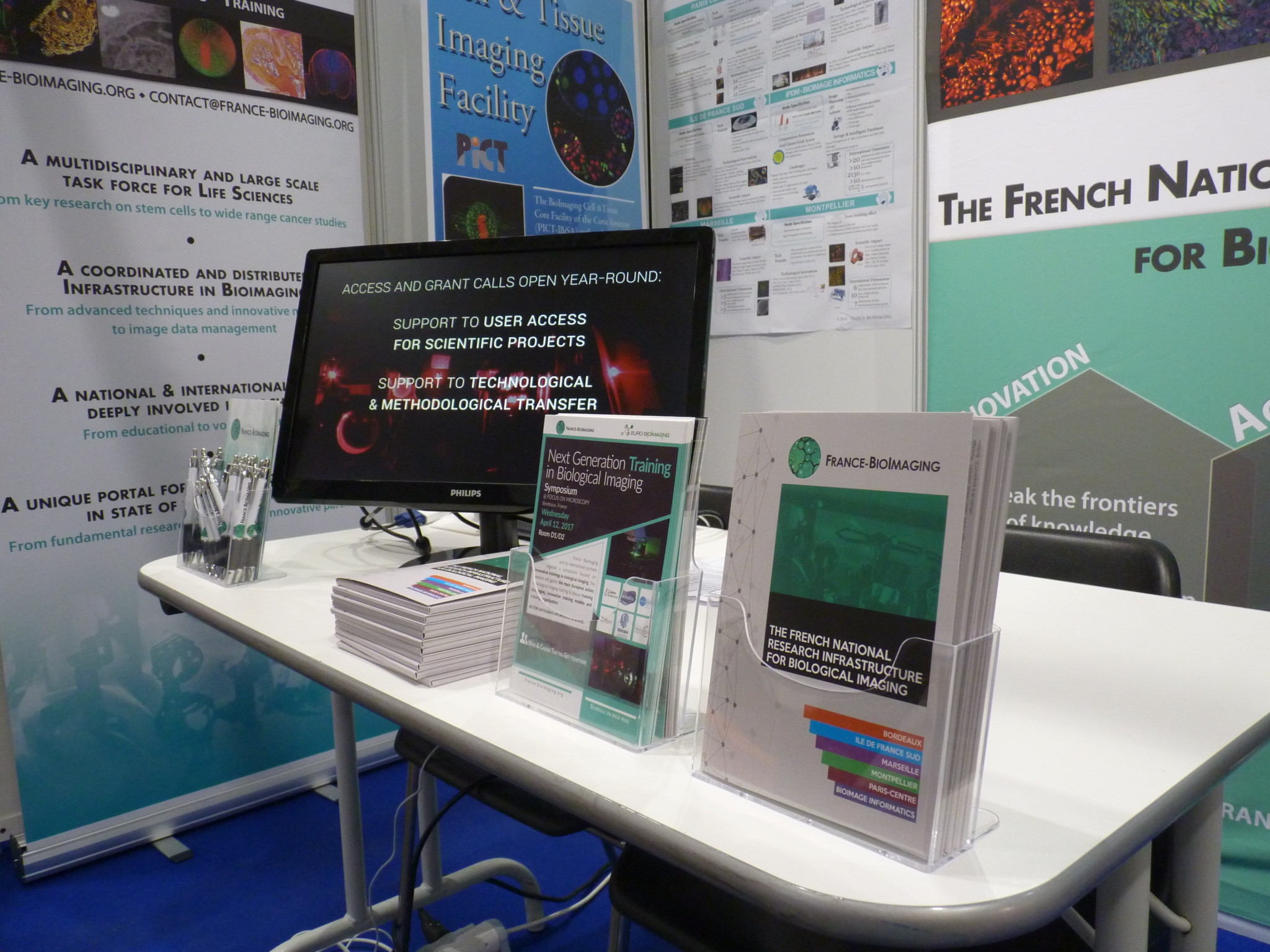

Volunteers from the Bordeaux Node gracefully assisted the coordination team in setting up & animating our booth.
Symposium on Training
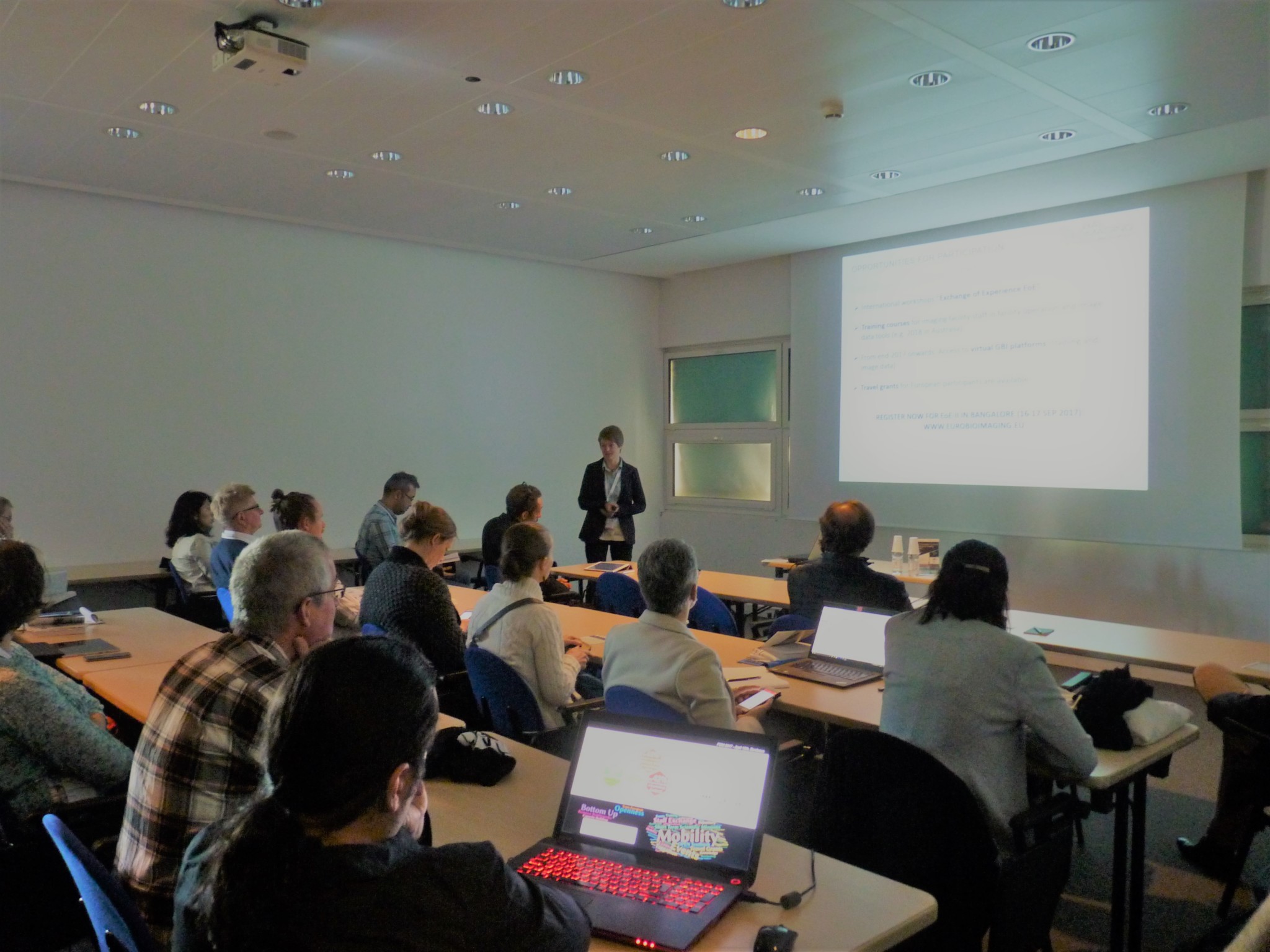 With more than 40 attendees, the Symposium on Training (Wednesday 12 April) was a great success. The public and speakers shared fruitful ideas about the past, present and future of biological imaging training at the European level.
You can find the program of the Symposium here.
We would like to thank the Focus on Microscopy organizers for their work in ensuring the success of the conference.
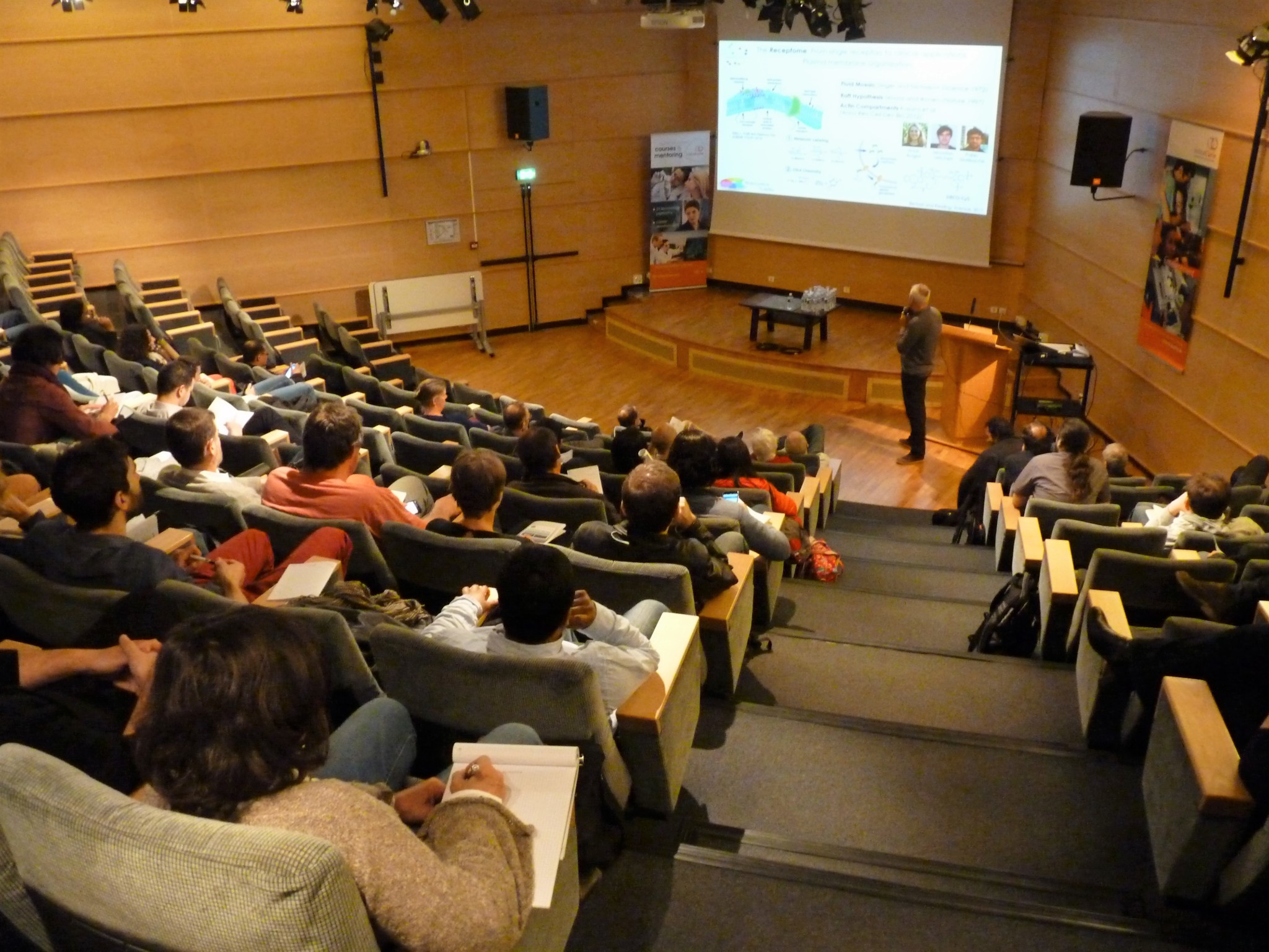
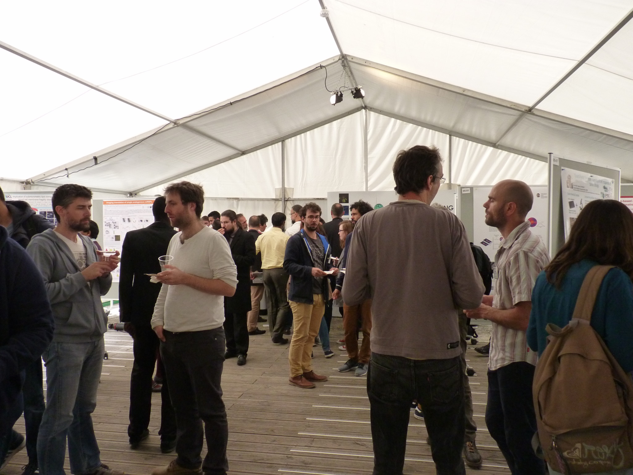
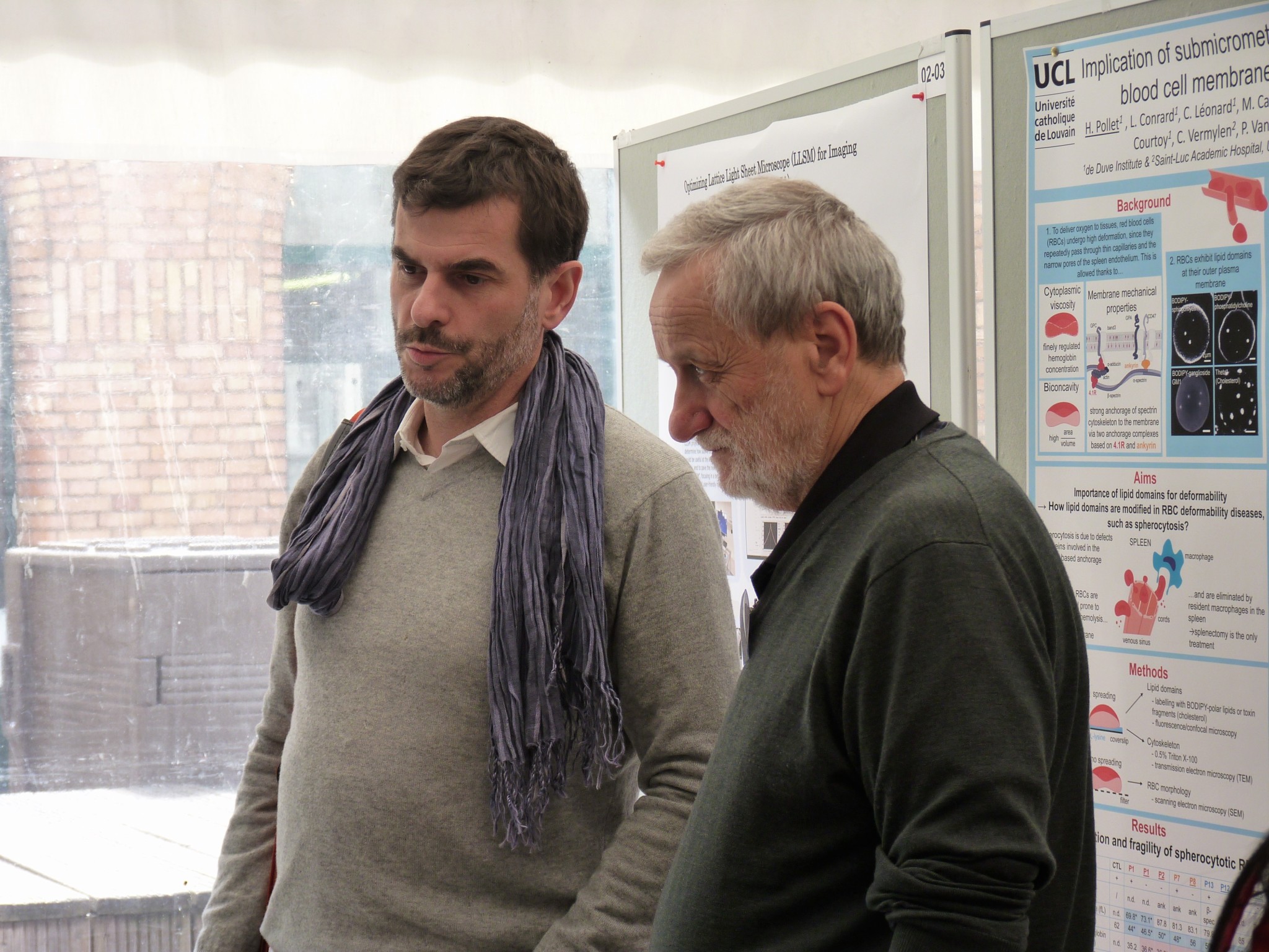

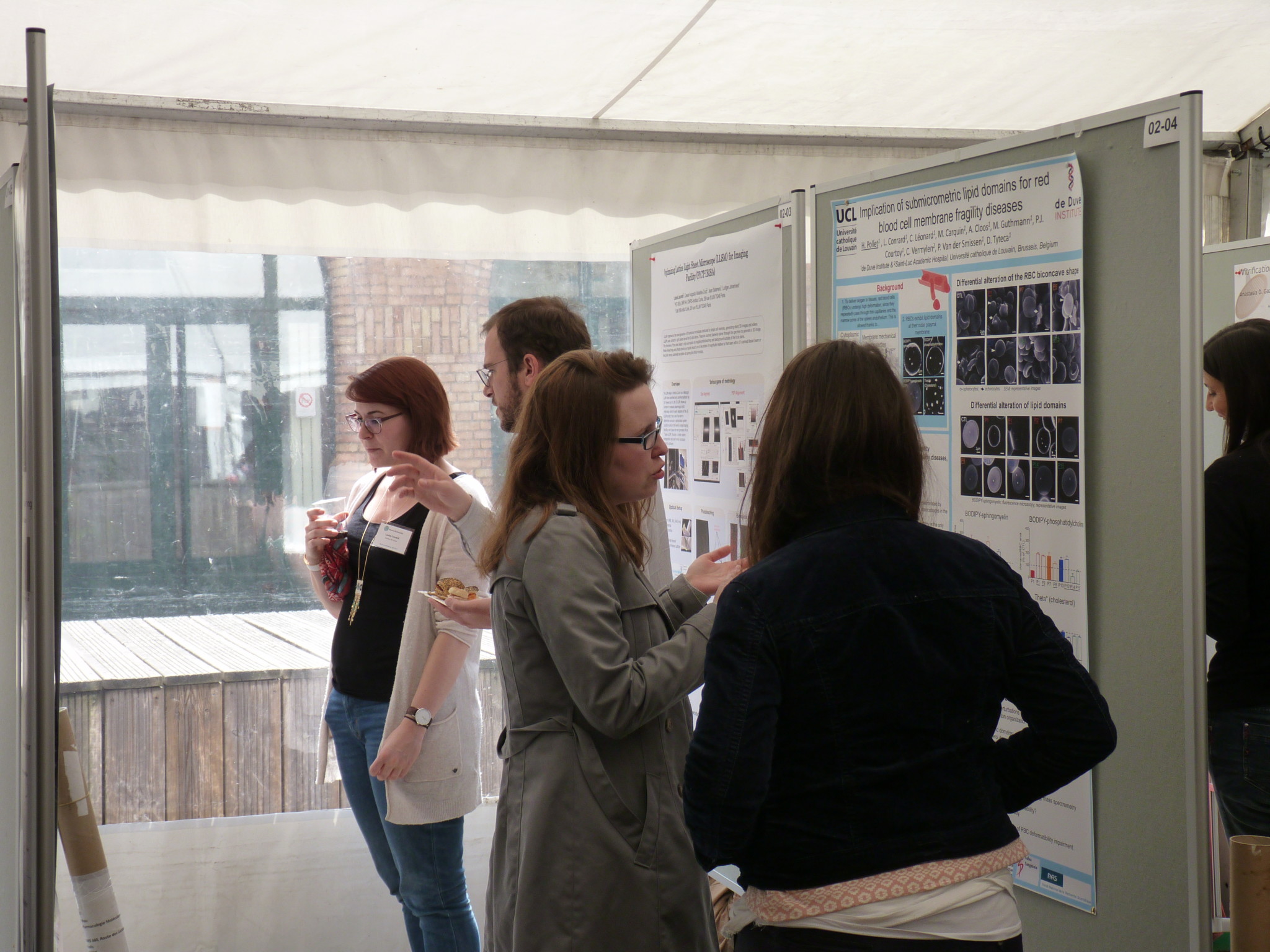
Our 4th Annual Meeting took place on Friday April 14th 2017. We had the pleasure of welcoming about 80 participants at the Curie Institute. This year, the meeting focused on the question of future challenges in biological imaging.
The four sessions of the program were centered around the following topics:
- Quantification of the molecular dynamics and coordination in cells and small organisms, including at the nanometer scale
- Imaging architectures and processes of life, from molecular complexes to multi cellular systems
- New frontiers for imaging, sensing, and controlling biomolecules
- Bioimage informatics, image processing and microscopy data management
At lunch time, the room was buzzing during the poster session.
We would like to thank our invited speakers, Teng-Leong Chew, Enrico Gratton, Markus Sauer & Jean-Christophe Olivo-Marin, as well as our chairs & all participants who shared their work with their peers through a talk or a poster.
Find all the details of the organization & program here.
We are happy to announce the 4th Mini-symposium on Bioimage Informatics, which will take place the 27th of June 2017 in Rennes. This event is organized by the GDR ImaBio and France Bioimaging.
The idea of this annual workshop on Bioimage Informatics is to bring together experts from the fields of image analysis and biology, to promote exchanges between the scientists in these fields and to contribute to the creation and maintenance of a French community in the field of Bioimage informatics, which is continuing in gaining importance in the field of Bioimaging. This year, the workshop is organized as a one-day satellite of the “Imaging the cell” conference, held in Rennes the 28th-30th of June 2017 (http://www.atoutcom.com/imagingthecell2017/).
Details & registration: http://gdr-miv.fr/en/events/bioimageinformatics2017/
Abstract submission deadline extended to May 26th
After 14 years, the Multinational Congress on Microscopy will again be organized in Croatia on September 24-29, 2017. In its 13th issue, the traditional conference series is returning to Istria, this time to the beautiful coastal town of Rovinj.
MCM2017 is jointly organized by 8 societies: Austrian Society for Electron Microscopy (ASEM), Croatian Microscopy Society (CMS), Czechoslovak Microscopy Society (CSMS), Hungarian Society for Microscopy (HSM), Italian Society of Microscopical Sciences (SISM), Serbian Society for Microscopy (SSM), Slovenian Society for Microscopy (SDM) and Turkish Society for Electron Microscopy (TEMD).
MCM2017 will bring together leading experts and young researchers that develop microscopy methods and apply them in the fields of life and material sciences. It will also include a trade exhibition in order to encourage exchange between the producers of microscopy-related equipment and researchers.
MCM conferences have always been an excellent opportunity for microscopists to exchange ideas and experience and to establish new cooperations and joint projects. Our aim is to provide an optimal balance between talks given by world-renowned scientists and a possibility for talented young scientists to introduce themselves to an international audience.
We believe this conference will be a highly rewarding professional and networking experience for all. Additionally, we encourage you to take this opportunity to explore the highlights of coastal town Rovinj with its beautiful surroundings and to experience the unique local blend of nature, culture and gastronomy.
NEUBIAS, the network for bioimage analysts, is organizing a second training school for facility staff. Training is open for all staff scientists, graduate students, post-docs or faculty who work in the context of Bioimaging facilities and provide assistance and training to users in need of Bioimage Data Analysis.
Topics, requirements, program and more: http://eubias.org/NEUBIAS/training-schools/staff/ts5-gothenburg2017/
Extended deadline for abstracts submission: May 26, 2017