This 2 day annual confocal course from the RMS utilises many different sample types and fluorescent probes (DNA stains, classic antibody labels and fluorescent proteins) which are chosen to best demonstrate particular problems and techniques. Focus is always on the techniques they enable and the problems they generate, which will be applicable to any sample types. The 2-days consist of short tutorials followed by hands-on practice.
Day 1 takes participants through the basic principles of confocal microscopy and then trains through hands-on practice how to configure and image multicolour, multidimensional samples using a confocal microscope.
Day 2 builds on the experiences of day 1 and enables participants to try FRAP and spectral profiling.
The series of Abercrombie meetings have been held since the death of Michael Abercrombie in 1979. Michael was a pioneer in the field of investigating cell behaviour using timelapse microscopy. Abercrombie meetings are held only every five years and therefore offer an excellent opportunity to review the major advances in our understanding of cell motility and look to the new emerging concepts in the field.
The previous 7 Abercrombie meetings have been extremely successful and have attracted significant numbers of attendees from the world-leading labs in the adhesion, migration and cytoskeleton fields to present their work in an extremely interactive and collaborative environment. The meeting provides an excellent platform for open and constructive discussions between researchers from world-leading labs focused on complementary areas of study using different types of methods and approaches from advanced microscopy to computational modelling.
The 8th Abercrombie meeting will be held from 11-14th September 2017 in Oxford and will address the key, exciting new findings and emerging approaches in the study of cell migration across a range of biological contexts, both in vitro and in vivo. Abstract Submission is currently open and will close on 20 July 2017.
The programme is not yet available but you can get an idea of the meeting from the 2012 programme displayed here.
NEUBIAS, the network for bioimage analysts, is organizing a second training school for early career reserchers. This year, training is open for all researchers interested in acquiring knowledge about image analysis, with no prerequisite required.
Topics, requirements, program and more: http://eubias.org/NEUBIAS/training-schools/training-school-2-bias-ecis/ts4-gothenburg-sept-2017/
L’objectif de cette formation est de donner aux ingénieurs des plates-formes d’imagerie cellulaire et aux personnes qui utilisent ces technologies, des outils statistiques adaptés aux problématiques du domaine. Dans le but de traiter les grandes séries d’images acquises selon les diverses modalités, il est proposé de renforcer la maîtrise des outils statistiques pour une analyse pertinente mais aussi pour une optimisation des acquisitions.
La formation se déroulera du lundi midi au mercredi après-midi pour 2 demi-journées de cours (rappel des notions de bases, modélisation, plans d’expériences) et 3 demi-journées de travaux pratiques/dirigés.
Préinscriptions en ligne jusqu’au 12 Mai à :
https://www.vjf.cnrs.fr/limesu
The course will be an introduction to image processing and analysis, with a focus on biologically relevant examples. The attendees will learn the fundamentals of image analysis including how to do basic Macro programming in Fiji (ImageJ) for automated batch analysis of images, use different software solutions for image analysis, and will be introduced to visualisation and explorative data analysis after extraction of numerical data from images.
The teachers are Simon Nørrelykke and Szymon Stoma (IDA-ScopeM, ETH, Zurich, Switzerland), and Chong Zhang (SIMBioSys, University Pompeu Fabra, Barcelona, Spain).
The course is suitable not only for beginners in image analysis with no experience, but also for those who want to extend their knowledge of basic principles and more specialised tools.
The Executive Board of France-BioImaging has decided to support the following projects:
AAP Support to Events
- Training on Metrology in photonic microscopy, Rouen, November 7-10, 2017 – by Damien Schapman, Université de Rouen, ANF. Funded by Bordeaux Node.
AAP User Access
- Direct visualization of the post synaptic density, by Nicolaj RIIS CHRISTENSEN (University of Copenhagen), hosted by Bordeaux (IINS, BIC).
- Phosphorylation serving as a regulator of Arc function on AMPA-receptor trafficking, by Maria Steene ERIKSEN (University of Bergen), hosted by Bordeaux (IINS, BIC).
The Correlated Multimodal Imaging Node Austria (CMI) is the official Austrian Euro-Bioimaging initiative of leading imaging experts in Austria. It was established as a consortium of eight universities and research institutes in and around Vienna (www.bioimaging-austria.at) to foster and pioneer correlated multimodal imaging across scales in life sciences. CMI offers more than 35 state-of-the-art imaging technologies from preclinical imaging to atomic resolution, numerous multimodal correlated imaging pipelines, and various support services, such as data and image analysis, to researchers on a national and international level.
This year’s Imaging in the Life Sciences Meeting is organized by CMI and hosted by the Ludwig Boltzmann Institute for Experimental and Cinical Traumatology.
Please register by May 5th at www.bioimaging-austria.at/web/pages/cmi-imaging-meeting.php. Please use the same link to submit your poster abstract.
En juillet prochain (4 au 7 juillet 2017) sera organisé à Bordeaux, le 15ème colloque de la Société Française des Microscopies. Le comité d’organisation a le plaisir de vous accueillir, à nouveau, 30 ans après le colloque de 1987, dans la cité culturelle et historique de Bordeaux, classée au patrimoine mondial de l’Unesco. Bordeaux vit actuellement un renouveau extraordinaire avec l’impressionnante transformation des berges de la Garonne, l’inauguration de la Cité du Vin, et la future ligne LGV. Ce sera en 2017, l’une des villes les plus « attractives » du monde sur le plan touristique.
Ce congrès a pour objectif de réunir une large communauté de scientifiques autour du thème des microscopies électronique, optique et à champ proche et imagerie vibrationnelle. Le comité scientifique a construit un programme attractif avec 15 sessions couvrant les applications et développements en science de la vie, science des matériaux, instrumentation et les méthodes. Les symposiums communs et les sessions en parallèle Matériaux et Biologie permettront de faire un point notamment sur les avancées dans les domaines de la caractérisation des matériaux à différentes échelles, de la matière molle, de la superrésolution, de la cryomicroscopie haute résolution, de la microscopie corrélative….
Le congrès se déroulera à l’école d’ingénieur Bordeaux-INP ENSEIRB-Matméca à Talence, accessible facilement depuis le centre-ville de Bordeaux par la ligne de Tram B. Dans ce lieu convivial, les participants et exposants auront la possibilité d’échanger librement. L’exhibition d’instruments, de techniques et accessoires sera importante. De plus, en partenariat avec l’école d’été du GN-MEBA, des microscopes de pointe pourront même être accessibles pendant la période du congrès.
Apres le vif succès de Nice en 2015, nous vous encourageons chaleureusement à soumettre vos contributions pour poursuivre cette dynamique. A très bientôt.
The 3rd Course on High Content Screening and Image Analysis for Biosciences is a comprehensive and intensive course that aims to provide the basis of image-based screening. It is designed to graduate students, early career investigators and everyone willing to enter in the multiplex era. The course includes lectures given by specialists in the field and hands-on sessions with HCS instruments and open-source software for image and data analysis.
Cryotechnologies in electron microscopy
The FBI Marseille node is composed of the PICsL light and electron microscopy core facility and three associated research and development (R&D) labs. The FBI-PICsL facility is located on the Marseille Luminy campus and hosted by two institutes: the Centre d’Immunologie de Marseille Luminy (CIML) and the Institut de Biologie du Développement de Marseille (IBDM). The R&D labs comprise two labs of these institutes (Lenne lab at IBDM and Marguet lab at CIML) and the biophotonics lab of the Fresnel institute (Mosaic group led by H. Rigneault). The FBI-PICsL facility via its microscopy resources is dedicated to help its users deciphering cellular mechanisms in the fields of cell biology, immunology and developmental biology.

In 2017, the FBI-PICsL facility comprises 40 setups. Altogether, these microscopes cover a wide range of temporal and spatial scales from the second to milliseconds and from single molecules to entire organisms. This repertoire of microscopes is composed of cutting-edge light microscopes (FCS, FCCS, STED 3D, PALM/dSTORM, Fast polarization-resolved spinning disk, Light sheet microscopes) among which 8 are homemade and 3 electron microscopes (two TEMs and one SBF-SEM). Ten staff members manage the facility under the scientific supervision of Pierre-François Lenne (IBDM) and Didier Marguet (CIML). The FBI-PICsL facility staff is also involved in teaching microscopy techniques to the facility users and within courses open to external academic and industrial users.
New set-ups on the FBI-PICsL facility
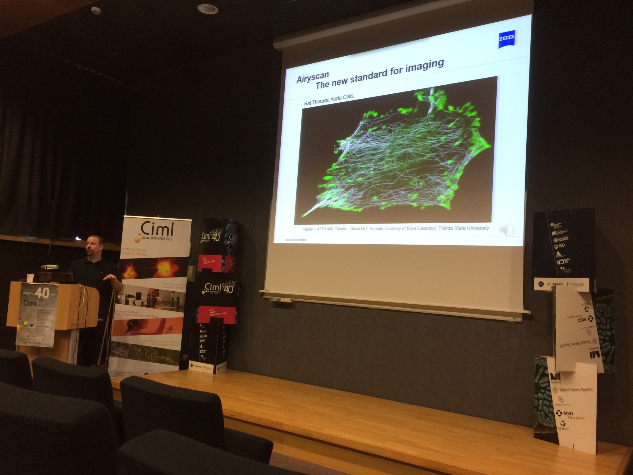
LSM 880 Fast-Airyscan

A new confocal microscope using the technology Airyscan (LSM 880 Fast-Airyscan, Zeiss) has been purchased through a co-funding FBI/Inserm/CNRS/Region PACA. The Airyscan improves spatial resolution (1.7 x) by imaging the Airy disk onto a concentrically arranged hexagonal detector array. Its detection area consists of 32 single detector elements, each of which acts like a very small pinhole. The confocal pinhole itself remains open and doesn’t block light – thus all photons of the whole Airy disk are collected. The signals from all detector elements are then reassigned to their correct position, producing an image with increased signal-to-noise ratio and resolution.
FEI Teneo VS
The FBI-PICsL facility also recently homed an electron microscope dedicated to automated 3D imaging.
Revealing the complex 3D architecture of cells and tissues is crucial for the structure-function correlation in biological systems. However, in most of the electron microscopy experiments, only a small portion of the sample is imaged. For instance, one 70nm-section of an average vertebrate cell only represents 0.4-1.0% of its volume. Beyond the fact that this tiny volume might not be representative to account for the entire cell ultrastructure, the sectioning also avoids a definite visualization of the cellular organization.
Until recently, the imaging of big volumes (tens to hundreds of cubic micrometres) by electron microscopy was tedious because it involved manual serial sectioning. Beyond the need of super-skilled people, manual serial sectioning has a limited z resolution of 70nm and is a time-consuming process at the sectioning, imaging and post-processing steps. Within the last 10 years, the Serial Block-Face Scanning Electron Microscopy (SBF-SEM) has emerged as the method of choice for the imaging of big volumes by electron microscopy.
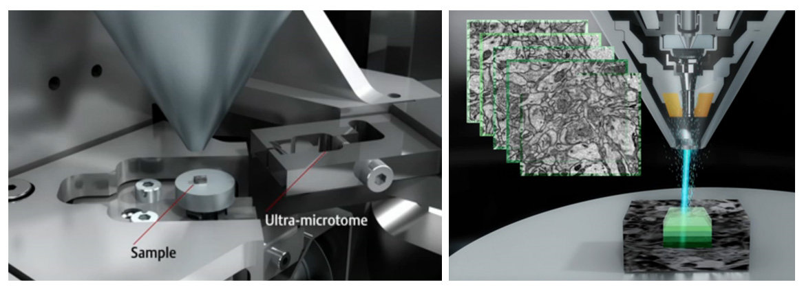
The concept behind SBF-SEM is to have a microtome within the chamber of a scanning electron microscope (see Figure). This set-up enables the iterative sectioning and imaging of the sample block-face for hundreds of microns in a fully automated way (Figure). When an electron beam hits a sample, it interacts with it and yields several signals that can be detected and assigned back to the scanned positions on the sample surface. Among these signals, the backscattered electrons can be collected to retrieve information on the sample electron density at a given point. Routinely, a working pixel size in SBF-SEM datasets is ±5nm and the slicing can be as thin as 20-30 nm.
In addition to be an excellent microscope in classical SEM mode, the Teneo VS microscope by FEI is currently the most advanced microscope to carry out SBF-SEM. It does not only allow you to cut and image the sample in a fully automated way but it also gives the possibility to avoid charging artefacts during imaging by putting the sample in low vacuum conditions. By limiting the charging, the microscope gives you the opportunity to retrieve more information from the sample. For instance, the sample can be scanned with different acceleration voltages in order to retrieve ultrastructural information at different depths (10nm – 20nm – 30nm – 40nm). When this multi-energy imaging is combined to a 40nm mechanical slicing, it gives rise to previously inconceivable datasets of hundreds of cubic microns of tissue with a voxel size of 5x5x10nm !
Since the delivery of the FEI Teneo VS microscope at the FBI-PICsL facility in December 2016, we tested the microscope on a broad range of biological samples in classical SEM mode (fly egg-laying apparatus, Xenope embryo, mouse face…) and in SBF-SEM mode (cell pellets, Drosophila muscle, mouse brain, marine sponges, Xenope embryo…). In particular, the group of Laurent Kodjabachian (IBDM), working on multiciliated cells, wanted to image the same cell type in both classical SEM mode and in SBF-SEM. We could 1. assess the penetrance of a mutant phenotype in classical SEM mode and 2. localize the basal bodies of these multi-ciliated cells in SBF-SEM (see Figure below).
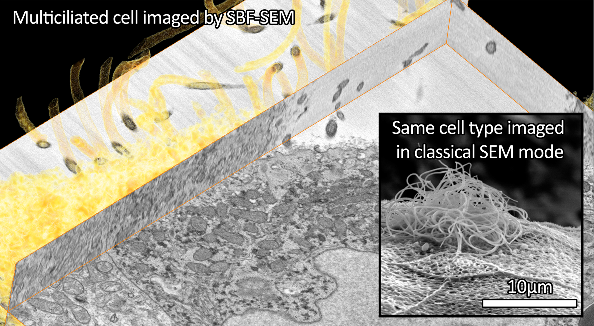
Photo credits: Virginie Thome and Nicolas Brouilly
Major implications of the FBI-PICsL facility
Spot variation fluorescence correlation spectroscopy (svFCS) (Hai-Tao He and Didier Marguet’s team)
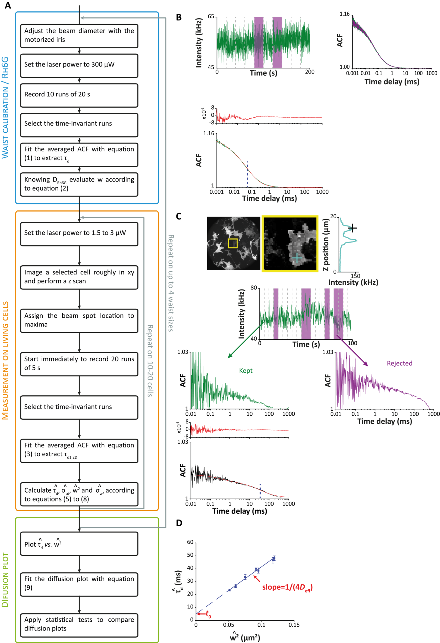
In collaboration with Nicolas Bertaux (Institut Fresnel, AMU, Centrale Marseille), members of the Hai-Tao He and Didier Marguet team published a user guide for characterizing plasma membranes subdomains in living cells by sv-FCS2. This book chapter aims to serve as a guide for setting and applying the svFCS methodology to study the plasma membrane of both adherent and nonadherent cell types.
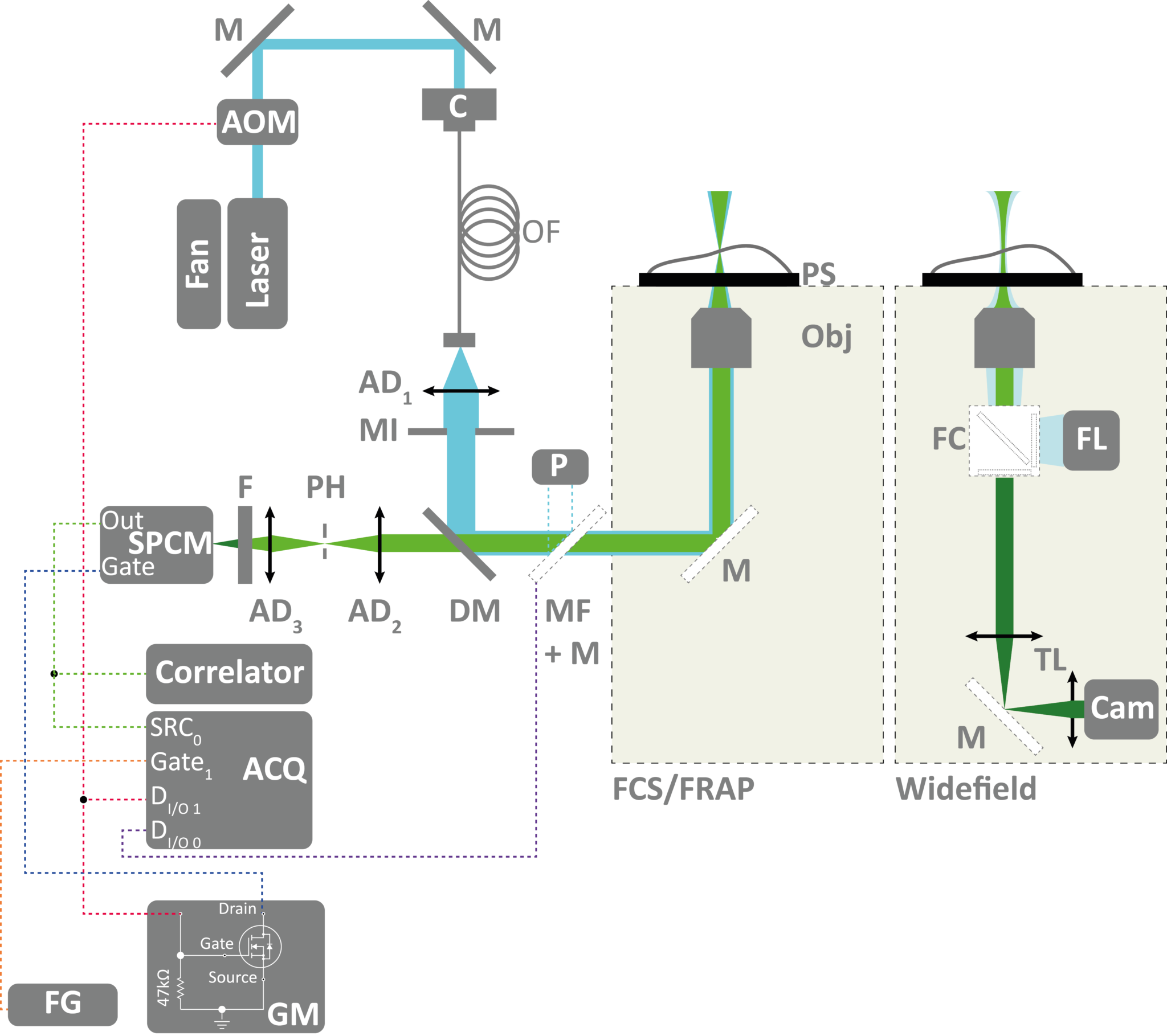
recovery after photobleaching (FRAP).
(Click to view the image in full size)
This technique has been used to understand a genetic disease by studying molecular diffusion at the plasma membrane. In collaboration with Christophe Lamaze (Institut Curie) and Céline Galès (Institut des Maladies Métaboliques et Cardiovasculaires de Toulouse), Hai-Tao He and Didier Marguet’s team worked on decoding the dysfunction of molecular mechanisms involving interferon-γ (IFN-γ), key protein for immune defense. This protein is not able to fulfil its natural function as a protective cytokine when its receptor is “blocked” in the wrong place on the cell membrane: a modification to the immune response causing severe infections sometimes even life-threatening for young children.
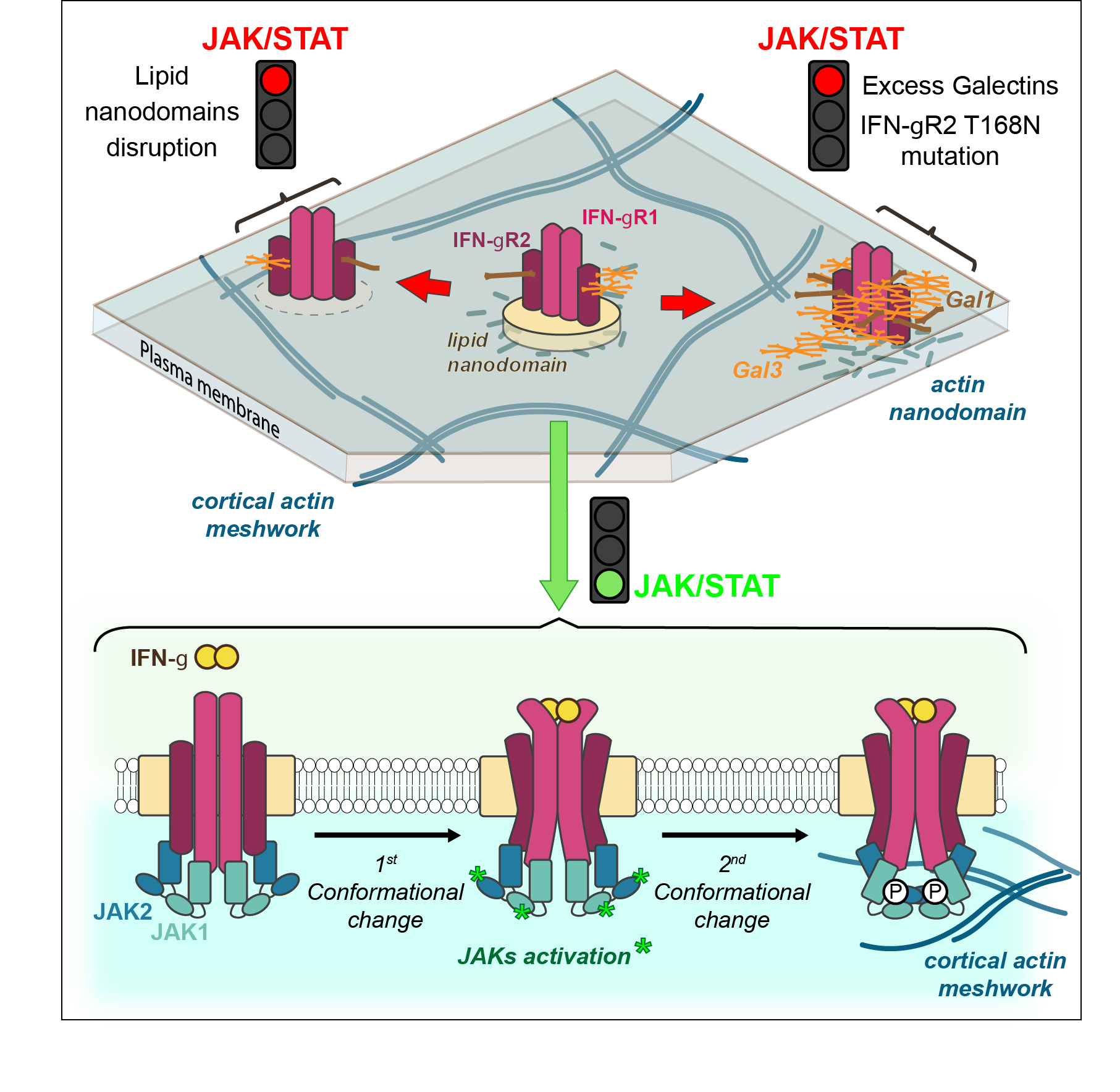
Lipid and Actin Nanodomains Is Critical for JAK
Activation
IFN-γ receptor is composed of two protein chains to which are generally associated six sugars. However, some patients diagnosed with Mendelian susceptibility to mycobacterial diseases syndrome (MSMD) have seven sugars. This simple gain of glycosylation affects greatly the immune response against mycobacteria.
Using their expertise in biophysics and more particularly in svFCS, this CIML team showed the existence of nanodomains and their fundamental role in signaling activity. “It was completely unexpected that only one additional sugar could modify the way the receptor is localized on the surface of the cells, affecting greatly the later signaling events”, explained Hai-Tao He. “IFN-γ receptor is then delocalized towards another membrane nanodomain populated by galectin proteins, that bind to this additional sugar” added Yannick Hamon2 (cf Figure)
As therapeutic target, galectins are an appealing perspective, particularly as turning off their expression canceled in vitro the pathologic effect linked to the glycosylation gain of the IFN-γ receptor. However, therapeutic programs towards patients requires the exploration and understanding of molecular mechanisms by uniting expertise of biologists and physicians but also mathematicians and physicists as it was the case in this discovery4.
News
In March 2017, a workshop on super-resolution is organized in Marseille by Sébastien Mailfert, Jean-Bernard Fiche and Orestis Faklaris on multicolor STORM an dSPT-pointillism data analysis, this workshop is dedicated to advanced researchers in this field. This event is supported by FBI.
The FBI-PICsL facility is also homing the 14th RTmfm convention from the 20th to the 22nd of March 2017. This convention is a great opportunity for imaging facilities staff to share their experiences and ideas on the past, present and future of their work (technical development, big data issues and solutions, link with industry).
Our facility also yearly organizes the following course: Scientific platform, instrumental sharing, how to build up and develop a service. The aim of the course is to acquire, by alternating courses, exercises and practical conditions, methods and constraints to the development of a provision of scientific equipment service. We insist on how to develop a contributory model for managing a scientific platform.
References
- Mailfert, S., Hamon, Y., Bertaux, N., He, H-T. & Marguet, D. Methods in Cell Biology, 139, 1-22 (2017)
- Blouin, C., Hamon, Y. et al. Cell, 166, 920-934 (2016)
- http://www.cell.com/cell/fulltext/S0092-8674(16)30909-6
The RMS Light Microscopy summer school is an annual, residential three day course held at the University of York covering the principles of light microscopy. Participants are also trained in practical issues surrounding light microscopy. After introductory presentations, the course is taught predominantly through hands-on practical sessions.
The course is suitable for both novices and more experienced users wanting to gain a greater understanding of the microscope and feedback every year is always fantastic. Students came from a range of backgrounds in 2016, within both research and commercial organisations. All benefited greatly from the Course and left with increased understanding and skills.