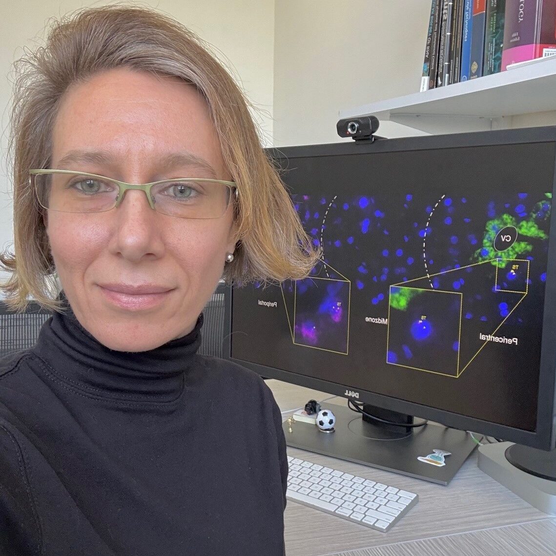
We are delighted to introduce Carolina Eliscovich, one of the awardees of the “FBI Call for User Access Projects 2024.” Carolina was granted access to one of the France-BioImaging facilities to advance her research using cutting-edge imaging technologies.
In this interview, she shares insights into her scientific journey and her current project, which explores how mRNAs are spatially organized and regulated in the liver of a living organism using advanced RNA imaging techniques.
To start, could you tell us a bit about yourself? What has been your academic journey so far, and what is your current role or area of work?
I am originally from Buenos Aires, a cosmopolitan and multicultural city in Argentina; and it is in this city located in the southern region of planet Earth where my academic journey starts inspired by the legacy of multiple local Nobel Laureates in Biomedical Sciences surrounded by yerba mate drinking and fútbol. Encouraged by my high school teachers to follow my curiosity to understand how living things function, I studied and graduated in molecular biology at the Faculty of Exact and Natural Sciences of the University of Buenos Aires.
Shortly after that, I moved to Europe to continue my scientific formation and completed a PhD in Biological Sciences at the Pompeu Fabra University and Centre for Genomic Regulation in the vibrant city of Barcelona, Spain. My PhD thesis work, under the mentorship of Dr. Raúl Méndez, focused on the study of how maternal mRNAs encoding for proteins required for spindle formation and chromosome segregation are locally translated by the RNA-binding protein that promotes cytoplasmic polyadenylation to ensure proper meiosis progression in Xenopus oocytes.
I subsequently crossed the Atlantic Ocean back again but this time I headed to the North, to the greatest New York City in the USA, where I joined, as a postdoctoral fellow, the laboratory of Dr. Robert H. Singer at Albert Einstein College of Medicine. During my training in his lab, I acquired unique professional expertise in RNA biology by using single-cell and single-molecule fluorescence microscopy technology. We developed a robust methodology, named super registration, using single-molecule fluorescence in situ hybridization in combination with immunofluorescence (smFISH-IF) to determine the physical interaction between individual mRNA molecules and their binding protein(s) within single neuronal cells using standard wide-field microscopy. As a result of a successful collaboration with other colleagues in the lab, we also developed a novel imaging technology to visualize and quantitate the mechanism of translation of single transcripts in real time in living cells.
I have always been interested in the cell biology of mRNAs since my first days at the lab bench; and now my lab also at Einstein expands on the study of these versatile macromolecules by watching them within cells in tissues. Most of the knowledge we have about RNA regulation comes from studies in cells in culture, but little is known about how RNAs behave in the native context of the tissue under physiologic conditions, and this is the knowledge we would love to contribute to the field.
What are you currently working on in your research? What is the main topic or challenge you’re exploring?
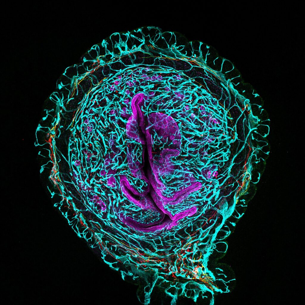
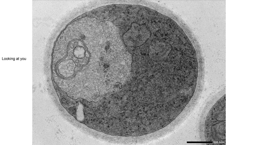
I recently became interested in the biology of the (mouse) liver; and my current research program at Einstein focuses on the development of RNA imaging technology using fluorescent-labeled probes to visualize the spatial organization of mRNAs in the healthy and regenerating liver. The architecture of the liver is unique and what I find fascinating the most is that gene expression of the hepatocytes-the dominant cell type in this organ- are spatially regulated depending on their distance to the vasculature.
This organ is also remarkably resilient and capable of regenerating itself upon a variety of injuries or surgical removal of a significant portion of the organ mass. This process has biological and clinical relevance because it is the foundation of liver transplants and, unfortunately, organ transplantation is the only treatment for liver failure. What we found is that during this incredible regeneration response, hepatic gene expression is dynamically reprogramed, and therefore, it is an ideal model system to study mechanisms of mRNA regulation in time and space in a living organism.
A current ongoing challenge we face in our studies is to examine individual cells not only at tissue-scale but also with single molecule sensitivity while preserving tissue morphology. Also, and importantly, high-throughput and multiplexing has been always challenging in imaging-based technologies due to a limited number of different fluorophores that can be simultaneously detected because of spectrally overlapping wavelengths. Luckily, we were able to overcome these difficulties thanks to the France-BioImaging available technology!
At what point did you come across France-BioImaging, and what made you want to use its services or connect with the infrastructure?
The France-Bioimaging infrastructure came along in my research thanks to Dr. Edouard Bertrand (Institute of Human Genetics, Montpellier, France). I had the privilege of working with Edouard at Einstein while he was doing a sabbatical year in the Singer lab a few years ago. Later, he introduced me to the mission of the infrastructure and how the state-of-the-art imaging technologies and resources were bringing to all investigators across France that could not access the equipment otherwise.
I remember that I felt a bit jealous of the platform; my lab was located overseas, how would I get access to the France-BioImaging facilities that I needed?
And the answer came last year when France-BioImaging launched for the first time the call for external users (both national and international). It was a great opportunity to finally gain access to the platform and get training on the technology that was missing in our studies: to visualize, simultaneously, multiple individual mRNA molecule species in the intact mouse liver tissue during regeneration. And I submitted my application.
Could you walk us through your experience accessing France-BioImaging? Which facility did you work with, how did the process go, and what stood out to you during your time there?
The experience at the France-BioImaging facility was incredible!
My application included access to sequential FISH methodology developed by Dr. Davide Normanno, postdoc in the Bertrand lab, thus, I visited the Bertrand laboratory and the Montpellier Resource Imaging (MRI) facility in the Institute of Human Genetics in Montpellier for two weeks. Before my arrival to Montpellier, we worked virtually on the design of the fluorescent probes, protocols and details on sequential FISH methodology and equipment. Everything was planned before my arrival.
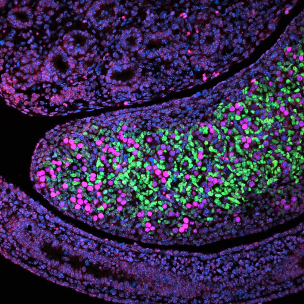
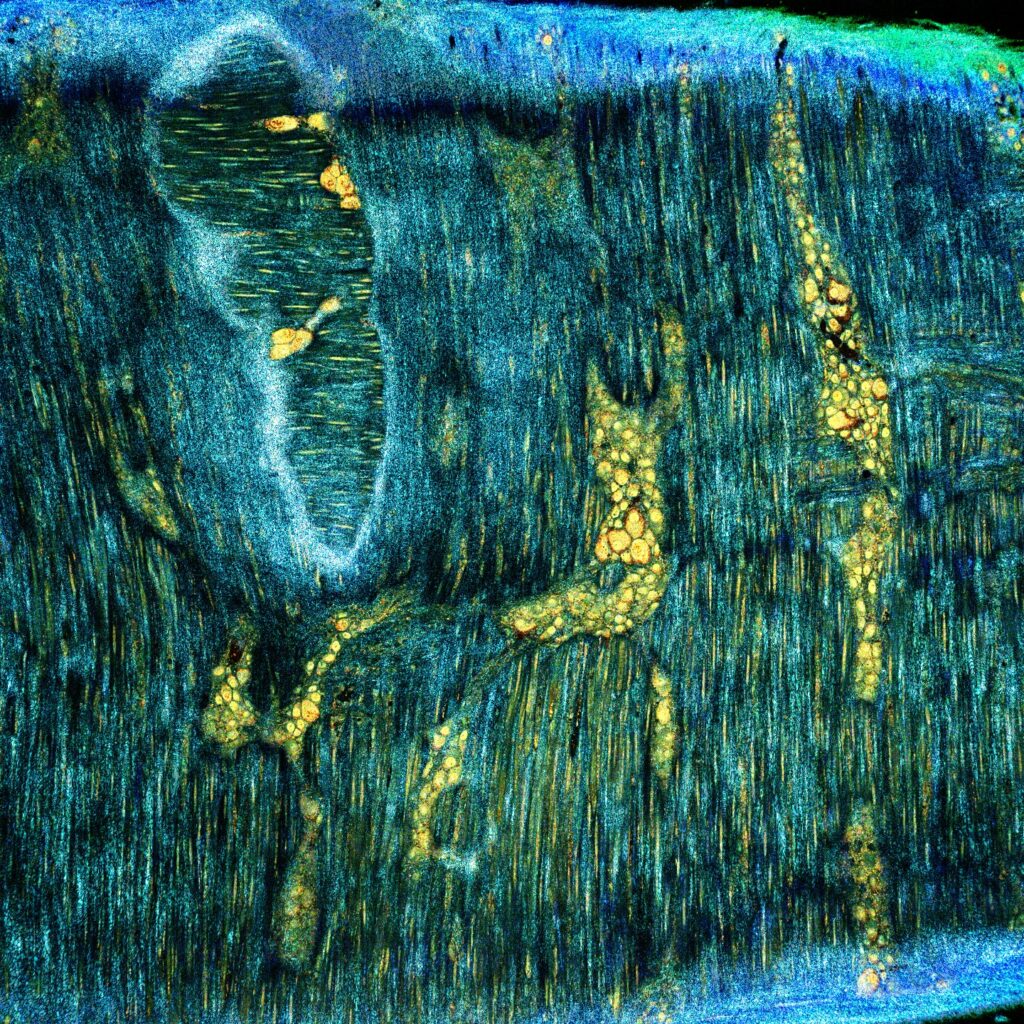
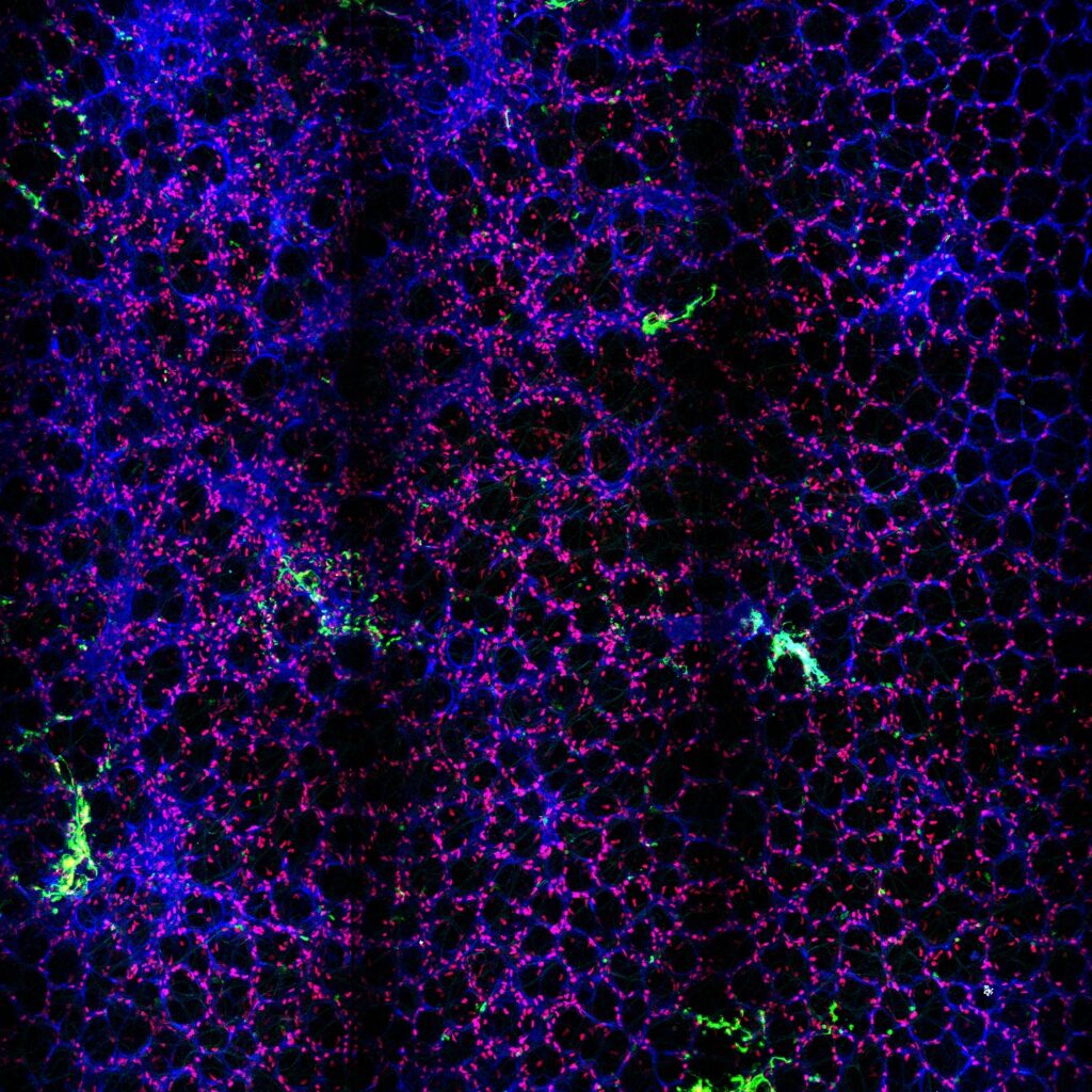
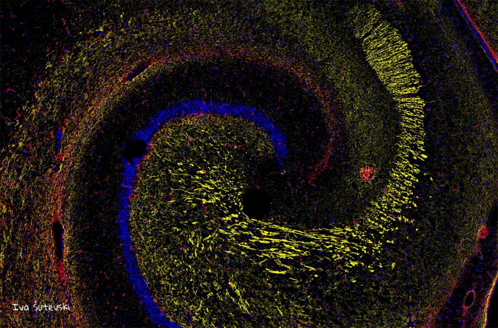
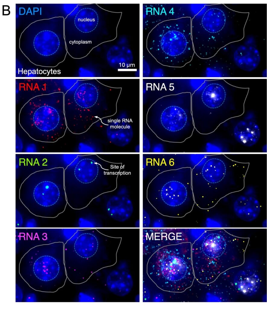
But life is certainly filled with unexpected events and on my very first days in Montpellier, Davide went suddenly on a medical emergency that required surgery and was not able to be in the lab the subsequent days*; and it was thanks to the collegiality and generosity of the investigators at the France-Bioimaging infrastructure that I was lucky to meet and count on Drs. Marcelo Nollmann and Jean-Bernard Fiche at the Center for Structural Biochemistry, just across the street, who rapidly jumped in and helped to finish the training on the sophisticated automated microscope with a microfluidic perfusion system to perform sequential FISH. We were running against the clock and against all odds our sequential FISH pilot experiment on liver tissue worked beautifully and we were able to visualize, for the first time, multiple mRNAs within hepatocytes in the intact liver tissue during regeneration, something we could not have done without accessing France-BioImaging. Thanks to all!**
I will never forget how I felt while looking at the first images we took; it was one of those unique moments in the lab where we are fortunate to see the unseen.
*Davide recovered well, and he is currently healthy and working in the lab.
**I am deeply thankful to Edouard, Davide, all the people in the Bertrand lab, Marcelo and Jean-Bernard for their kindness and generosity during my visit. I could not have done anything without them!
What did microscopy bring to your project specifically? Were there insights or results you couldn’t have obtained otherwise?
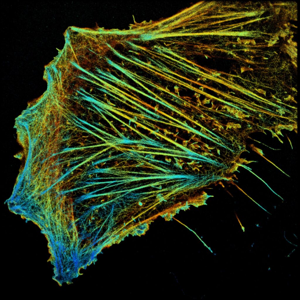
My lab is, in essence, an RNA imaging lab. Single-cell and single-molecule microscopy allow us to use the power of quiet observation and understand what every single cell is doing in its preserved microenvironment within the tissue, giving us insight about the heterogeneity present in a biological sample that is usually masked in other methodologies.
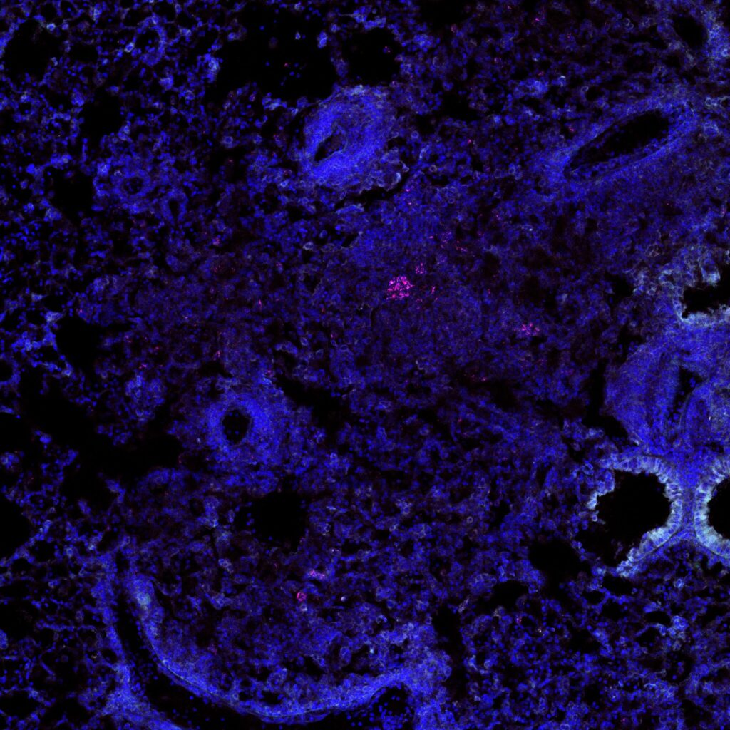
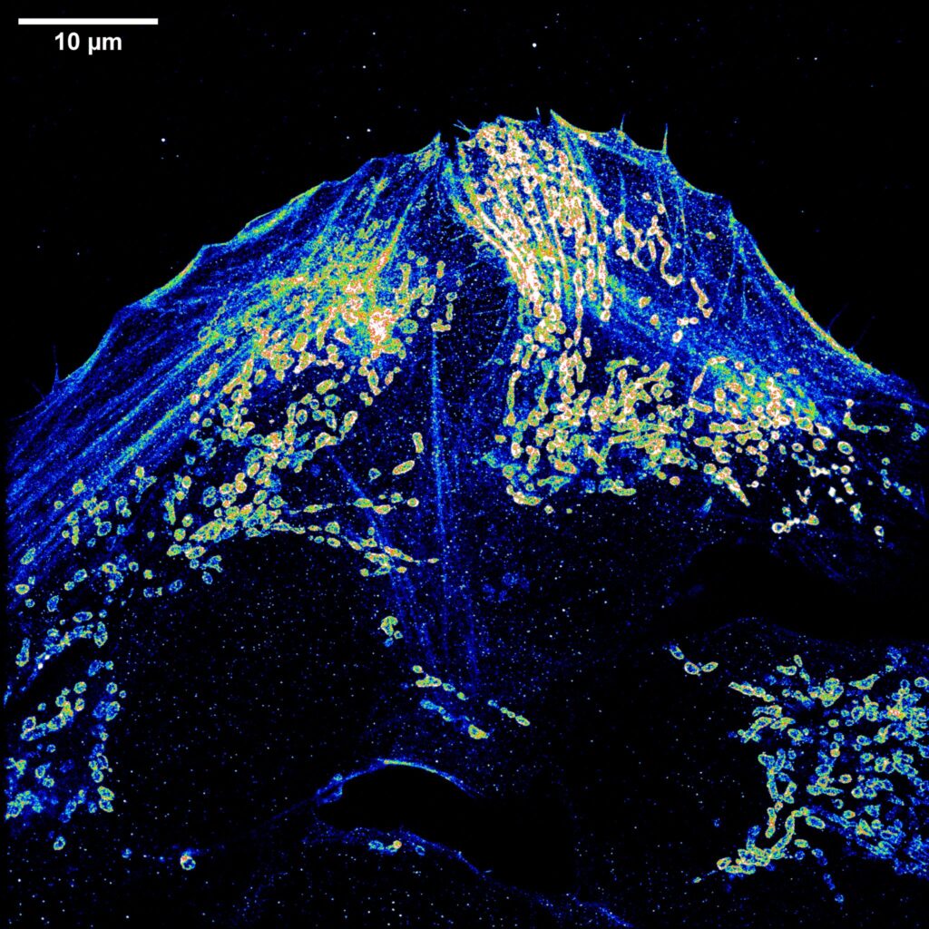
Even though we perform standard smFISH (up to 2-3 different RNA species) in liver tissue in our lab in a routinely manner, we could not have been able to advance the method to image multiple RNA species (multiplexing; up to 16 different RNA species) and in a high-throughput way (by the use of an automated microfluidic system) without accessing to the France-BioImaging facility at the MRI-IGH in Montpellier.
Sequential smFISH imaging technology applied to liver tissue allows us to study the simultaneous expression of multiple mRNAs within the same hepatocyte giving us unprecedented gene expression information of a regenerative liver cell in vivo.
The analysis of these images will give us understanding about mechanisms that the liver may employ to segregate different functions along the hepatic lobule and shed a light on the co-expression of genes that have been always thought not to be expressed at the same time in the same cell. This will allow us to explore in depth the relationship between tissue morphology and the molecular state of the cells and, therefore, cell function in vivo.
Looking back, would you encourage other researchers to use France-BioImaging’s platforms and access program? What would you say to someone considering it?
I would encourage everyone in the scientific community from students and postdocs to principal investigators to look for opportunities to access the France-BioImaging facilities. The first thing I would say to someone considering this is that they will never regret the experience because it is a great chance to learn and test the feasibility of a new imaging technology and/or methods applied directly to their specific research question, something that could take a lot of time (and funds) if they do not have the technology already established in their home institutions.
The France-BioImaging facility nodes include a great variety of state-of-the-art microscopes and imaging technologies available that I am sure each researcher can find the proper match for their scientific needs.
Finally, this access program would eventually help them to establish collaborations with the experts in the host lab/institution and even lead to the opportunity to reimplement the technology in their home labs or institutions where it would serve as the foundation for future directions of research in the upcoming years.
If I had the opportunity to apply to the France-BioImaging call for user access program again, I would do it without a doubt!