France-BioImaging in Beijing – Strengthening Sino-French cooperation in bioimaging
Category: Announcement
We were honored to represent France-BioImaging in Beijing during the Third Sino-French Joint Meeting on BioImaging, held from November 4 to 6, as part of the 30th anniversary celebrations of the CNRS China Office.
This landmark event gathered nearly 100 scholars, researchers and industry representatives, including a delegation of 10 French experts and over 20 leading Chinese specialists led by CAS academician Cheng Heping. The meeting was co-hosted by the Biophysical Society of China (BSC), the CNRS International Research Network for BioImaging (IRN BioImage), the CAS Institute of Biophysics, the Beijing Laboratory of Biomedical Imaging, and the CNRS China Office.
Fostering scientific cooperation through technological innovation
Under the theme “Innovation in Biomedical Imaging Technologies and Facility Development”, the forum featured:
plenary talks and scientific workshops,
technical sessions on advanced multimodal imaging,
facility visits of the National Biomedical Imaging Center (NBIC), showcasing China’s research infrastructure dedicated to biomedical imaging.
We explored major advances in MINFLUX, SIM, STORM imaging, correlative light and electron microscopy, miniature two-photon microscopy, in vivo neuronal activity recording, tissue clearing and data visualization, while also exchanging experience on the construction and operation of imaging centers.
Specialized training sessions combining theory and hands-on practice enabled Chinese participants to translate innovation into real research capabilities, ashared priority for the IRN BioImage community.
Strengthening strategic partnerships
Within the broader CNRS strategy in China, this mission reaffirmed the central role of the IRN BioImage as a structuring instrument for long-term scientific cooperation. By uniting leading research infrastructures, training initiatives, and technological development efforts in bioimaging, the IRN is not only advancing joint research but also building a shared vision for future large-scale scientific partnerships. As part of France-BioImaging, we see this network as a strategic driver, enabling France and China to jointly shape the next generation of biomedical imaging innovations, facilities and expertise.
Looking ahead: Bordeaux 2026
The meeting concluded with the announcement that the Fourth Sino-French Joint Meeting on BioImaging will take place in Bordeaux, France, in October 2026, a milestone we are proud to help deliver.
For France-BioImaging, this mission was a powerful opportunity to deepen our shared research ambitions, strengthen our international network, and accelerate innovation in multimodal bioimaging.
A big thank you to the organizing teams, our Chinese partners, and all CNRS and France-BioImaging colleagues who contributed to these rich exchanges and to the strengthening of our cooperation.
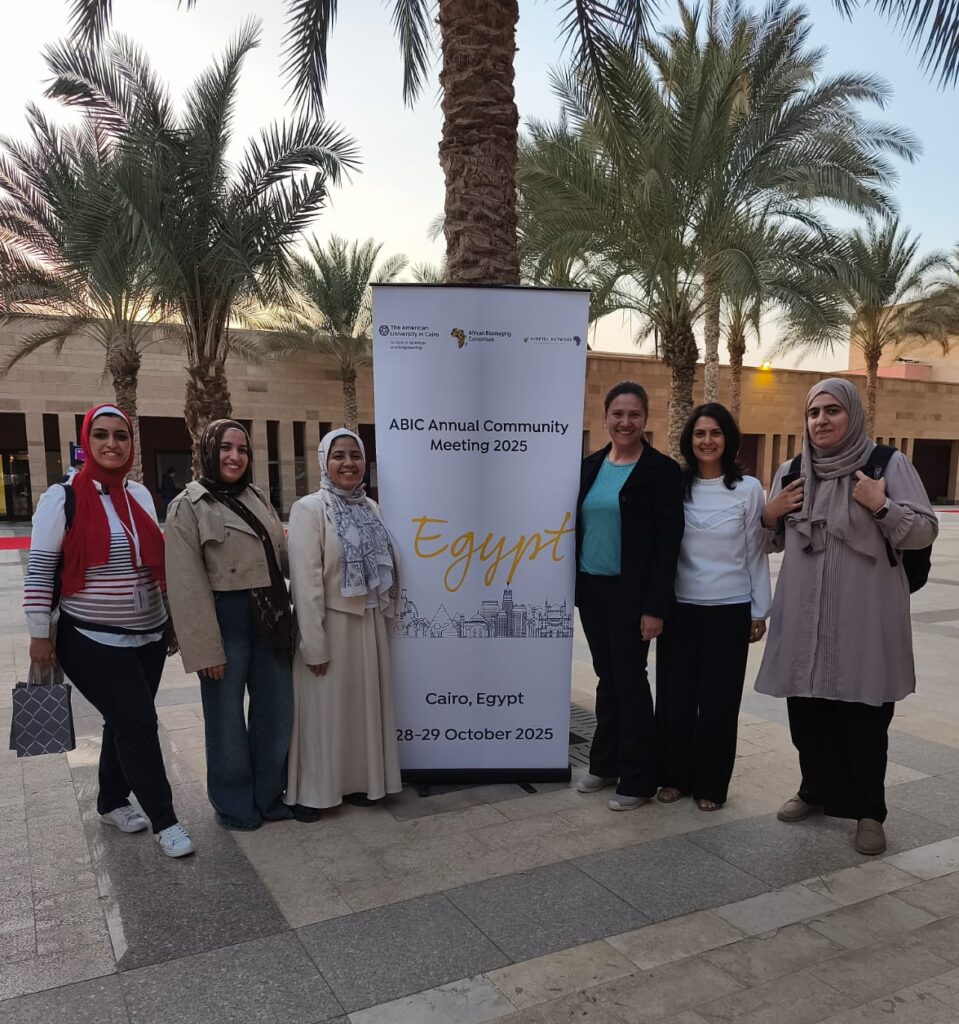
Last month, Samira Benadda, Head of the core imaging facility at IBENS (France-BioImaging Paris Centre node) was invited to the 4th Annual ABIC Meeting, held in Cairo and bringing together the African community specialized in biological imaging. Samira co-coordinates the Africa division of France-BioImaging with Jean-Luc Verdeil, Researcher at CIRAD. The event highlighted the Africa-France initiative, supported by France-BioImaging, which is committed to promoting and strengthening access to imaging technologies through expertise sharing, training, and the development of sustainable collaborations.
Working closely with ABIC, Samira presented the activities and achievements of the Africa division, including the grants obtained by Global BioImaging which have enabled the hosting of African researchers within the France-BioImaging nodes, facilitating access to imaging technologies for both research projects and professional training. The meeting also provided a major opportunity to discover and better understand imaging infrastructures in Africa, their needs, their organization, and the prospects for collaboration. Exchanges with local platforms emphasized the importance of supporting capacity building and fostering broader access to advanced imaging technologies.
This participation has thus contributed to strengthening existing partnerships and encouraging the emergence of new collaborations.
France-BioImaging and all the French community aims to develop and promote innovative imaging technologies and methods. But microscopy images can also take an artistic, creative look and make the invisible world beautiful, allowing people to see the visual appeal of the life sciences.
We enjoyed the diversity of the images submitted with many different microscopy techniques, models and applications represented. A big thank you to all the participants!
The National Coordination Team and the Executive Board are proud to announce the winners of the FBI Image Contest 2025:
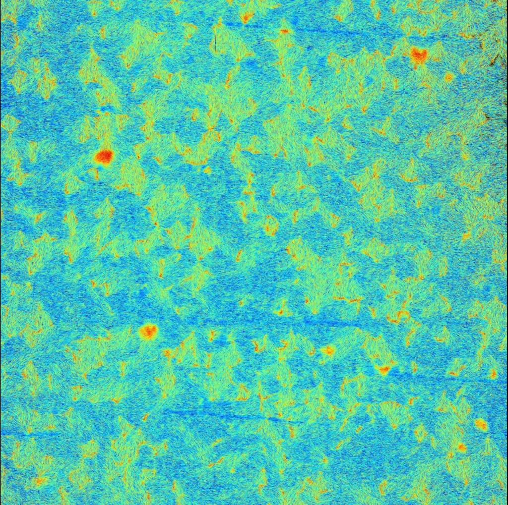
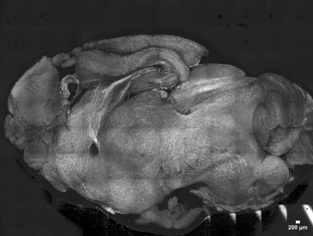
1st Place: Nicolas Barois, BioImaging Center Lille (BICeL)
Gut Flower-Flora
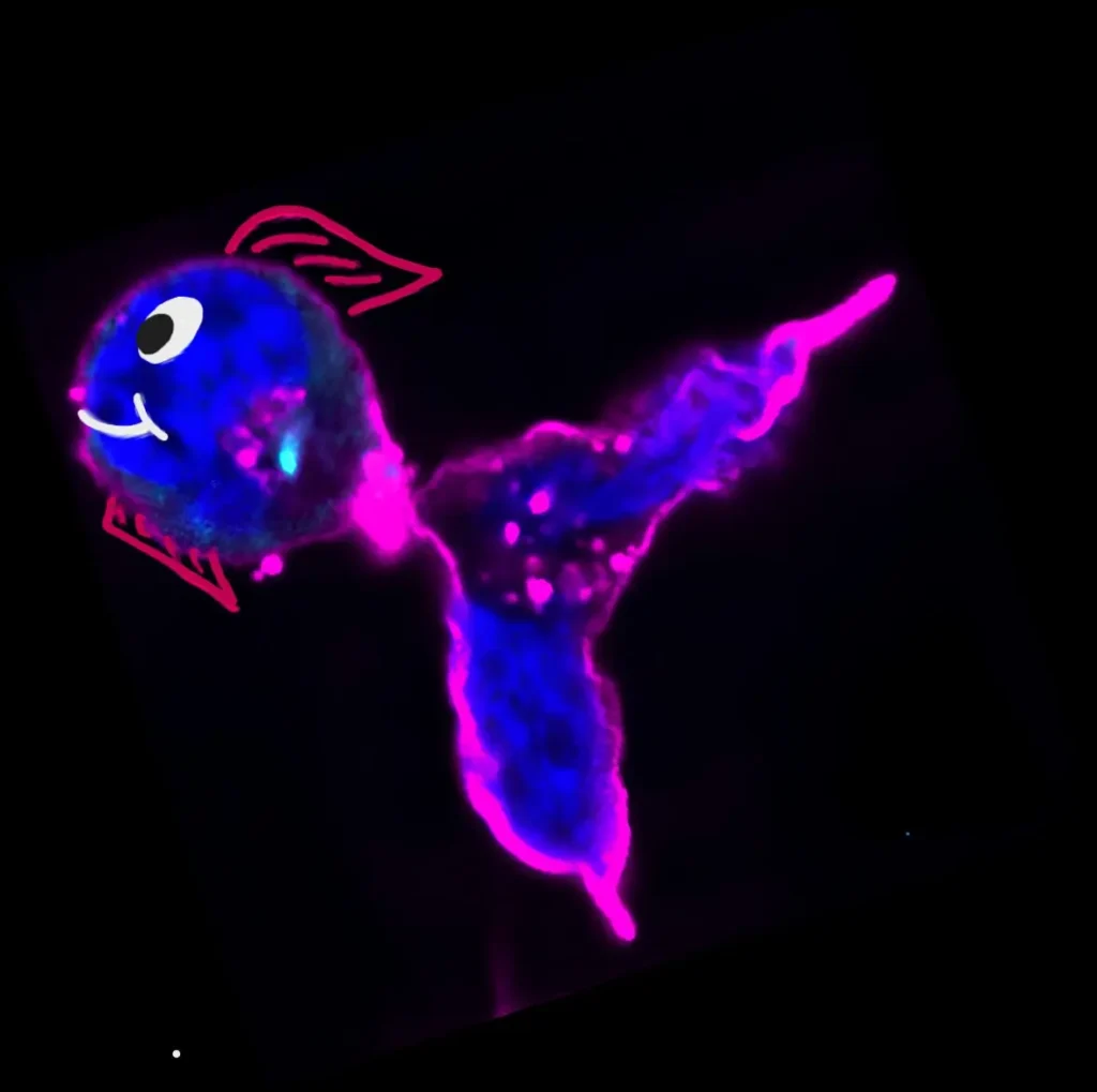
Cross-sectional view of a thin slice of mouse gut. By cutting a thin slice of the gut, the inner went out. Normally I cut the gut in small tubes, which are cut in two longitudinal pieces. I kept this piece because I was curious to see it with the SEM. Scanning Electron Microscopy
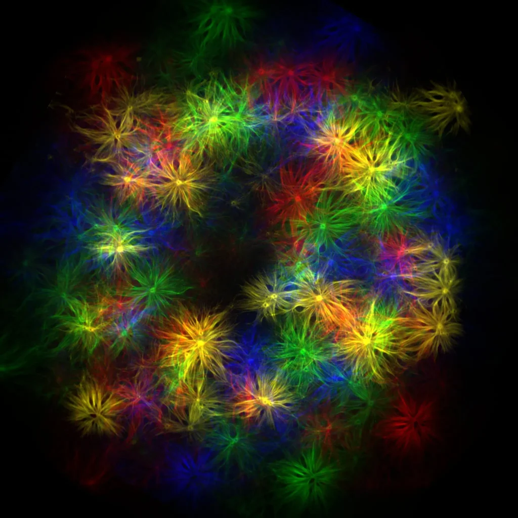
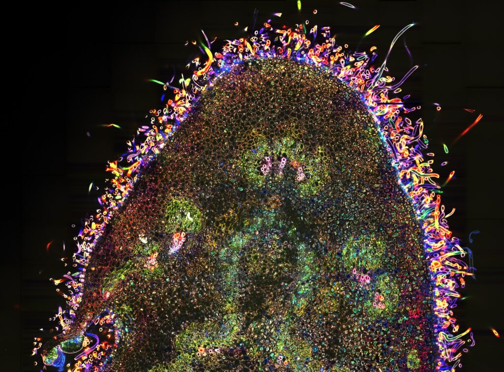
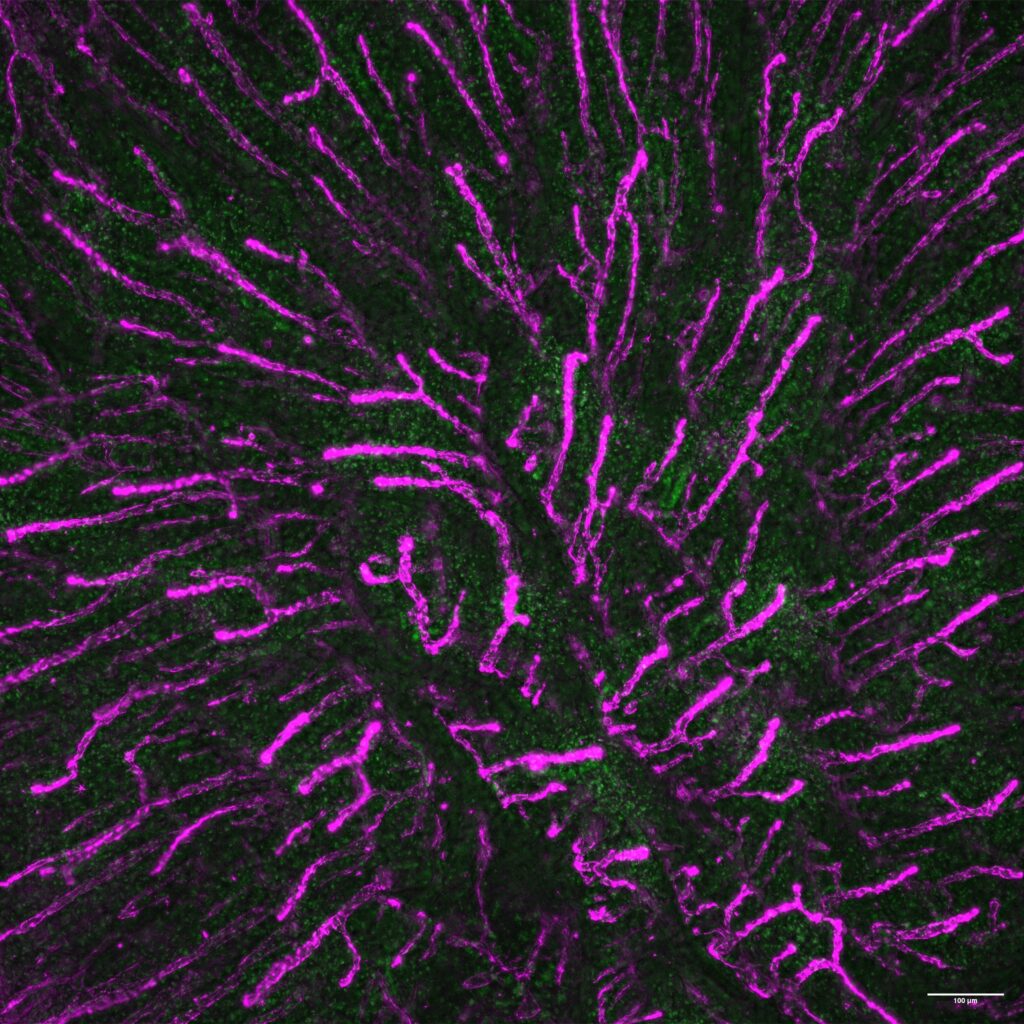
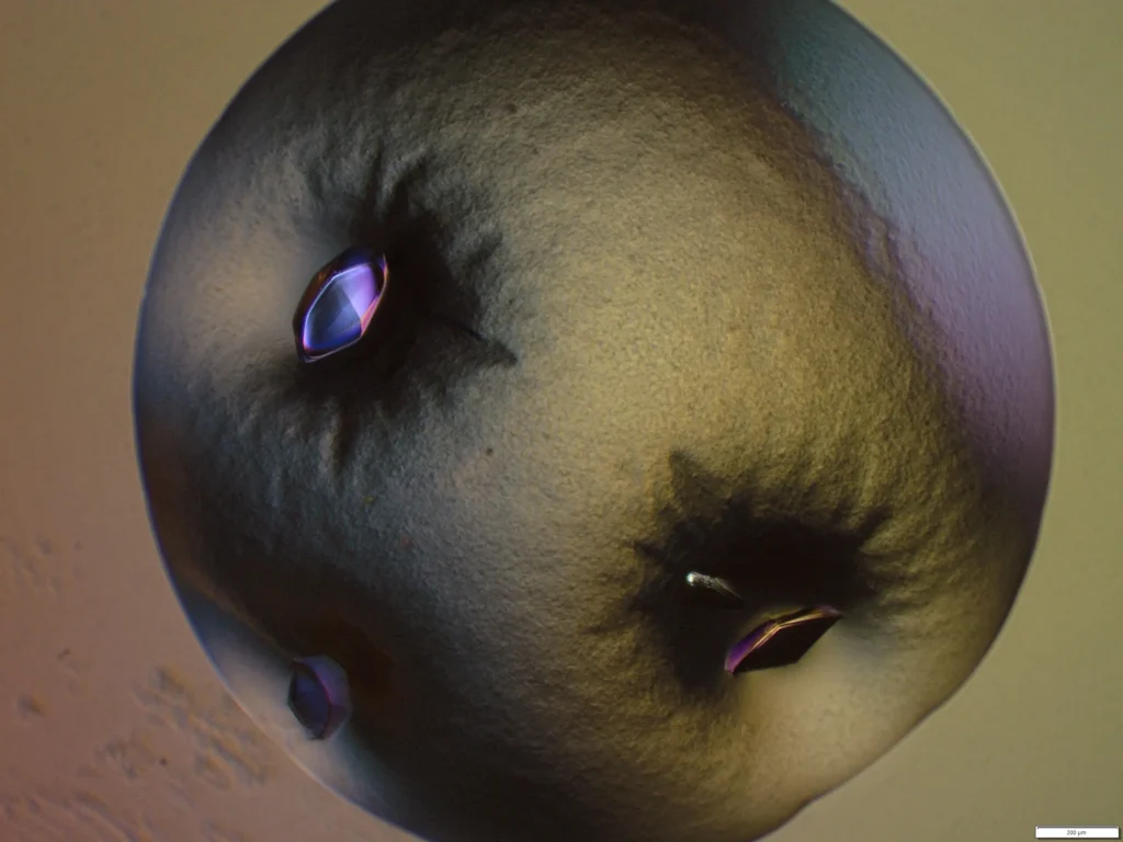
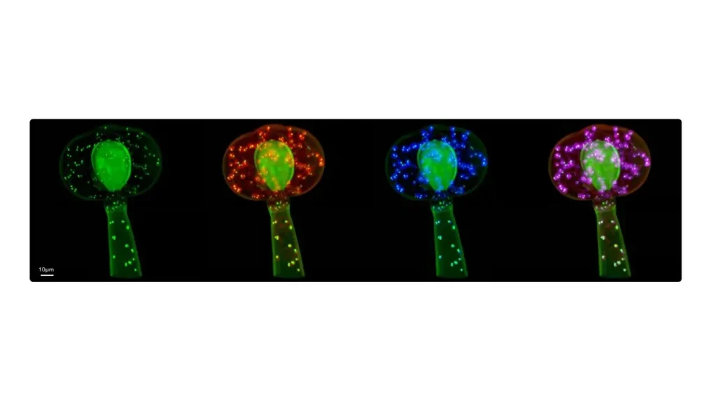
2nd Place: Vishwadeep Mane, Plant Reproduction and Development Laboratory (RPD, ENS Lyon)
The Puzzled Awakening
Cotyledons, the first leaves of a plant, break free from the seed to launch life after germination. Emerging as a pair, they unfold into a nearly perfect, circular lamina that captures light for photosynthesis. At the microscopic scale, their surface reveals a mosaic of interlocking puzzle-shaped cells, dotted with stomata. These intricate cell shapes are nature’s solution to balancing internal pressure, relieving mechanical stress, while guiding growth into a robust and harmonious form. Between the cotyledons rises the first genuine leaf, a quiet promise of the plant’s future. Cotyledons mark the awakening of life, and in their puzzled cells, we see both resilience and beauty. Confocal Laser Scanning Microscopy
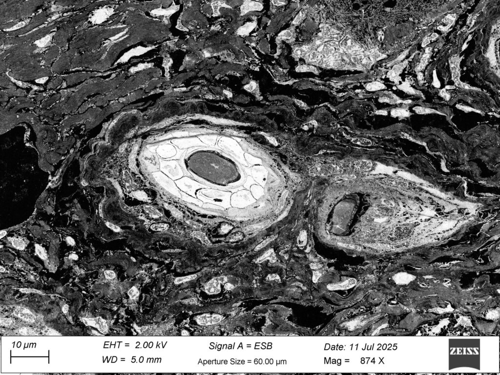
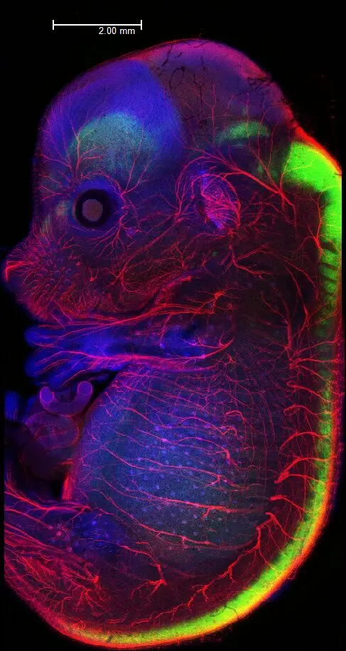
A recent study published in The Journal of Neuroscience highlights the successful collaboration between the Normandie and Bordeaux nodes of France-BioImaging. Researchers from UMR INSERM 1245 (Rouen), in partnership with the Rouen University Hospital, UMR INSERM 1237 (Caen), the Institut des Neurosciences des Saints-Pères (Paris), the PRIMACEN imaging platform (Rouen) and the Bordeaux Imaging Center (BIC), investigated the role of the endothelial NMDA receptor in the positioning and differentiation of cortical oligodendrocytes.
Using a conditional gene knockout model, the team demonstrated that the absence of this endothelial receptorleads to impaired myelination during development. These defects include reduced myelin sheath thickness and an increased number of axonal mitochondria, as observed by transmission electron microscopy (TEM) performed at the BIC. For TEM, the project received support from France-BioImaging through the Euro-BioImaging user access portal.
Importantly, these structural alterations were associated with long-lasting motor deficits in adult mice. The findings provide new insights into how disruptions in oligodendrocyte-vascular interactions contribute to white matter lesions associated with prematurity, which in severe cases can lead to cerebral palsy.
cdnkfndznfznfz
A: 3D model visualizing oligodendrocyte precursors (green) migrating along cortical vessels (red) in 2-day-old mice. B: Electron microscopy images of the corpus callosum of wild-type (left) and endothelial NMDA receptor knockout mice (right) at 15 postnatal days. Receptor knockout results in reduced myelin sheath thickness (arrowheads) and increased number of axonal mitochondria (arrows). C: Impairments of the myelination process are associated with motor disorders persisting in young adult mice (P45).
This collaboration illustrates how complementary expertise across France-BioImaging nodes can advance the understanding of complex neurodevelopmental mechanisms.
DNA-PAINT is a super-resolution imaging technique that relies on the transient binding of short fluorescent DNA “imager” strands to complementary “docking” strands attached to the target structure. Each binding event produces a localized burst of fluorescence that can be precisely detected and accumulated to reconstruct the image at nanometer resolution.
However, one major limitation remains: imager strands that are not bound continue to diffuse in the sample and emit fluorescence, creating background signal. This prevents researchers from using high imager concentrations and significantly slows down the acquisition process.
To overcome these limitations, a research team led by Yves Mely at the Laboratory of Bioimaging and Pathology (Strasbourg University) in collaboration with a team led by Alain Burger (Nice Institute of Chemistry) developed a new approach that incorporates a dark donor dye into the imager strand. A dark donor is a dye that remains almost non-fluorescent on its own but can transfer its energy to a nearby fluorescent acceptor when the two are brought together. In this system, the modified nucleobase X acts as the dark donor: it stays essentially dark in solution, but when the imager hybridizes with the docking strandlabelled with ATTO 647N, X activates the acceptor’s fluorescence. As the signal appears only during true binding events, this fluorogenic behaviour markedly reduces background noise and enables the use of higher imager concentrations.
Schematic of the DRET-DNA PAINT concept. Oligonucleotides containing the dark donor X as a nucleoside substitute act as imager strands and transiently bind to the docking strands labeled with the acceptor dye ATTO 647N. This leads to DRET from X to ATTO 647N and thus to the turn-on of the acceptor emission
Single-molecule experiments confirm that the system maintains binding kinetics compatible with DNA-PAINT, and that fluorescence increases roughly 50-fold upon duplex formation. The method was then applied to fixed HeLa cells: microtubules were reconstructed in around 30 seconds, with a resolution of ~50 nm and a median localization precision of 18 nm. By comparison, classical DNA-PAINT required 30 minutes to reach a similar result.
When compared to FRET-PAINT, a variant of DNA-PAINT in which fluorescence is generated through energy transfer between a donor and an acceptor dye brought together during hybridization, the dark-donor strategy showed a clear advantage. FRET-PAINT can suffer from signal leakage, as the donor dye may emit light in the acceptor detection channel. In contrast, the dark-donor system produced far less leakage, leading to cleaner images while preserving a similar acquisition speed.
Composite of TIRF projection and super-resolution image reconstruction of microtubules in HeLa cells. a) DRET-PAINT with 100 nM S-Im imager strand and 30 seconds of imaging time. b) FRET-DNA PAINT with 100 nM S-Im imager strand and 30 seconds of imaging time c) DNA-PAINT with 1 nM of imager stand and 30 min of acquisition time. d) DNA-PAINT with 100 nM S-Im imager strand and 30 seconds of imaging time. Scale bar is 5 µm.
The main limitation of this first-generation system lies in the photobleaching of ATTO 647N, which shortens the usable imaging time. The authors suggest possible improvements, including the use of more photostable acceptor dyes or the development of new donor–acceptor pairs with enhanced brightness to support longer and higher-resolution acquisitions.
Overall, this work provides the first proof of concept that dark-donor DNA-PAINT can deliver fast, low-background super-resolution imaging and could become a valuable addition to the growing set of DNA-based nanoscopy tools.
F-BIAS is a professional network that brings together bioimage analysts across France. Hosted within France-BioImaging core facilities, its mission is to support researchers with high-quality expertise in image processing and analysis. Created in 2021, the network provides analysts with a strong community where they can share technical and methodological knowledge, and collaborate on innovative solutions.
F-BIAS also offers a monthly Open Desk in bioimage analysis: short sessions with imaging experts where you can ask any question related to image processing challenges you encounter in your research projects. If you need guidance in bioimage analysis, this is the perfect place to start!
No Open Desk available, or need more time to address your issue? F-BIAS also provides collaborative projectsupport for more complex requests that require customized tools and a significant time commitment from analysts.
Join the network, discuss the challenges you face with your microscopy data, and let our experts help you find the best solutions!
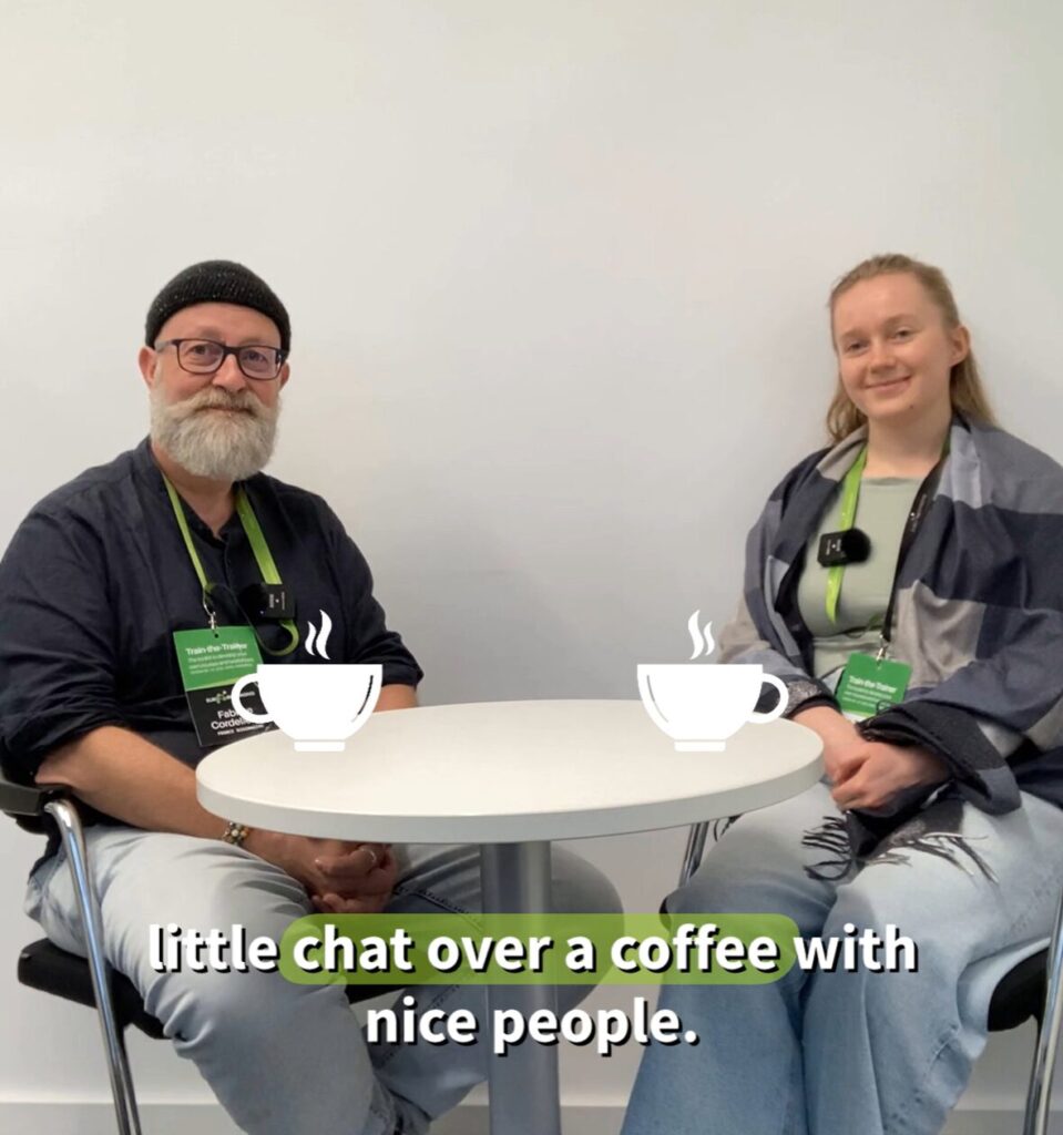
Last month, several members of France-BioImaging took part in the opportunities offered by Euro-BioImaging’s EVOLVE project, enabling valuable exchanges and inspiring experiences.
Through the Job Shadowing initiative, Guillaume Gay, Data Engineer for the FBI.data mission and Research Software Engineer at LIRMM (Montpellier), welcomed Kenneth Ho, Big Data Systems Engineer at the Francis Crick Institute (London).
They discussed their shared challenges and solutions in microscopy data management, and this week-long visit gave them the time to delve deeply into several technical topics.
Caroline Thiriet, Deputy Administrative Director for International Relations and Industry at France-BioImaging, hosted Virginia Pierini, Service Manager at the EMBL Imaging Centre. Their discussions focused on how France-BioImaging coordinates its distributed national infrastructure, which brings together 10 Nodes and more than 30 imaging facilities across France.
Finally, Fabrice Cordelière, Head of Training for France-BioImaging and Bio-Image Analyst at the Bordeaux Imaging Center, took part in the Train-the-Trainer event, where he mentored Iva Švecová from the Light Microscopy Facility at the Institute of Experimental Medicine of the Czech Academy of Sciences in Prague. Eva benefited from Fabrice’s extensive experience in team management, user training, coding practices, and user-driven data backup workflows.
We are delighted to contribute to the EVOLVE programme, which fosters collaboration, peer learning, and mutual inspiration among Euro-BioImaging Nodes. These initiatives reinforce our shared commitment to open, coordinated, and high-impact scientific services for the life science community.
The microscopy platform of Gustave Roussy is organizing a webinar dedicated to Huygens Software on Tuesday, November 25th, at 14:00 (CET).
This event will introduce the theory of deconvolution and image restoration through three main parts:
Principles of Deconvolution:Dive into the theoretical foundations to better understand this key process in microscopy
Demonstration of Deconvolution and Image Restoration: Discover the essential features of deconvolution and image restoration, and learn how to automate these processes
Benefits, Limitations, and Quality Control: Identify the strengths and limitations of deconvolution while ensuring the quality and robustness of your results
You can share your own images for the demonstration and receive a direct feedback on the improvements in your restored images!
Mitochondria, often described as the bioenergetic powerhouses of our cells, and more broadly of our entire organism, play a central role in metabolism. These organelles are involved in multiple metabolic processes (including carbohydrate and lipid degradation) and produce ATP (adenosine triphosphate), an essential molecule for a wide range of biochemical reactions.
Maintaining mitochondrial integrity is therefore crucial, as dysfunctions can lead to severe pathologies such as myopathies, neurodegenerative diseases or metabolic conditions like diabetes. Better understanding mitochondrial function and dysfunction is key to addressing these challenges. This understanding partly relies on the observation of mitochondria and the analysis of their morphology under different conditions.
Electron microscopy (EM) is the gold-standard technique for visualizing mitochondria, as it provides high-resolution imaging of cellular ultrastructure. However, segmentation and morphological analysis remain challenging due to the lack of contrast and color information in EM images. Existing pipelines to overcome these limitations are often time-consuming or too complex for users without experience in advanced deep-learning models.
To tackle this challenge, a research team from the Restore Institute in Toulouse, led by Mathieu Vigneau and Jean-Philippe Pradère, has developed EMito-Metrix, a computational tool designed for the automatic segmentation and analysis of mitochondria from 2D EM images.
The team created six species-specific models and one generalist model by training a segmentation algorithm with their own annotated EM images. Their results demonstrate that the tool enables highly specific detection of mitochondria according to their species of origin. With its user-friendly interface, EMito-Metrix allows users to easily visualize and analyze 26 mitochondrial metrics, presented through automatically generated graphs. In addition, EMito-Metrix includes a machine learning module that provides predictive analytical capabilities to assess how experimental factors, such as genetic mutations or drug treatments, may affect mitochondrial morphology and ultrastructure.
To validate their tool, the researchers analyzed mitochondria across the entire tree of life. More than 35 000 mitochondria were processed, with over 800 objects per species. The results obtained with EMito-Metrix are compelling, enabling precise segmentation of mitochondria by species and efficient quantitative analysis of their metrics.
The AI algorithm is capable of accurately detecting mitochondria (in colour) from tissues from different species imaged in ME (left). For each segmented mitochondrion, the tool extracts 26 morphology and ultrastructure metrics that can be displayed using graphs. The radar plot (centre) illustrates striking differences in metrics between vertebrates and invertebrates. Based on these metrics, the neural network is able to predict the class to which each mitochondrion belongs with 94% accuracy (right).
In conclusion, EMito-Metrix supports mitochondrial research by simplifying morphological analysis, saving researchers valuable time and reducing the risk of bias.
The RIC Paris-Saclay is pleased to announce the conference “AI & Image Analysis”, which will take place on Thursday, November 20, 2025, from 1:00 p.m. to 4:30 p.m. at the Paris-Saclay Faculty of Medicine (Research Building).
Organized in collaboration with the Interdisciplinary Object “BioProbe” (University Paris-Saclay), this half-day event is open to researchers, engineers, clinicians, faculty members, PhD studentsand students.
It will highlight recent advances in Artificial Intelligence and Deep Learning applied to cellular and tissue imaging, through presentations by both academic and industrial experts (University Paris-Saclay, University of Nantes, Institut Pasteur, École Polytechnique, Sartorius, and Nanolive).
The event will be recognized as doctoral training for the graduate schools BioSign, Cancerology (CMBS), and Therapeutic Innovation (ITFA) of UPSsaclay.
Invited speakers and titles
Perrine Paul-Gilloteaux (BioCore – University of Nantes) – AI in Correlative Microscopy: Unlocking the Potential of Multimodal Imaging.
Olivier Schwartz (Institut Pasteur) & Mathieu Fréchin (Nanolive SA) – Life in Motion: From AI-Enhanced Imaging to Cellular Process Simulations.
Organization committee: Larbi Amazit, Régis Bobe, Laurent Combettes, Cécile Denis, Marie Erard, Evelyne Ferrary, Isabelle Garcin, Anne Guiochon-Mantel, Aude Jobart-Malfait, Valérie Nicolas, Oliver Nüsse.
As the 2025 edition of the France-BioImaging Image Contest is still running, we wanted to highlight our previous winners and their projects. Here is a quick throwback to our 2024 winners.
Before getting to the heart of the matter, we want to remind you that you still have time (before November 14th) to submit your best images and try to win your registration fees for one 2026 microscopy-related event! Please make sure you upload your images on the following link:
Dans le cadre du programme “Imaging 4 All – Access Track”, le Dr Brice Tonfack, de l’Université de Yaoundé (Cameroun), a bénéficié d’un grant pour collaborer avec Jean-Luc Verdeil, le responsable scientifique de la plateforme d’imagerie MRI-PHiV du CIRAD à Montpellier. Ce séjour allant au-delà de l’aspect technologique, a permis de structurer une collaboration scientifique…
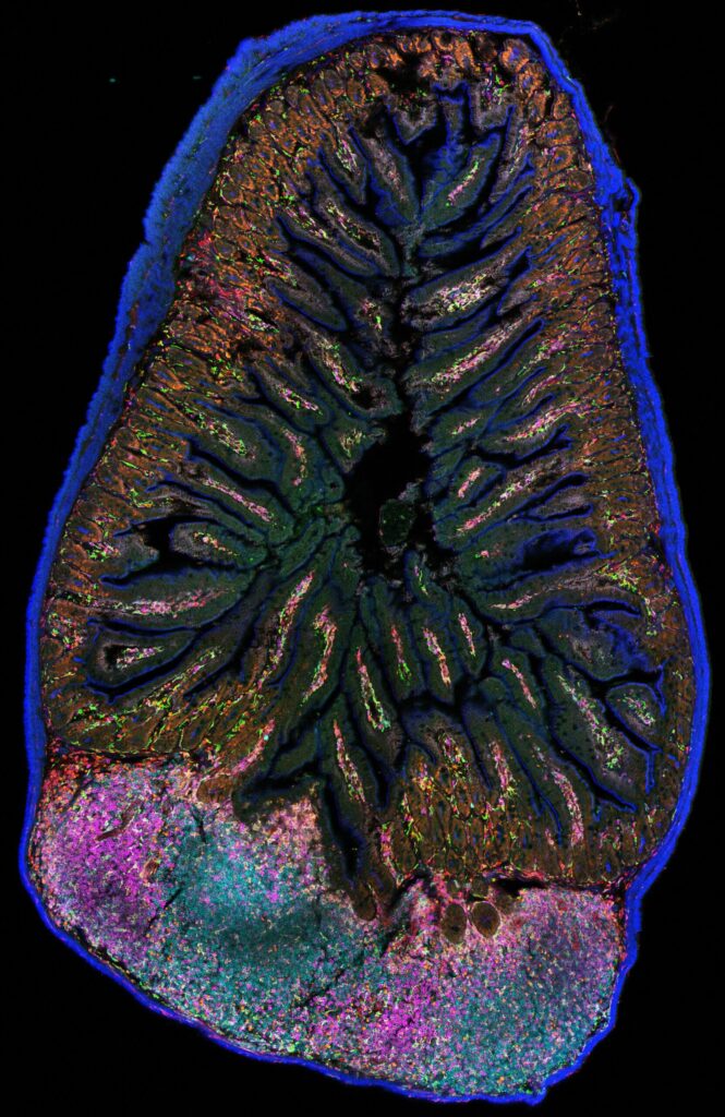
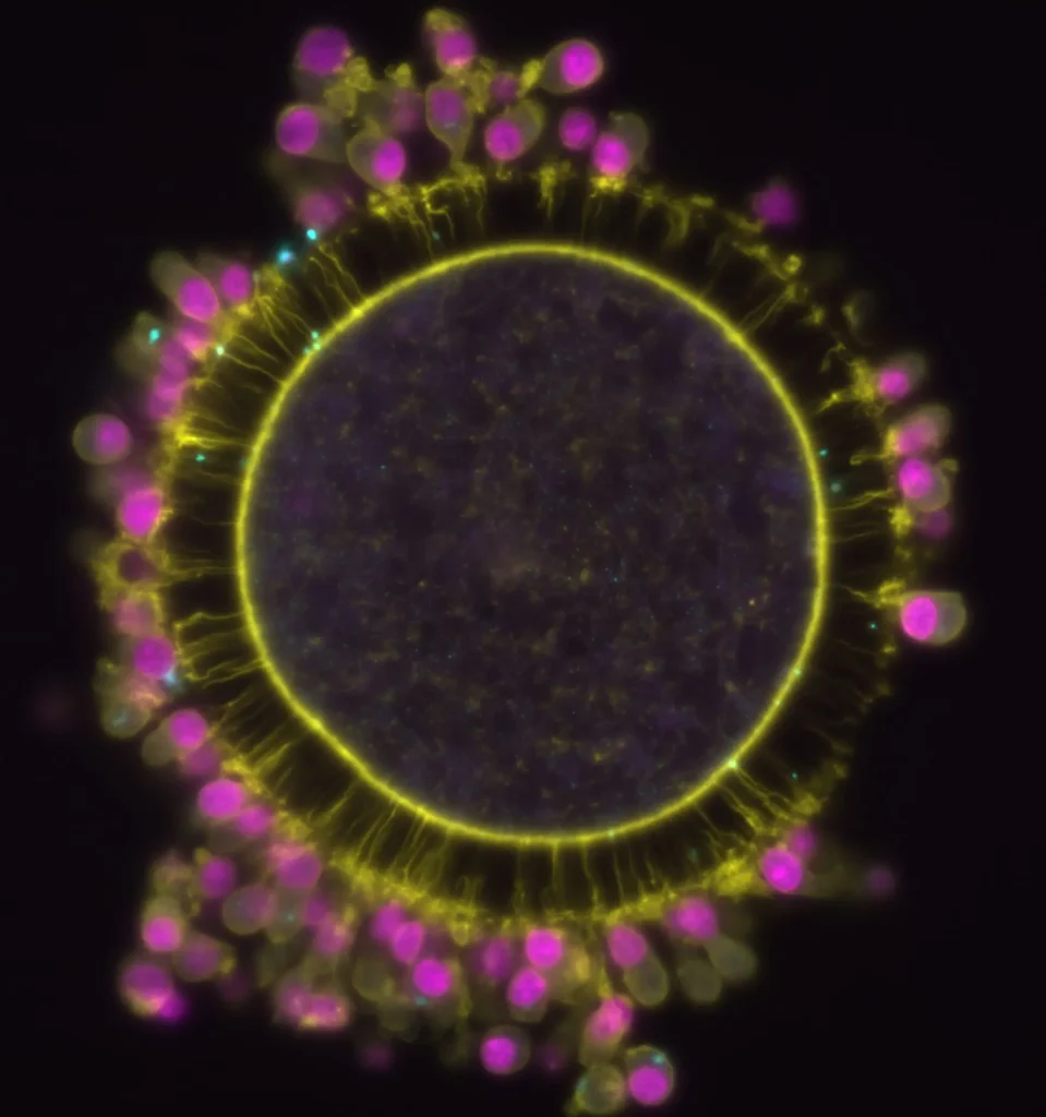
Last year, we enjoyed the winning images submitted for their artistic take and their quality. Thanks to Vanessa WEICHSELBERGER , Dalia EL ARAWI and Frédéric FERCOQ for their beautiful images!
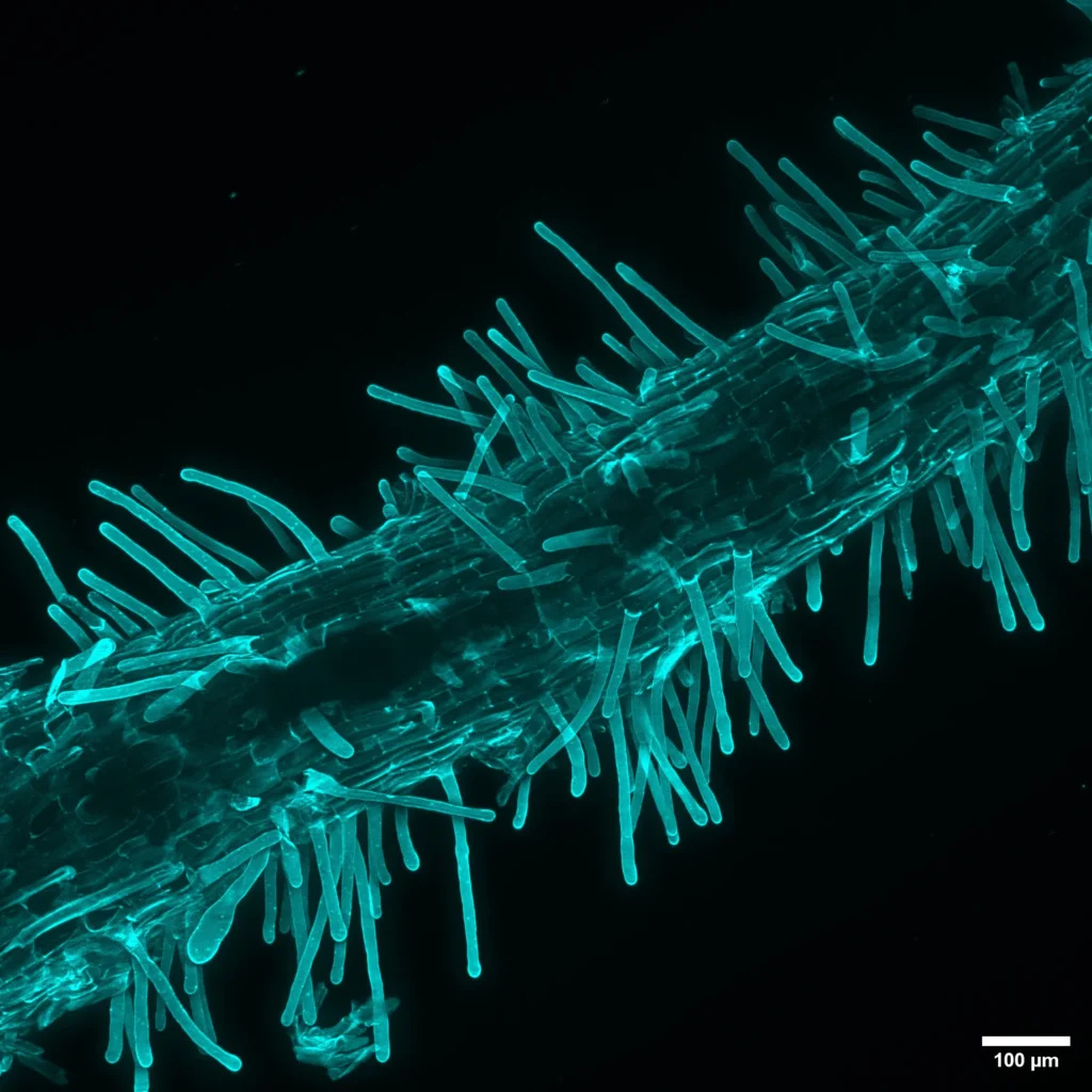
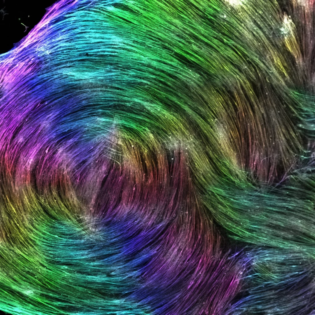
1st Place: Vanessa WEICHSELBERGER
Pierre-François LENNE team, Institut de Biologie du Développement de Marseille (IBDM)
“Rays of repetitive beauty”
Marine plant collected in Mediterranean Sea, stained for Actin using Phalloidin.
2-photon microscope
Vanessa WEICHSELBERGER is a shared post-doctoral researcher between the lab of Pierre-François LENNE at IBDM in Marseille and Vikas TRIVEDI at EMBL in Barcelona. She is a developmental biologist and interested in how cells coordinate morphogenetic processes with each other.
The image she submitted isn’t related to her research proejct. It is of a sample that they took from the Mediterranean Sea on a lab day out, it is part of an underwater plant. She believes it stands for their interest in shape and form beyond their own research projects. It represents their broad curiosity in biological structures.
They stained the sample for Actin and Nuclei and just admired its structure, pattern and periodicity.
“It is a great example of how amazing all kind of life is!“
By multiplying and arranging the image into a periodic wheel, the periodicity of the sample itself is even more highlighted.
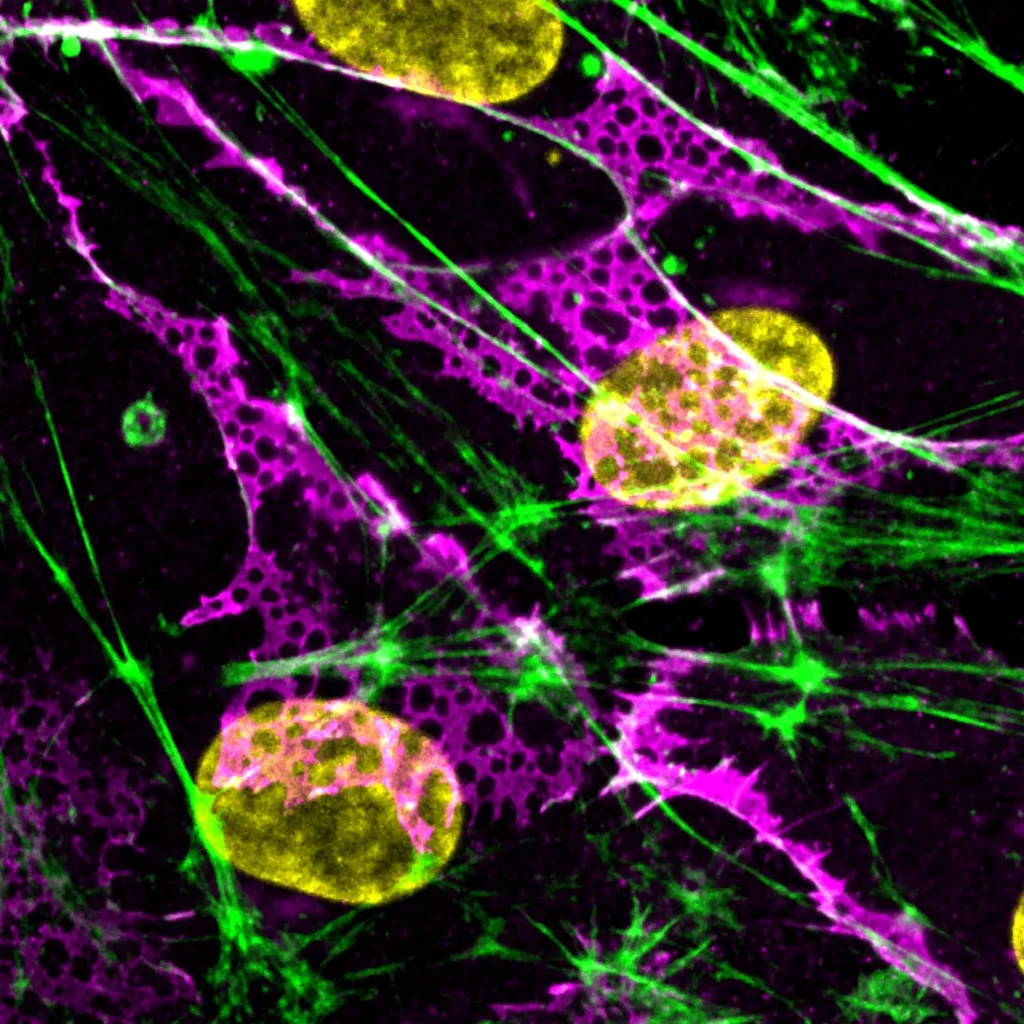
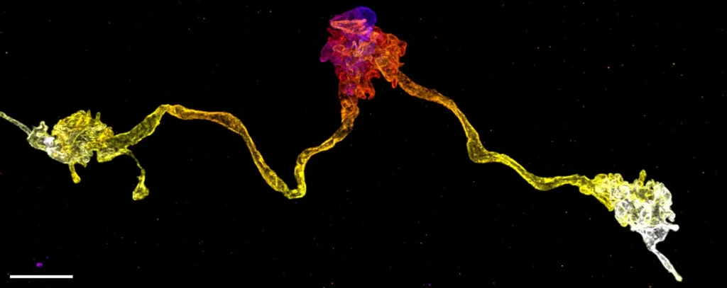
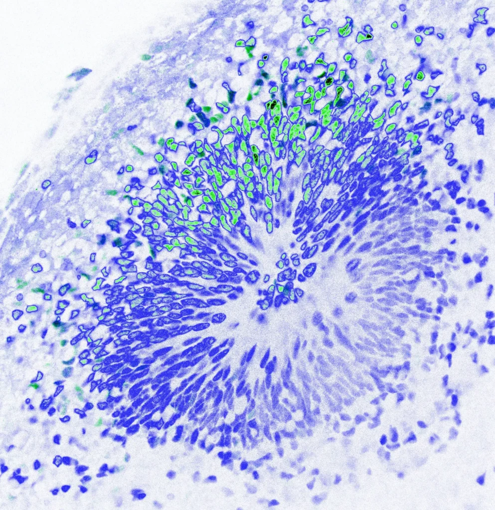
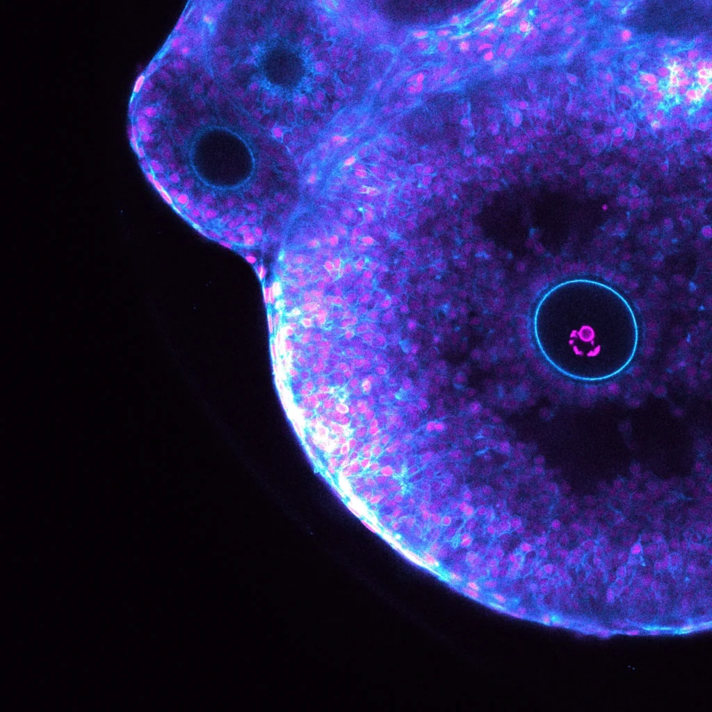
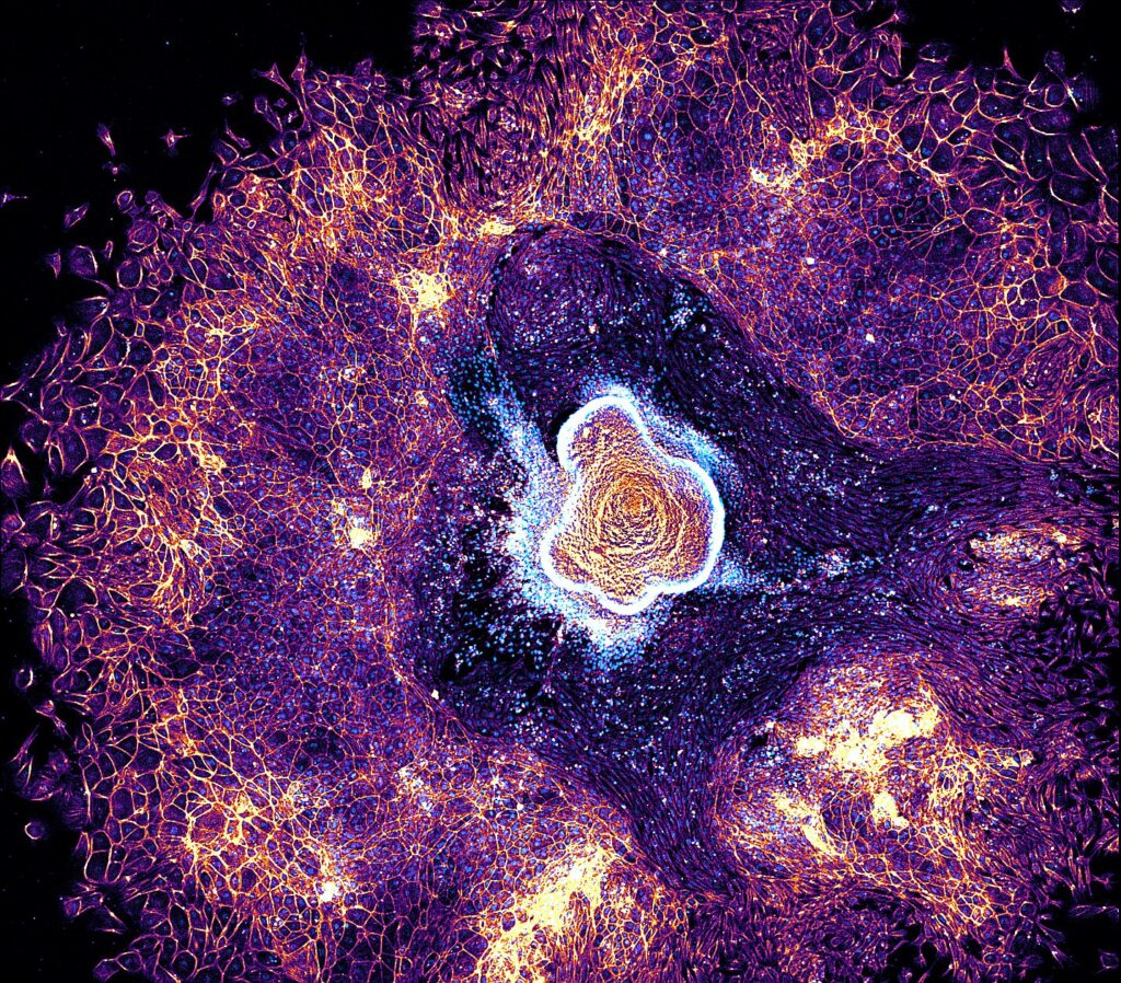
2nd Place: Dalia EL ARAWI
Pierre-François LENNE team, Institut de Biologie du Développement de Marseille (IBDM)
“Gastrula Nebula”
Murine embryonic organoid illustrating cells collective migration on a laminin-coated surface. Phalloidin-labeled Actin filaments and Hoechst-stained Nuclei highlight detailed cellular architecture and a remarkably structured tissue organization.
Zeiss LSM 880 Confocal Microscopy
Dalia EL ARAWI is a post-doctoral researcher in biophysics, currently working at the IBDM in Marseille. Her research focuses on studying Gastruloids, 3D aggregates of mouse embryonic stem cells.
Gastruloids have emerged as a powerful in vitro model to study early embryonic development and tissue patterning. Within a few days, Gastruloids undergo symmetry breaking, axial elongation and germ layers specification, forming structures that closely mimic embryonic tissues both genetically and morphologically. Despite their ability to self-organize into complex architectures, 3D Gastruloids face developmental limitations over time, including tissue surface tension, apoptosis, and high phenotypic variability. To address these challenges, she used a hybrid 2.5D Gastruloids approach: first, generating 3D Gastruloids with proper differentiation and antero-posterior symmetry breaking, then transferring them onto a 2D extracellular matrix that mimics extraembryonic tissues, promoting more advanced morphogenesis.
This is illustrated in the submitted image, where a 2.5D Gastruloid spreads across a laminin-coated surface, revealing detailed cellular architecture and tissue organization. By applying different labeling strategies, she uses this approach to gain insights into vascular and cardiac structure formation. Her findings suggest that substrate-guided Gastruloid models may potentially recapitulate key aspects of cardiovascular development, offering new insights into tissue self-organization and vascularization.
For Dalia, being awarded second place in the FBI Image Contest was very rewarding. The recognition increased the visibility of her work, sparked discussions with colleagues inside and outside the lab, and motivated her to present her research more widely. It also gave her confidence to pursue future opportunities related to microscopy and imaging!
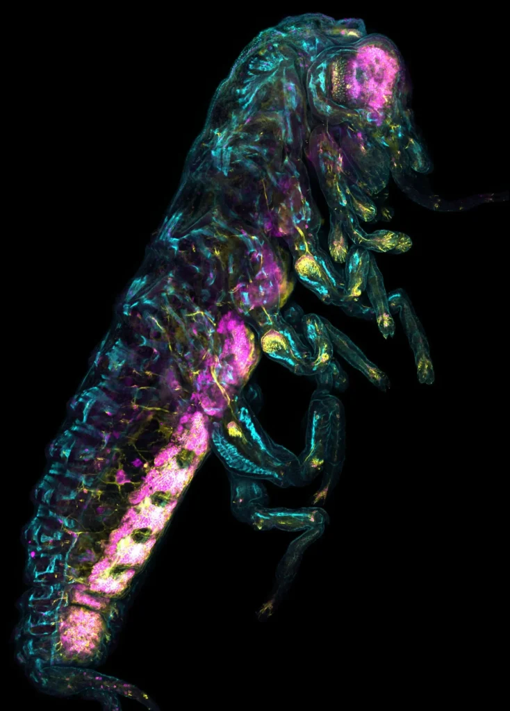
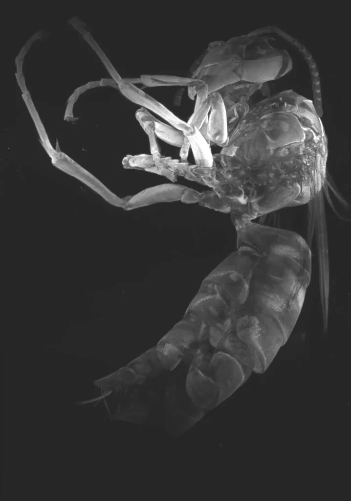
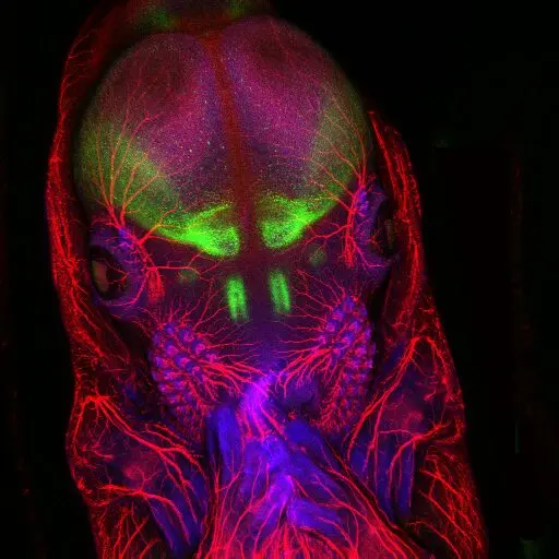
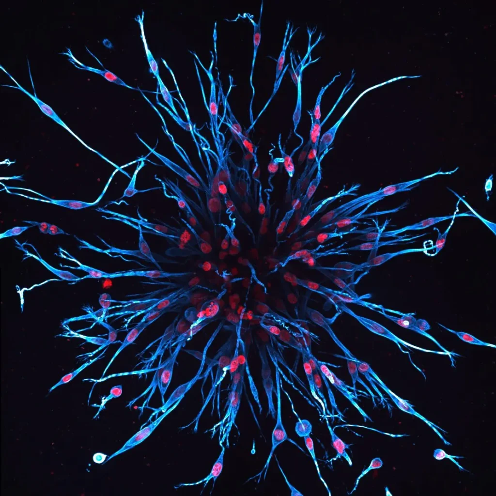
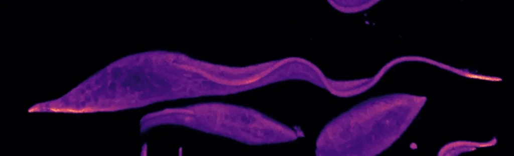
3rd Place: Frédéric FERCOQ
Molecules of Communication and Adaptation of Microorganisms team, Museum National d’Histoire Naturelle (MNHN)
“Explosion filarienne“
Internal architecture of Litomosoides sigmodontis, a parasitic nematode used as a model to better understand filarial infections. Under significant internal pressure to maintain its structure, this nematode experienced a cuticle rupture during handling, leading to the expulsion of some organs, including the ovary and intestine. The cytoskeleton appears in orange, and the DNA in cyan.
Confocal microscopy
Frédéric FERCOQ is a Maître de conférences in the UMR 7245 “Molecules of Communication and Adaptation of Microorganisms” at the MNHN in Paris. He works in the “Parasites and Free-Living Protists” team, where his research focuses on host–parasite–symbiont interactions during filarial infections. He studies how anti-parasite immune responses and bacterial endosymbionts such as Wolbachia influence parasite development, fertility, and tissue pathology. Microscopy is central to his approach, enabling him to visualize parasite structure, host tissue organization, and cellular interactions in detail.
The submitted image shows the internal anatomy of a female Litomosoides sigmodontis filarial worm after mechanical rupture. Due to the high internal pressure required to maintain its structure, the cuticle burst during manipulation, causing the expulsion of internal tissues, here the ovaries and intestine. Tubulin staining (orange) highlights microtubule-rich structures, while DNA is shown in cyan.
Winning the FBI Image Contest allowed Frédéric to attend the Microscience Microscopy Congress (MMC Series) in Manchester in July 2024, where he presented both a poster and an oral communication. The MMC is one of Europe’s leading interdisciplinary conferences dedicated to advances in microscopy across the life and physical sciences. It was a great opportunity for him to share their most recent publication (https://doi.org/10.1371/journal.ppat.1013301) and to present it in the context of their broader, microscopy-driven research on filarial development and host–parasite interactions. The event also allowed him to connect with researchers from diverse imaging backgrounds and explore new techniques relevant to parasitology.
We use cookies on our website to give you the most relevant experience by remembering your preferences and repeat visits. By clicking “Accept All”, you consent to the use of ALL the cookies. However, you may visit "Cookie Settings" to provide a controlled consent.
This website uses cookies to improve your experience while you navigate through the website. Out of these, the cookies that are categorized as necessary are stored on your browser as they are essential for the working of basic functionalities of the website. We also use third-party cookies that help us analyze and understand how you use this website. These cookies will be stored in your browser only with your consent. You also have the option to opt-out of these cookies. But opting out of some of these cookies may affect your browsing experience.
Necessary cookies are absolutely essential for the website to function properly. These cookies ensure basic functionalities and security features of the website, anonymously.
Cookie
Duration
Description
cookielawinfo-checkbox-analytics
11 months
This cookie is set by GDPR Cookie Consent plugin. The cookie is used to store the user consent for the cookies in the category "Analytics".
cookielawinfo-checkbox-functional
11 months
The cookie is set by GDPR cookie consent to record the user consent for the cookies in the category "Functional".
cookielawinfo-checkbox-necessary
11 months
This cookie is set by GDPR Cookie Consent plugin. The cookies is used to store the user consent for the cookies in the category "Necessary".
cookielawinfo-checkbox-others
11 months
This cookie is set by GDPR Cookie Consent plugin. The cookie is used to store the user consent for the cookies in the category "Other.
cookielawinfo-checkbox-performance
11 months
This cookie is set by GDPR Cookie Consent plugin. The cookie is used to store the user consent for the cookies in the category "Performance".
viewed_cookie_policy
11 months
The cookie is set by the GDPR Cookie Consent plugin and is used to store whether or not user has consented to the use of cookies. It does not store any personal data.
Functional cookies help to perform certain functionalities like sharing the content of the website on social media platforms, collect feedbacks, and other third-party features.
Performance cookies are used to understand and analyze the key performance indexes of the website which helps in delivering a better user experience for the visitors.
Analytical cookies are used to understand how visitors interact with the website. These cookies help provide information on metrics the number of visitors, bounce rate, traffic source, etc.
Advertisement cookies are used to provide visitors with relevant ads and marketing campaigns. These cookies track visitors across websites and collect information to provide customized ads.