New microscope in the Imagopole:
Live-cell super-resolution with BioAxial CODIM100
Open day in Pasteur the 24/09/2015

pseudo logo Imagopole

Dear friends of imaging,

The BioAxial company and the Imagopole (the imaging platform of the Institut Pasteur) has collaborated for several years around the development of an entirely new approach to super-resolution imaging based upon conical diffraction. It is now a microscopy system, available commercially (http://www.bioaxial.com/products/codim100/product-description), the CODIM100.

PastedGraphic-1 
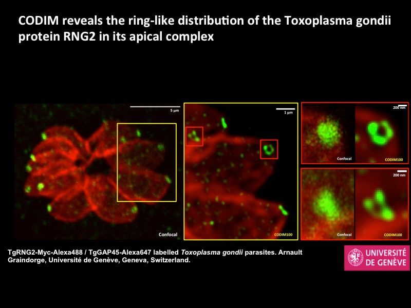
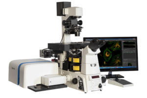
We host such a system, and it will be made available at the Imagopole to all scientists, following the Imagopole access policy (as any regular system) by the end of September 2015. The CODIM100 is very simple to use, and can be operated by autonomous users, following a short training session.

We organize an open day for the system Thursday the 24th of September. You are cordially invited to bring your sample and try the CODIM100. We will be there all day to help you setting up the microscope to harvest the best images.

If you are interested in coming to this open day, please write to Jean-Yves Tinevez (tinevez@pasteur.fr) and state your needs. We will allocate a slot depending on the time you need. We have the facilities to host and prepare live samples.

If you are interested in super-resolution microscopy, keep reading on.
While there already exist several super-resolution modalities, this system is distinguished by the unique combination of several performance features. In brief,:

    • it is ideal for live-cell super-resolution imaging (even long term time-lapse imaging);
    • it works with any fluorescent sample without any special preparation;
    • it has a very good sensitivity even to weakly labelled samples.

It is attached to a normal laser scanning confocal microscope. The typical usage is to image a sample with normal LSCM mode, then draw a ROI over the acquired image to rescan it in super-resolution. Here are some quick specifications of the super-resolution module:

    • Excitation lines: 488, 561 and 640nm
    • Objectives: 40x 0.95 air / 60x 1.2 water / 60x 1.49 oil.
    • Acquisition speed: a few seconds for a 25µm2 ROI.
    • Labels: any that can be excited with the wavelength mentioned above
    • Sample prep: any confocal or live sample with 0.17mm glass bottom coverslip. No restrictions on mounting media for fixed samples.
    • XY resolution: 110 nm with 488nm laser/0.95 NA, 90 nm with 488 nm laser/1.20 NA
    • Very low excitation power: less than 1 µW/µm2.

Some examples:
CODIM100_example1
CODIM100_example2

affiche 237 copie
New trends in super-resolution optical microscopy: unraveling the ultra-structure and dynamics of molecular assemblies in cells and tissues

PHASE I
September 30 to October 2, 2015
Bordeaux

WebsiteAfficheInscriptions

 
This Workshop will present the theoretical basis, the latest developments and the acquisition and data analysis procedures on super-resolution microscopy techniques used to investigate the ultrastructure of molecular assemblies and their dynamics in living cells and tissues.

Registration Deadline: March 27, 2015
Organizers:
Maxim DAHAN (Institut Curie, Paris), Orestis FAKLARIS (Institut Jacques Monod, Paris) and Marcelo NOLLMANN (Centre de Biochimie Structurale, Montpellier)
 

Phase I : critical assessment
  • Introduction to super-resolution techniques
  • Application and new developments in single-molecule super-resolution microscopy
  • Labeling, Theoretical and computational methods
  • In-depth super-resolution microscopy, STED, SIM microscopies
Phase II : technical workshop

18 trainees will be selected among phase I participants.
 

inserm.fr (2)
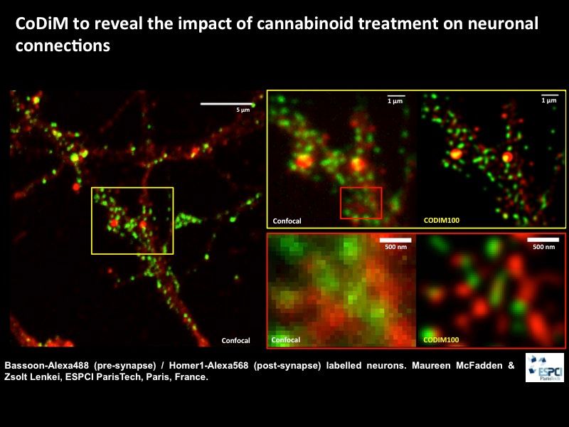
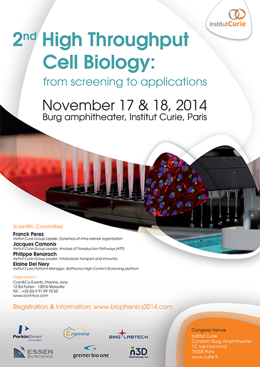
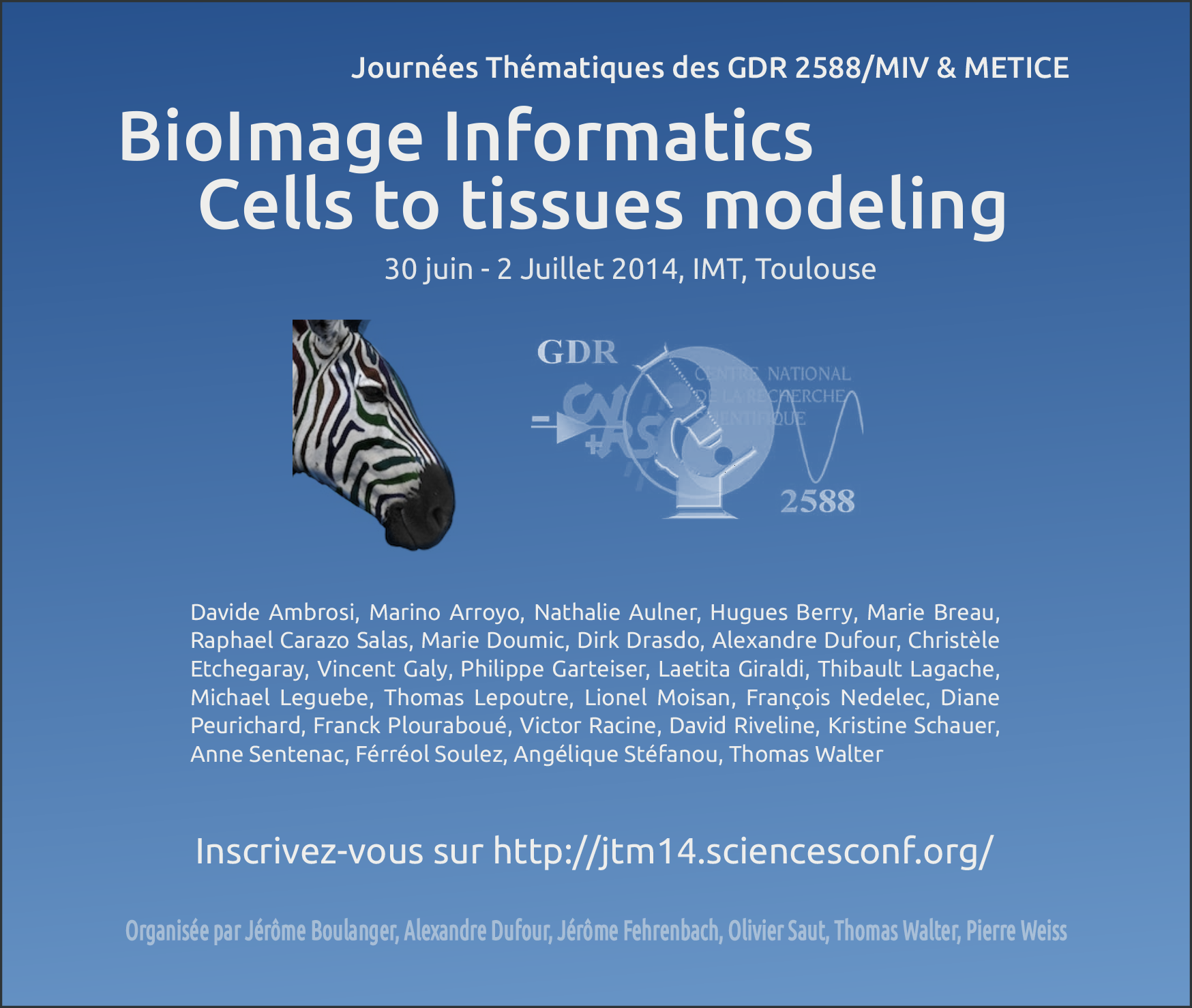
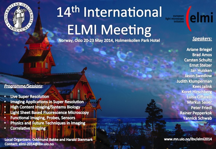
Renseignements et inscriptions
Pôle des Ateliers – Inserm
ateliers@inserm.fr
Tél : 33(0)1 44 23 62 03 ou 04

November 17, 2014, November 18, 2014 in Institut Curie Research Center

Workshop on Cell Biology, Biochemistry, Neurobiology, Physiology, Chemistry or Chemical Biology & Image Processing and Data Management


 Website, program and registration

Affiche-2nd-high-throughput-cell-biology-2014

The strength of this meeting lies in the mixture of scientific lectures on state-of-the-art, high-end microscopy combined with “hands-on” workshops and exhibition of the latest technology, organized by the leading companies in the field. This year we will maintain the format and focus on leading edge developments and its implementation to life science.

June 30, 2014, July 2, 2014 in IMT Toulouse

 
flyer_journee_thematique_2014
 

Download the programme here


 Website and registration


The strength of this meeting lies in the mixture of scientific lectures on state-of-the-art, high-end microscopy combined with “hands-on” workshops and exhibition of the latest technology, organized by the leading companies in the field. This year we will maintain the format and focus on leading edge developments and its implementation to life science.

14th international ELMI Meeting

20-23 May 2014 – Oslo, Norway


slide1

The 2014 ELMI meeting will be following the proven successful format of research talks, company workshops and exhibition.


Download the programme here


Website


ELMI was first set up in 2001 in order to promote the quickly developing field of light microscopy as a fundamental research tool for the life sciences and to strengthen the channels of communication between researchers and industry. The annual ELMI meeting, which has been running for the last 12 years at various venues across Europe, has an excellent reputation within the microscopy community, making this meeting a key event in the calendar of hundreds of scientists and developers.
 

The strength of this meeting lies in the mixture of scientific lectures on state-of-the-art, high-end microscopy combined with “hands-on” workshops and exhibition of the latest technology, organized by the leading companies in the field. This year we will maintain the format and focus on leading edge developments and its implementation to life science.

SummerSchool

The spirit of this Summer School is inspired by the most prestigious school ever founded in France, Saint Flour, the influence of which has spread to generations of researchers in Mathematics. Since its establishment in Brittany in 1994, this school has become a worldwide reference. It is resolutely international (participants from more than twenty countries have participated to the previous editions) and accessible to young scientists. It is an open yet privileged place for exchanges and discussions of major on-going work. Informal and warm, at a location where the sea and the land combine in a time varying relation, this school brings together, every two years for ten days, the world great teachers and researchers in Biomedical Imaging.

For its 11th edition in 2014, it relocates in the wonderful facility of Saint Jacut de la Mer close to Saint Malo and Mont Saint Michel.


Additional information


Extensive 6 hours lectures, seminars, and discussions are organized at the highest level, but with the freedom of spirit that is the tradition of Brittany. The school objective is to contribute without any exclusion to advances in a rapidly evolving field, and to foster participation in the adventure of research. It provides up-to-date, state-of-art knowledge on emerging areas and addresses important issues dealing with complex, multivariate systems, going from basic to applied research.

Audience: The Summer School is open to graduate students (M.S., PhD), post doctoral scientists, image processing scientists, radiologists, biologists, researchers and engineers in industry.


Lecture Program

Luis IBÁÑEZ, Kitware Inc., USA, “Open Science in Biomedical Image Analysis”
Aydogan OZCAN, UCLA, USA, “Computational Microscopy, Sensing and Diagnostics”
Rangaraj M. RANGAYYAN, University of Calgary, Canada, “Oriented Tissue Analysis in Medical Imaging”
Jocelyne TROCCAZ, UJF CNRS, France, “Computer Assisted Medical Interventions”
Dimitri VAN DE VILLE, Ecole Polytechnique Fédérale de Lausanne, Switzerland, “fMRI and EEG for Cognitive and Clinical Neurosciences”
Tony WILSON, University of Oxford, UK, “Advanced Microscopies for Biology”


Important dates

Candidature submission open from Dec., 2013 to March 15th, 2014 (Extended Deadline).

Contacts

Jean-Christophe OLIVO-MARIN, Institut Pasteur (jcolivo@pasteur.fr)
Oscar ACOSTA, LTSI, Université de Rennes 1, 35042 Rennes CEDEX, France
Fax: +33 223.23.69.17 – E-mail : oscar.acosta@univ-rennes1.fr

LogosSummerSchool

We are pleased to announce the EMBL Master Course Bioimage Data Analysis to be held from Monday, 12 May – Friday ,16 May 2014.

This course will focus on computational methods for analyzing images of proteins, cells and tissues, to boost the learning process of participants who have an immediate need to deploy image analysis in their own research. For this the course extends from basic principles in ImageJ, Matlab and R to the actual implementation of workflows using scripting. We expect that increasing the image analysis literacy of the participants will greatly enhance their scope, creativity and achievements of own research projects. We aim to gather expert knowledge to organize a world-leading course for image analysis in the field of biology.

The course will take place in Heidelberg, Germany at the EMBL Advanced Training Centre.

Registration and motivation letter deadline is February 25, 2014.

Please visit our course website for more details, including course schedule and list of confirmed speakers:

http://www.embl.de/bias2014/

You are welcome to circulate this announcement to interested members and groups within your institution.

We look forward to welcoming you to Heidelberg, Germany.

Scientific Organisers:

Kota Miura, EMBL Heidelberg, Germany

Sébastien Tosi, Institute for Research in Biomedicine – IRB Barcelona, Spain

Perrine Paul-Gilloteaux, Institut Curie, France

If you have any questions, please do not hesitate to contact:

Diah Yulianti

Conference Officer

European Molecular Biology Laboratory

Email: diah.yulianti@embl.de

Il reste des places pour cette formation d’analyse d’image spécialisée analyse dynamique (tracking, flux optique etc..)  du 4 au 6 décembre 2013 à Paris 7 organisée par la DR2 CNRS.

Inscriptions avant le 12 Novembre :  http://www.sg.cnrs.fr/bpc/pratique/RH/docs/PDF/F_RH_formation.pdf auprès de franck.elsherbini@dr2.cnrs.fr et olga.hodges@dr2.cnrs.fr.

Comprendre les différentes méthodes d’analyse de la dynamique en biologie et leurs paramètres

Objectifs :

– Comprendre les différentes méthodes d’analyse de la dynamique en biologie et leurs paramètres

– Savoir choisir le type d’étude dynamique la plus adaptée à la question biologique

– Savoir générer et interpréter des champs de vitesse

– Savoir générer et interpréter des trajectoires

Programme :

  • Définition des modes de dynamiques moléculaire et cellulaire et leurs caractéristiques mesurables,
  • Bases théoriques du flux optique, Création et Analyse de cartes de vitesse
  • Méthodes de post traitement basée corrélation de fluorescence (ICS-TICS-STICS-RICS)
  • Bases théoriques du suivi d’objets. Utilisation de tracking (de particule et de cellules) sous Fiji et ICY, choix des paramètres, Analyse du tracking (caractéristiques du mouvement mesurables : interprétation MSD, directionalité, temps de pause, etc…)
  • Compensation de mouvement et tracking
  • Conditions d’acquisitions (résolution spatiales et temporelles) pour l’analyse dynamique.
  • Les limites d’interprétation et les pièges.
  • Choix d’une méthode et essai par les participants sur leur problématique.

Intervenants :

Perrine Paul-Gilloteaux IR CNRS; Francois Waharte IR CNRS; Jérôme Boulanger CR CNRS; Patrick Bouthémy DR INRIA; Charles Kervrann DR INRIA

Dear Colleagues,


The next Winter Conference of the European Society for Molecular Imaging on “IMAGING THE DEVELOPMENT” might be of interest for you.


The one-week conference will take place from 19-24 January 2014 at the Ecole de Physique in Les Houches, France.


The set-up is very similar to the Gordon Conferences or the original Keystone symposia in the US. It would be great if you could join us in the French Alps.


Deadline for abstract submission is 8 October 2013 – see www.topim.eu!


Speakers (so far):

Yohanns Bellaïche – Paris, France

Peter Carmeliet – Leuven, Belgium

Mary Dickinson – Houston, USA

Peter Friedl – Nijmegen, The Netherlands

Mark Henkelman – Toronto, Canada

Michal Neeman – Rehovot, Israel

Lee Niswander – Colorado, USA

Nadine Peyriéras – Gif-sur-Yvette, France

Olivier Pourquié – Illkirch, France

Jens Schwamborn – Esch-sur-Alzette, Luxembourg

Mikael Tanter – Paris, France

Daniel Turnbull – New York, USA

Annemie van der Linden – Antwerp, Belgium

Magdalena Zernicka-Goetz – Cambridge, UK


Among all domains of biological science, development is one for which the quality and relevance of observations depend drastically on non-destructive methods. The secret life of embryos and fetuses remained terra incognita for scientists. In the last decades the progress of non-invasive imaging has completely changed our view – read more at www.topim.eu


Do not hesitate to contact the ESMI Office (office@e-smi.eu) for any further information you may need.

Bioimage analysis meeting in Barcelona, from 7th to 8th of October this year: EUBIAS 2013: European BioImage Analysis Symposium.

http://eubias2013.irbbarcelona.org

The Symposium aims at strengthening the image analysis community in Europe and at increasing the accessibility of image analysis tools for the community and end-users.

The 2-days Open Community Meeting (Oct. 7-8th) features an “Open Software Showcase” presentations contributed by developers, open discussions and debates, and oral and posters contributions, participation is open to everyone. This event will help you to get a concise overview of various image analysis software packages in only two days, and also to provide usage feedbacks directly to the developers.
Developers from following packages/librairies have confirmed so far and will present their current status, most recent updates and perspectives:
– ImageJ / Fiji
– ImgLib / ImageJ2
– ICY
– Ilastik, VIGRA
– OME (OMERO and Bio-Formats)
– KNIME
– CellProfiler
– BioImageXD
– CellCognition
– Mahotas
– EBImage
– Pink
– ITK /Farsight
– DIPImage

We welcome you to sign up to the BioImage Community Meeting now,
please visit the website for more details and for registration:

The Symposium also features a developers ‘Taggathon’ (Oct. 9-11th) event and a BioImage Analysis course (Oct. 9-12th) for european facilities members, please check the web for more details.

If BioImage Analysis is key to your research, do not miss this unique event to commit yourself in boosting the accessibility to BioImage Analysis tools and strengthening the community !

This event was conceptualized at the last ELMI meeting 2013 in Arcachon, is mainly sponsored by Euro-BioImaging (www.eurobioimaging.org) and OME, and by companies bridging open software. EuBIAS2013 is open to sponsor contributions and participation from the private sector. EuBIAS2013 is hosted by IRB Barcelona and is organized by many people from the University of Dundee, EMBL (Heidelberg), IRB Barcelona, CRG (Barcelona), EPFL (Lausanne), ETH (Zuerich), DZNE (Bonn), Institut Curie (Paris).


European BioImage Analysis Symposium – Barcelona 7 to12th of October
http://eubias2013.irbbarcelona.org

Organization Committee:
Jason Swedlow – University of Dundee,
Data Management and Analysis WP, Euro-BioImaging
Sébastien Tosi – IRB Barcelona – sebastien.tosi@irbbarcelona.org
Kota Miura – EMBL – kota.miura@embl.de
Julien Colombelli – IRB Barcelona – julien.colombelli@irbbarcelona.org
(tel: +34 93 4020451)

contact: eubias2013@gmail.com

 flyer

BioImage Informatics

8-9 Juillet 2013, Institut Curie, Paris

#keynotes

  • Bob Murphy
  • Jean-Christophe Olivo-Marin

#speakers

  • Yohanns Bellaiche
  • Laure Blanc-Féraud
  • Xavier Descombes
  • Dirk Drasdo
  • David Holcman
  • Charles Kervrann
  • Nadine Peyriéras
  • Vincent Studer
  • Alain Trubuil
  • Christophe Zimmer

Inscrivez-vous sur http://minilien.curie.fr/74wk7b

Organisée par

Jérôme Boulanger, Alexandre Dufour, Thomas Walter, Pierre Weiss