France-BioImaging and all the French community aims to develop and promote innovative imaging technologies and methods. But microscopy images can also take an artistic, creative look and make the invisible world beautiful, allowing people to see the visual appeal of the life sciences.
We enjoyed the diversity of the images submitted with many different microscopy techniques, models and applications represented. A big thank you to all the participants!
The National Coordination Team and the Executive Board are proud to announce the winners of the FBI Image Contest 2025:
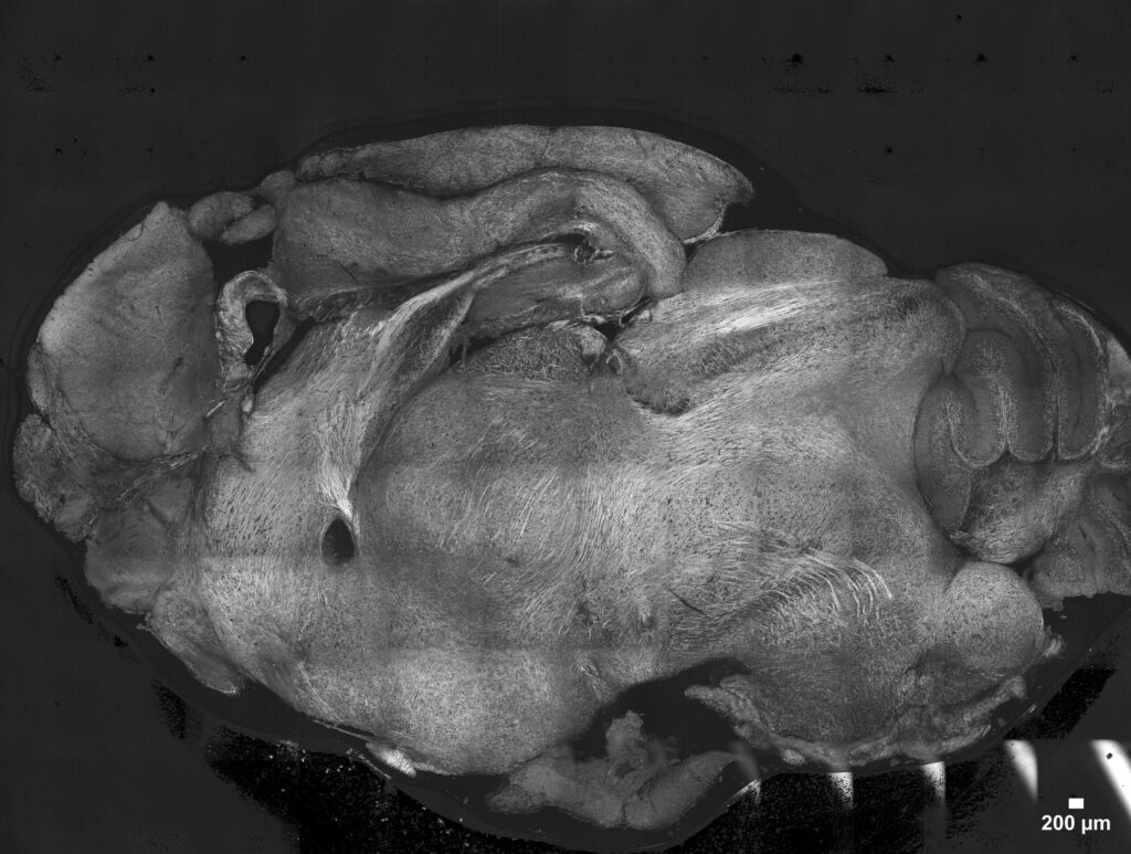
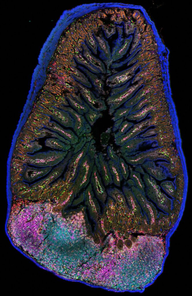
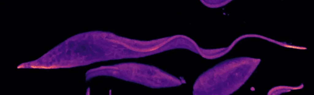
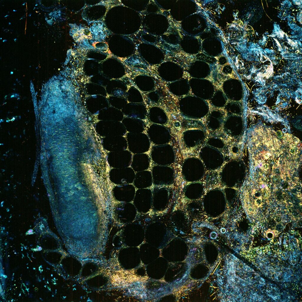
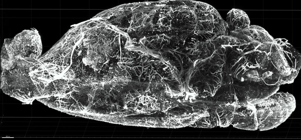
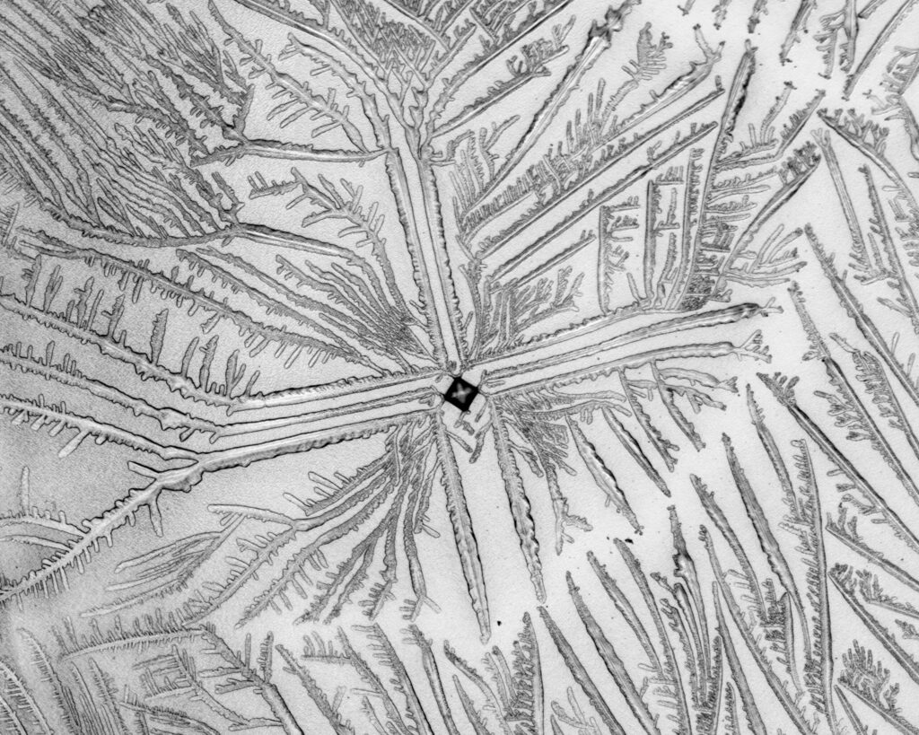
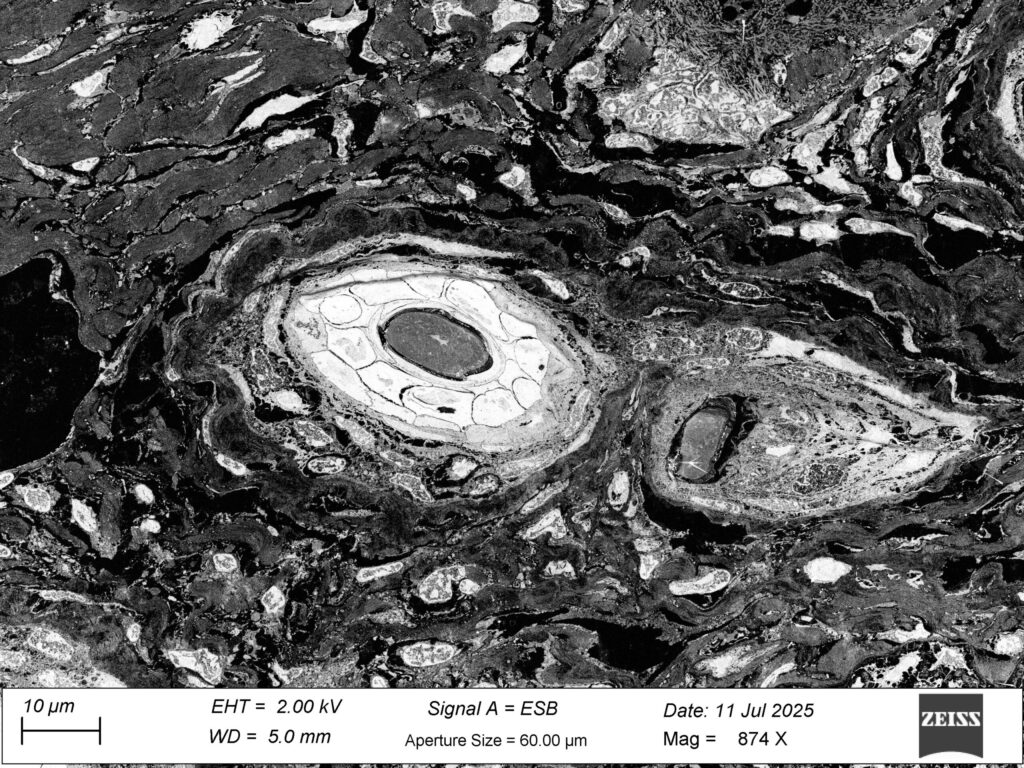
1st Place: Nicolas Barois, BioImaging Center Lille (BICeL)

Gut Flower-Flora
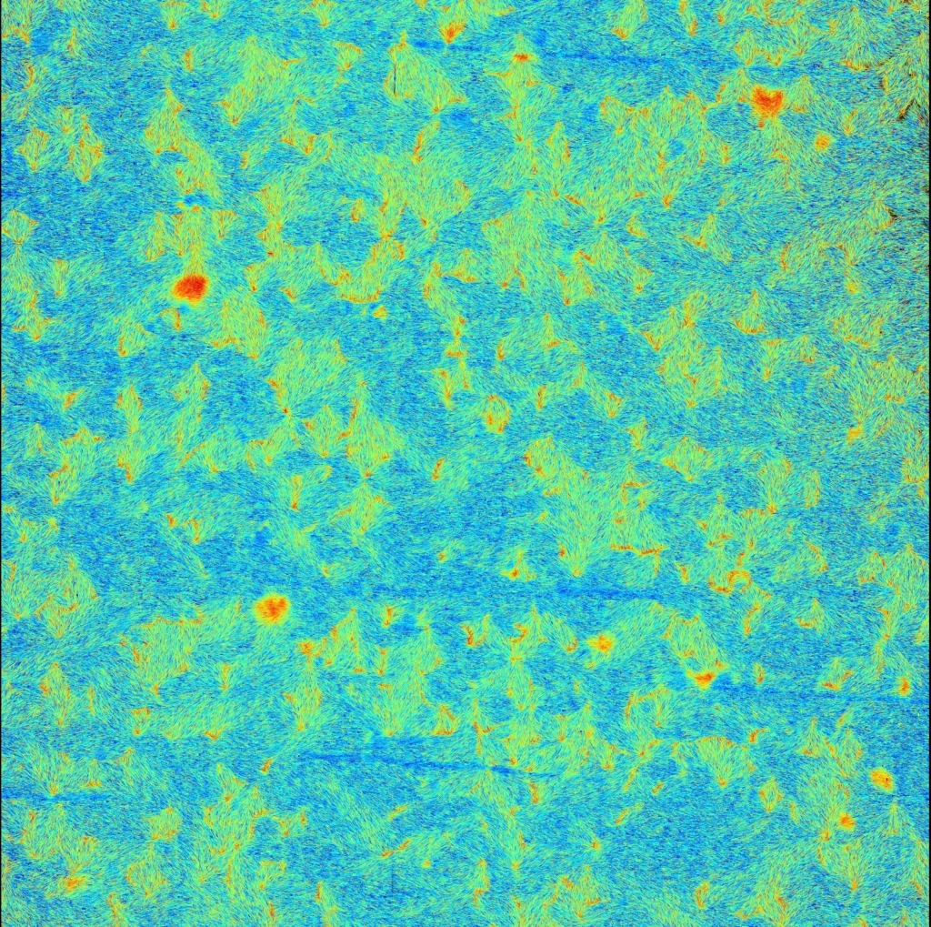
Cross-sectional view of a thin slice of mouse gut. By cutting a thin slice of the gut, the inner went out. Normally I cut the gut in small tubes, which are cut in two longitudinal pieces. I kept this piece because I was curious to see it with the SEM.
Scanning Electron Microscopy
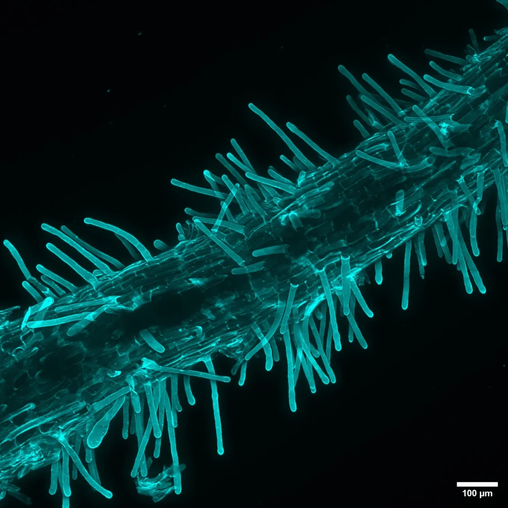
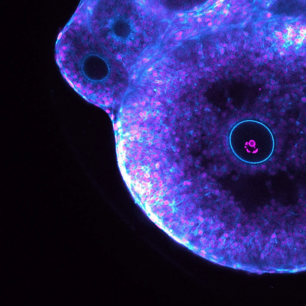
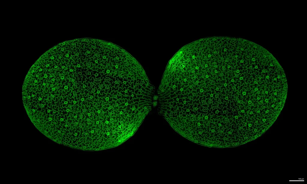
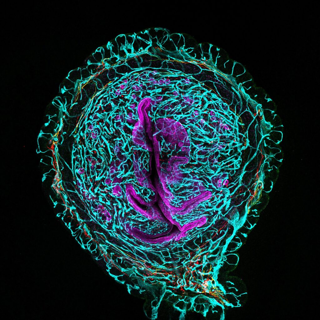
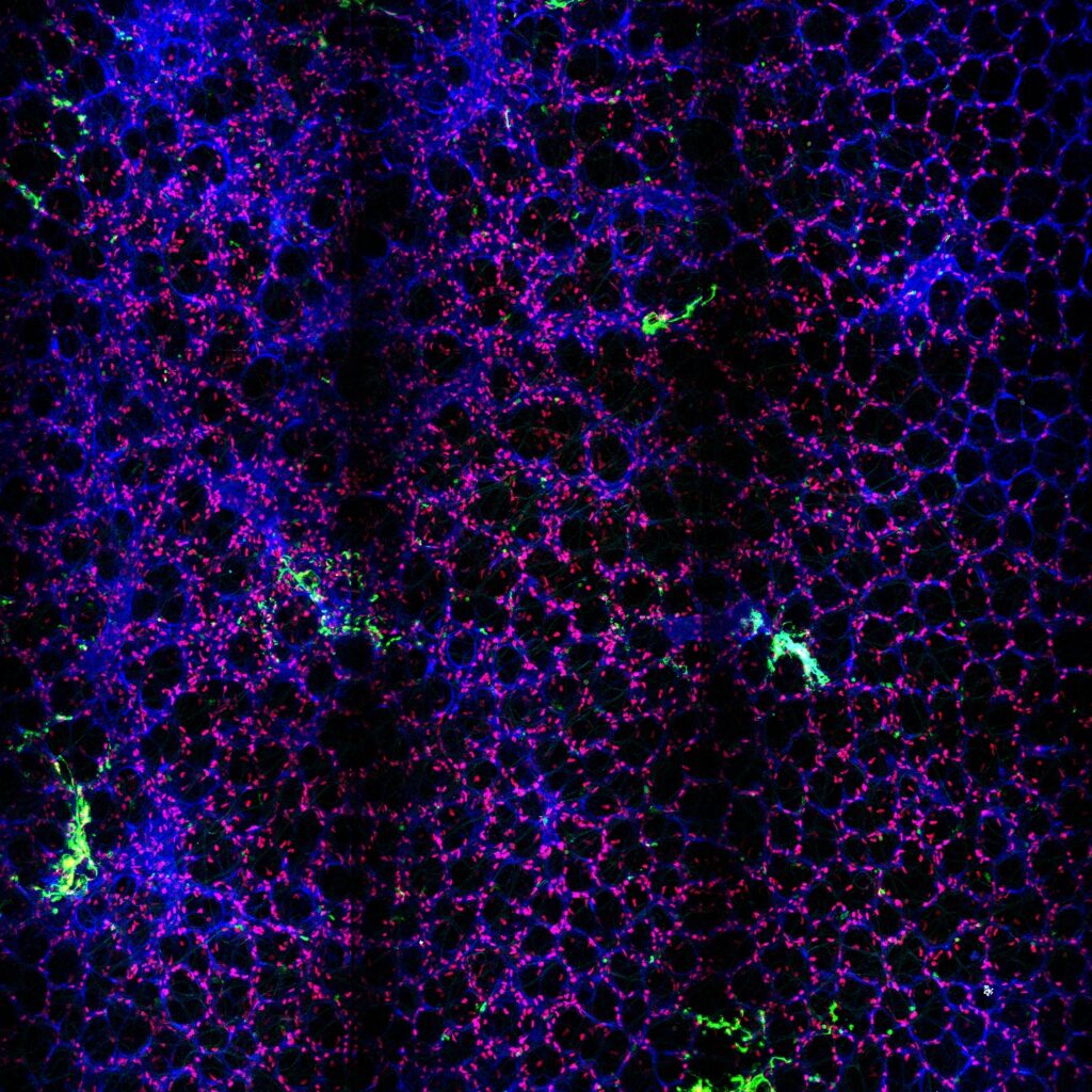
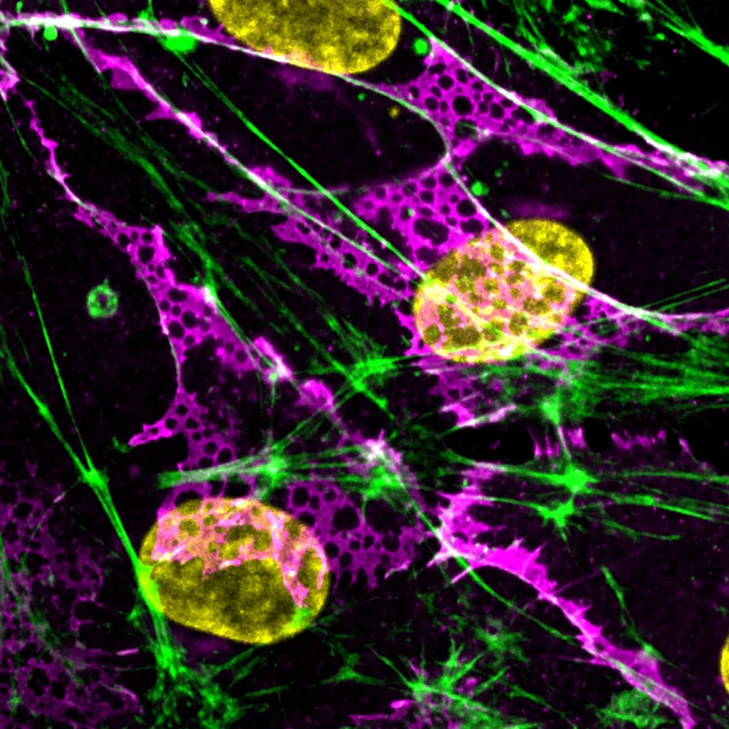
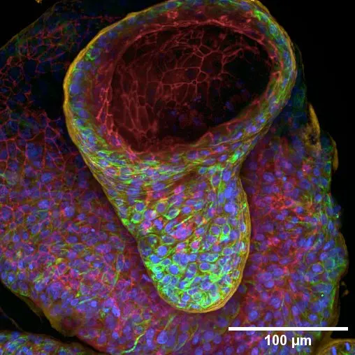
2nd Place: Vishwadeep Mane, Plant Reproduction and Development Laboratory (RPD, ENS Lyon)

The Puzzled Awakening
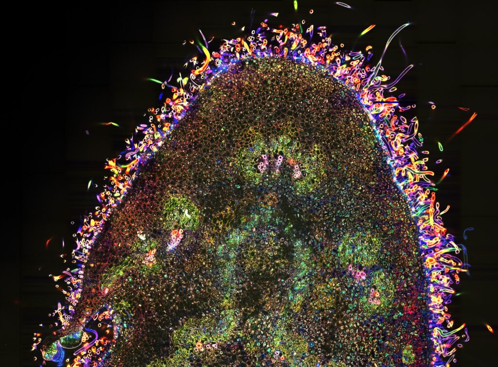
Cotyledons, the first leaves of a plant, break free from the seed to launch life after germination. Emerging as a pair, they unfold into a nearly perfect, circular lamina that captures light for photosynthesis. At the microscopic scale, their surface reveals a mosaic of interlocking puzzle-shaped cells, dotted with stomata. These intricate cell shapes are nature’s solution to balancing internal pressure, relieving mechanical stress, while guiding growth into a robust and harmonious form. Between the cotyledons rises the first genuine leaf, a quiet promise of the plant’s future. Cotyledons mark the awakening of life, and in their puzzled cells, we see both resilience and beauty.
Confocal Laser Scanning Microscopy
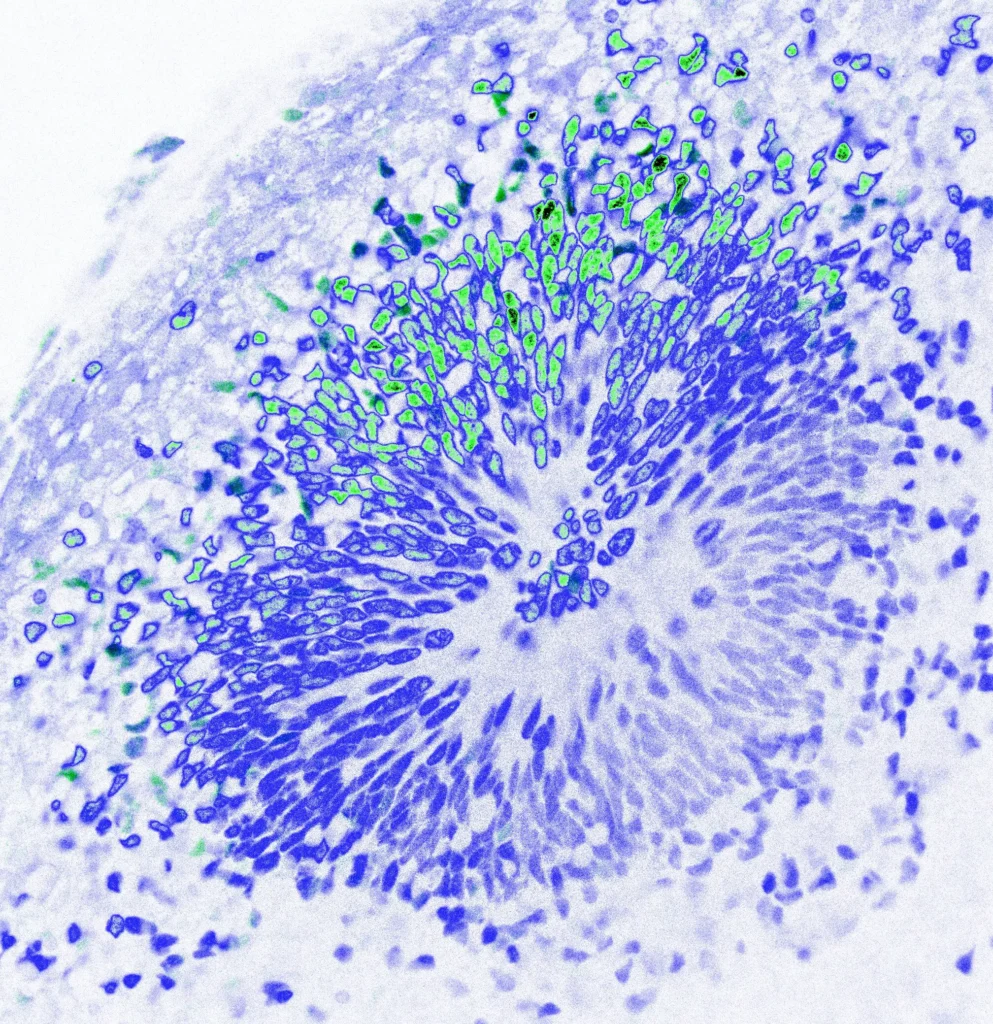
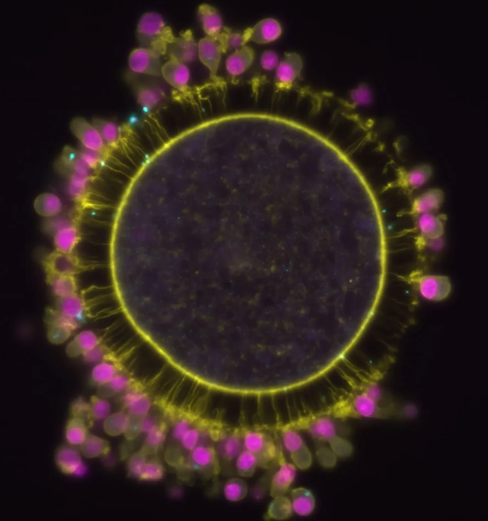
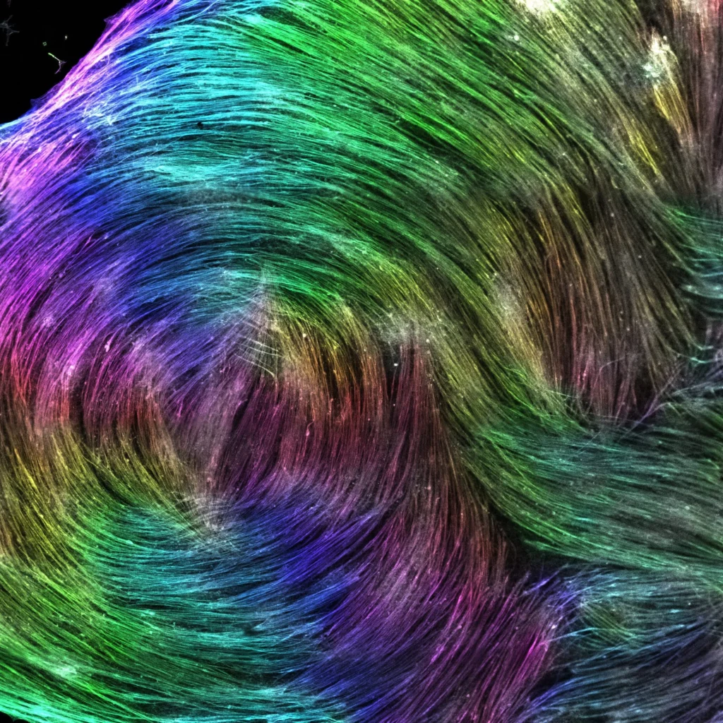
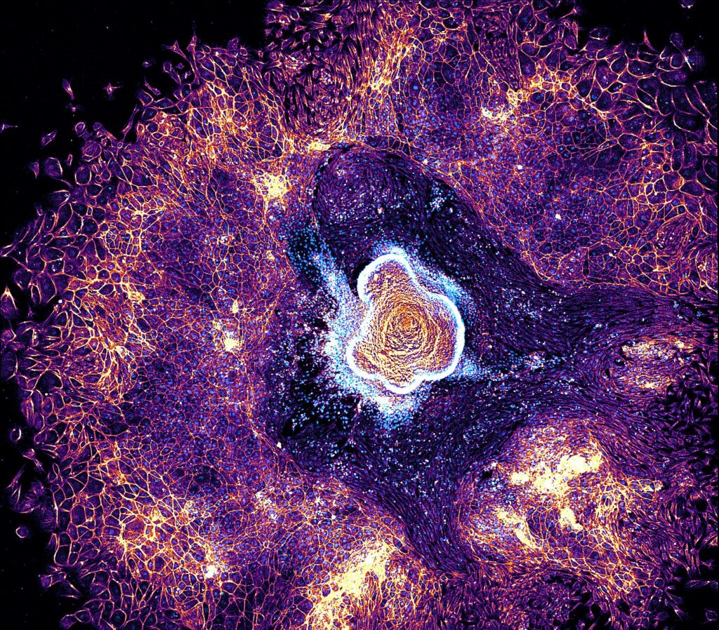
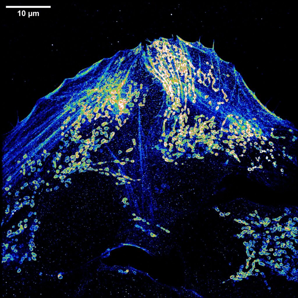
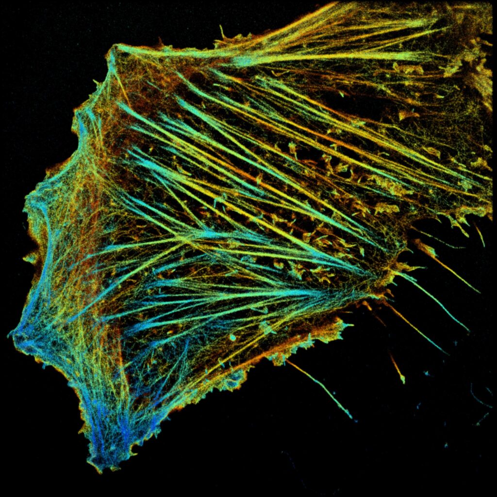
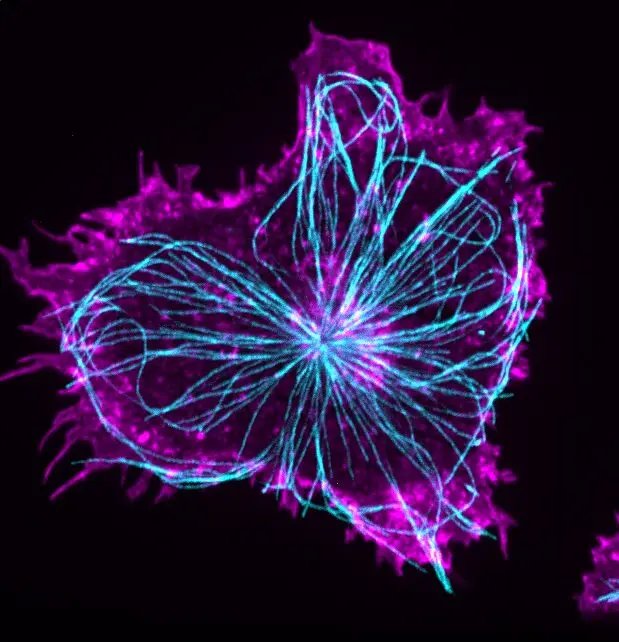
3rd Place: Simli Dey, Membranes and Cellular Functions Team, Institut Curie
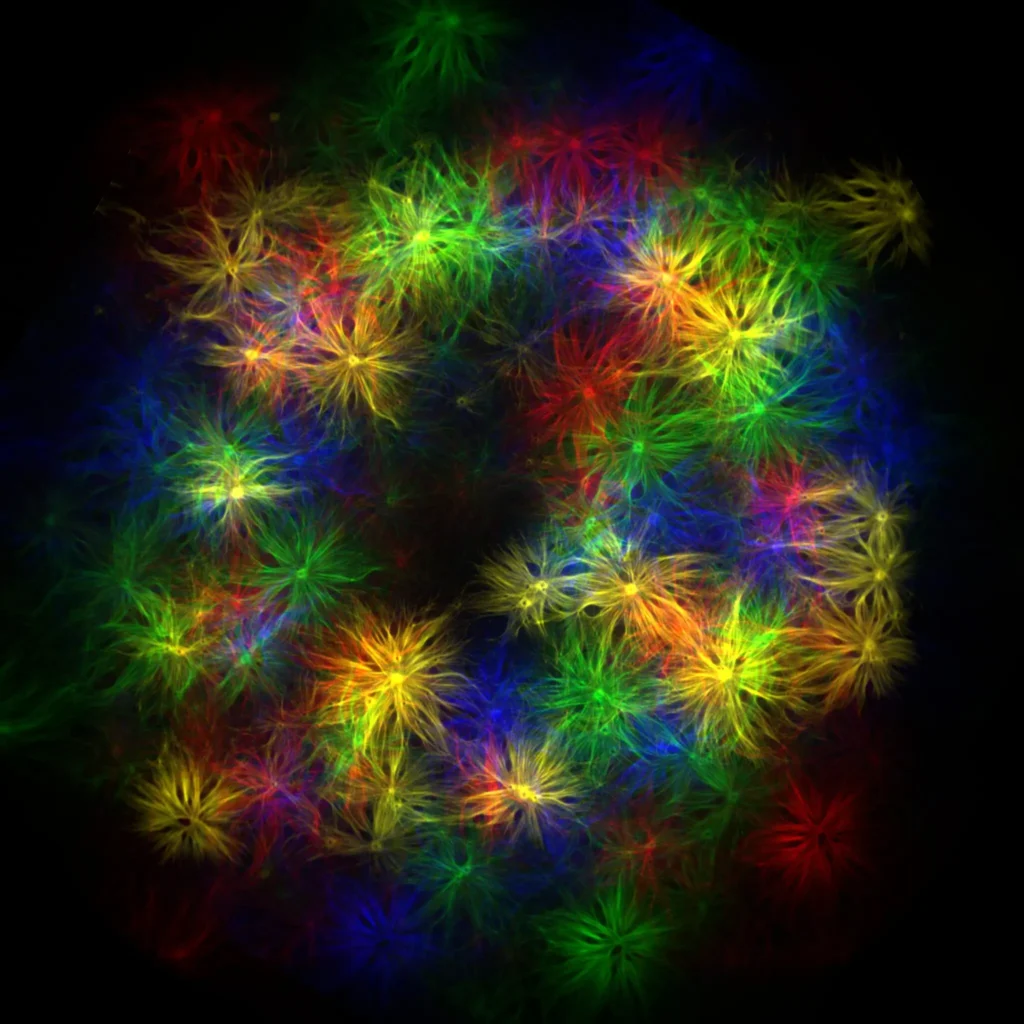
Kaleidoscope
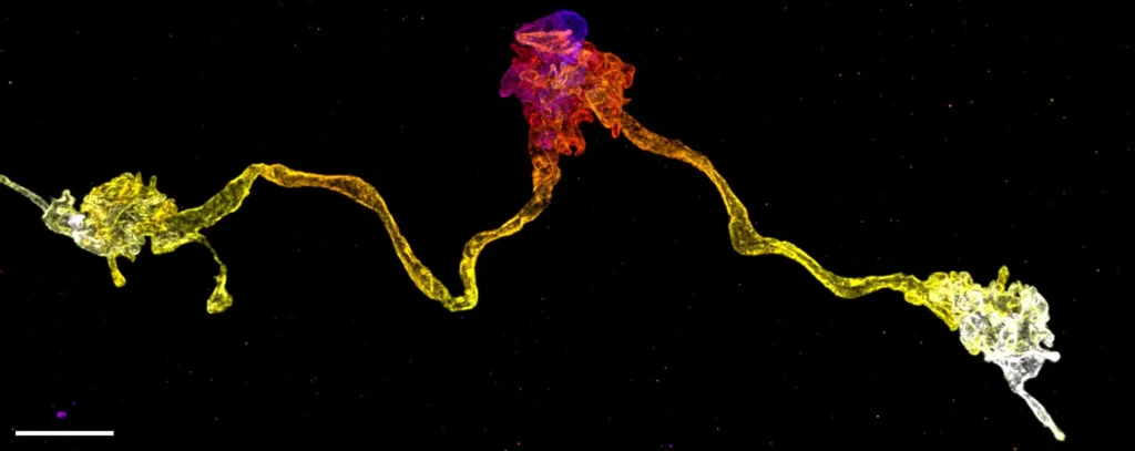
Directing branched actin filament growth from a curved lipid membrane site.
Total Internal Reflection Fluorescent Microscopy
Congratulations to the winners!
Explore all the images submitted here:


unit=\u00B5m

unit=\u00B5m


unit=micron