The Little Venice of Biology: What Lies Behind The Canals
Researchers and imaging engineers know better than anyone that an image is always more than meets the eye. Let us honor the winning image of our 2017 Image Contest by delving a little deeper into what lies behind these Venice canals.
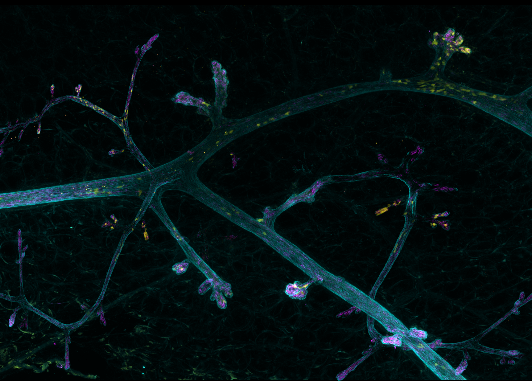
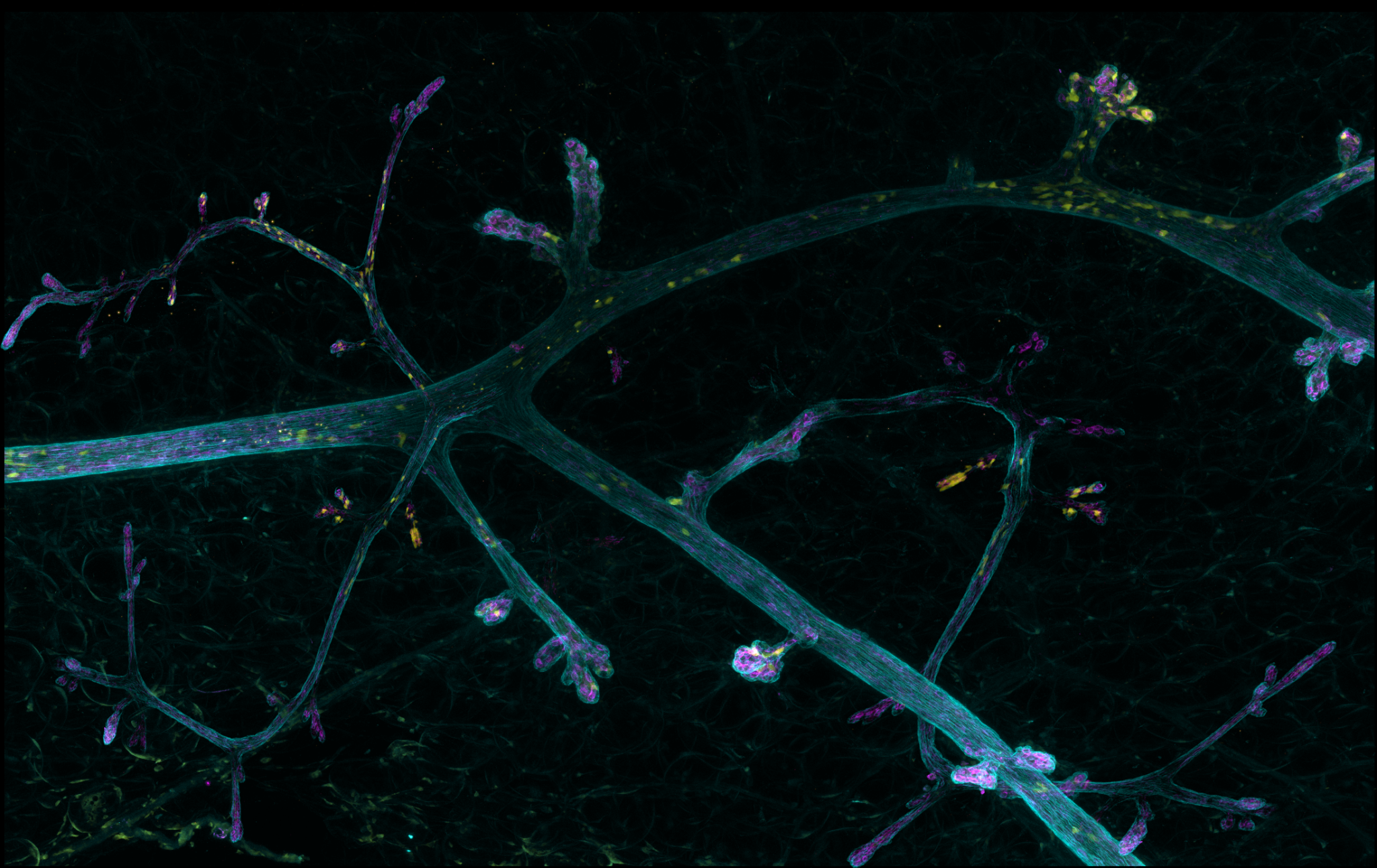
The image represents a large part of mice mammary gland. The canals spreading out like branches, are composed of two layers of cells: the epithelial epithelial cells – stained by anti-keratin 8 (in pink) – are surrounded by myoepithetial cells (in blue) stained by an anti-Smooth Muscle Actin. These cells, thanks to their contractile properties, participate in the? secretions from the gland. The yellow cells are modified cells, overexpressing tomato protein. The adipocytes of the mammary gland can be seen in the background.
The image was made by Marie Irondelle (PICT-IBiSA Biomaging Cell and Tissue Core Facility, Curie Institute), using a confocal microscope. It is a mosaic reconstruction – using the tile scan technique -from several depths of field with a range of 110 µm. The lighter areas are higher up in the sample, while the darker areas correspond to zones deeper in the tissue. The final projection measures 1.2 by 0.78 mm. The ducts shown in the picture have a diameter of 50 to 60 microns.
Carine Rossé and Emilie Lagoutte in the lab of Philippe Chavrier (membrane & Actin dynamics lab), from the Curie Institute, set out to study the behavior and movement of few cancerous cells inside the mammary gland. However, such a study required the development of alternative methods of observation, from the traditional methods of analysis, in order to observe the localization of the cells in the whole gland. The solution came from a 2016 Nature Methods paper entitled “Shrinkage-mediated imaging of entire organs and organisms using uDISCO”[1]. In this method, the researchers described the advantages of the uDISCO tissue-clearing protocol for the analysis of large samples, compared to other well-known methods. After adapting the protocol, Emilie Lagoutte was able to clear entire mice. She obtained and stained mammary glands that were shrunk by about 70% compared to their original size, cleared. The advantages of this technique are numerous; in particular, there is no more tissue loss due to slicing samples, the fluorescence can be maintained for a few weeks, and the reduced size of the sample allows to visualize the whole organ, leading to a higher chance to detect the zone of interest.
Imaging facilities and researchers have to work hand-in-hand to produce the best results possible. Marie Irondelle stressed the necessity for researchers to be educated about imaging technologies and their limitations; a state-of-the-art microscope will never be able to compensate for a low-quality sample. In the case of The Little Venice, the PICT-IBiSA Biomaging Cell and Tissue Core Facility collaboration with the Chavrier lab is undoubtedly a winning one.