The France BioImaging Image Contest is back for its 3rd edition!
This image contest is open to all within the imaging community: core facility staff and users, R&D labs teams and co-workers, students… Submit your best microscopy images for a chance to showcase your skills, research and creativity to the French bioimaging community and beyond, allowing people to see the visual appeal of the life sciences. Images from the contest will be featured on France BioImaging communication tools, online and in print.
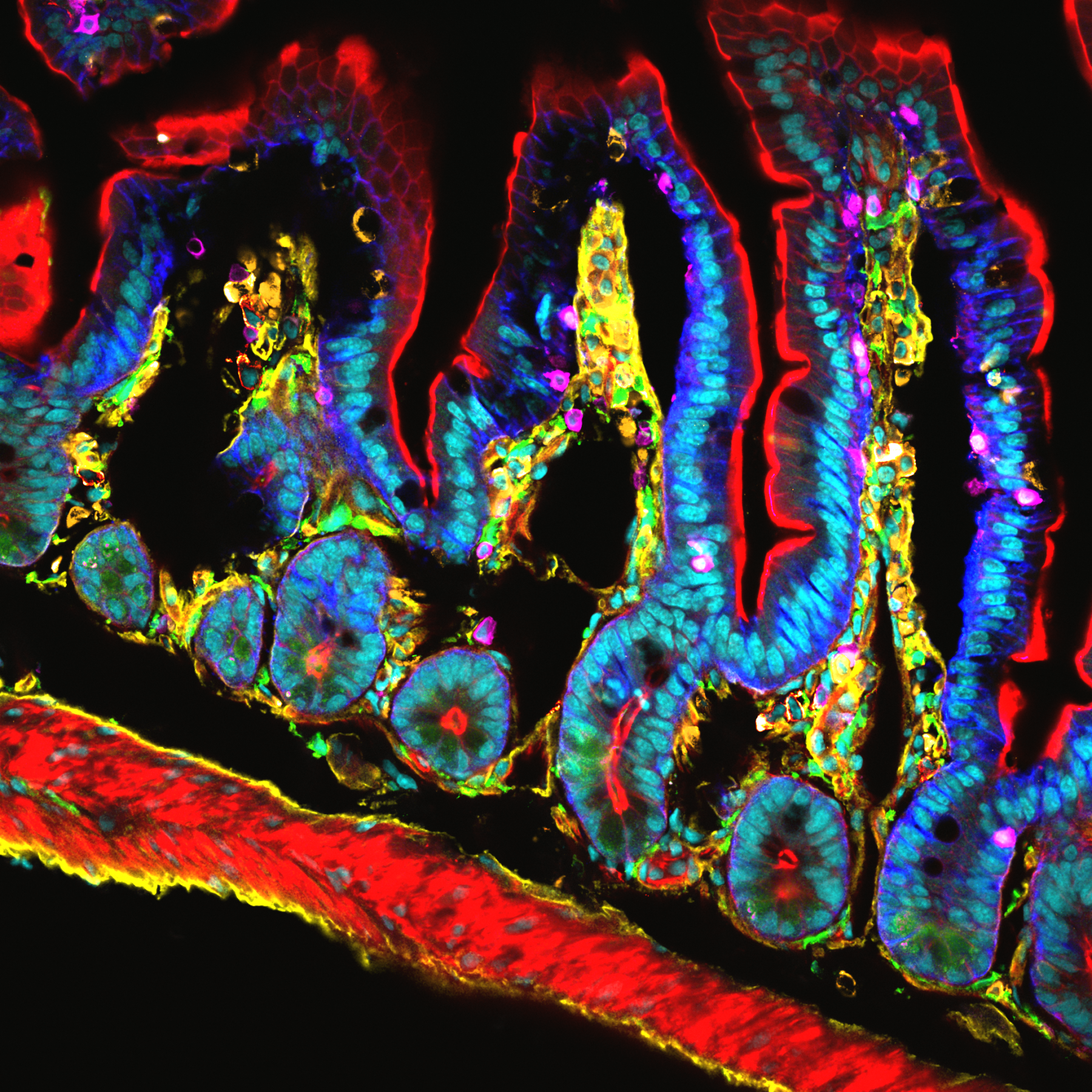
France BioImaging and all the French community aims to develop and promote innovative imaging technologies and methods. But microscopy images can also take an artistic, creative look and make the invisible world beautiful.
We are all eager to see your work !
Prizes
1 to 3 images will be awarded depending on the quantity and quality of the entries submitted. France BioImaging will cover the registration fees for one 2022 microscopy related event of the winners’ choice (FOM, ELMI, EMC, COMULIS conference, etc.).
Important: Only French or foreign participants affiliated to a French institution can enter the contest. Foreign participants non-affiliated to a French institution can submit images and will be featured in the gallery, but will not be evaluated as part of the contest.
Submission deadline: Friday, October 15th, 2021, 23h59 UTC+2.
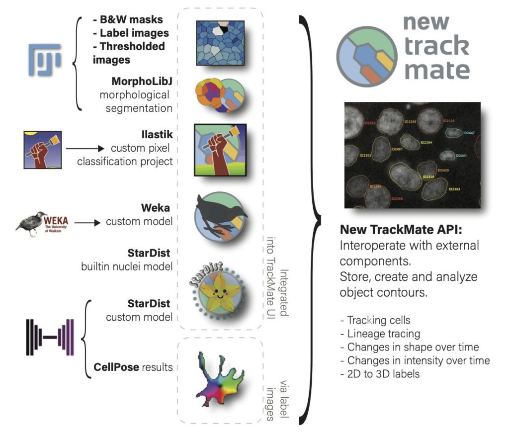
A new version of TrackMate is available now, with major changes that improve its versatility. TrackMate now integrates state-of-the-art segmentation algorithms from machine-learning and deep-learning such as StarDist, Ilastik and Weka.
TrackMate[1] is a Fiji plugin that address cell or organelle tracking in Life-Science microscopy images. Its main goals are to be user-friendly, interoperable and to serve as a platform to accelerate the development of novel tracking algorithms and analysis pipelines.
With this new version we rewrote almost entirely TrackMate so that it can integrate state-of-the-art segmentation algorithms and benefit from their output. For instance, TrackMate can now store, display, save, load and exploit object contours in 2D.
We also made a new application programming interface that will facilitate and accelerate reusing TrackMate in other analysis pipelines and allow 3rd party contributors to add new segmentation algorithms in TrackMate in an easy way. We used this API ourselves to add 7 new segmentation algorithms to TrackMate:
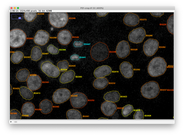
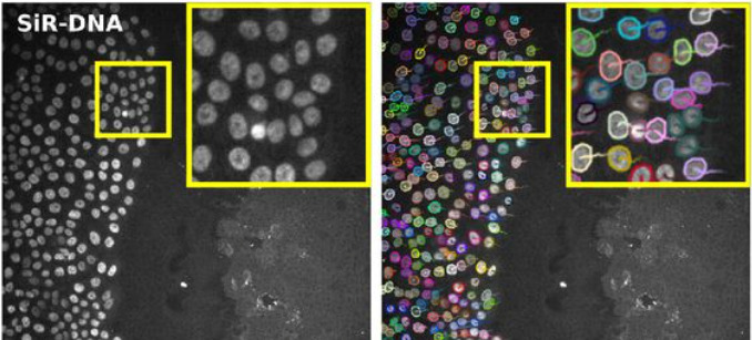
For instance, the StarDist[2] algorithm is integrated as two different detectors. The first one uses the built-in deep-learning model that can segment cell nuclei in fluorescence image in a wide range of situation. The robustness of the StarDist algorithm in turn positively impacts the robustness of tracking and allows for better detection of cell divisions with TrackMate tracking algorithms. This will facilitate cell migration studies.
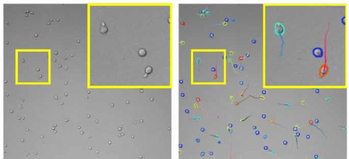
The TrackMate StarDist integration also allows for specifying and using a custom deep-learning model. For instance, we trained a specific model to detect T-cells imaged in bright-field microscopy and track them over time. Before the emergence of such detection algorithms, the tracking of label-free cells was difficult.
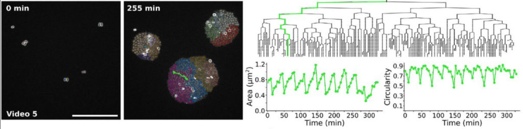
We also integrated the ilastik[3] segmentation software. A TrackMate user can input an ilastik classifier to detect objects then track them. We used them to study the bacterial growth of Neisseria meningitidis clones. The output of this analysis pipeline offers the lineage of each single cell along with its morphology and how it evolves across cell divisions.
The new capabilities of TrackMate can be used to address applications beyond tracking. For instance, it is now possible to use TrackMate to perform the segmentation of 3D objects using a slice-by-slice approach. This approach consists in segmenting objects in each 2D section of a 3D stack, then merging the segmentation results along Z in a subsequent step. This can be done in TrackMate, using the tracking step for merging. We implemented a novel tracking algorithm to foster this application, the overlap tracker. We could use this approach combining the cellpose[4] algorithm in 2D to segment 3D images of Arabidopsis thaliana floral meristem.
There are several other algorithms that are now offered to the TrackMate user, within a user-friendly software meant to interoperate with the key software of bioimage analysis. More importantly, TrackMate is an open-source academic software, and its new API will foster the development of new analysis pipeline with TrackMate and the integration of new algorithms by other developers, increasing the breadth of applications it can address for Life-Science researchers.
Bringing TrackMate in the era of machine-learning and deep-learningDmitry Ershov, Minh-Son Phan, Joanna W. Pylvänäinen, Stéphane U. Rigaud, Laure Le Blanc, Arthur Charles-Orszag, James R. W. Conway, Romain F. Laine, Nathan H. Roy, Daria Bonazzi, Guillaume Duménil, Guillaume Jacquemet, Jean-Yves TinevezbioRxiv 2021.09.03.458852; doi: https://doi.org/10.1101/2021.09.03.458852
[1] https://imagej.net/plugins/trackmate/
[2] Alejandro F. Frangi, Julia A. Schnabel, Christos Davatzikos, Carlos Alberola-López, and Gabor Fichtinger Uwe Schmidt, Martin Weigert, Coleman Broaddus, and Gene Myers. Cell detection with star-convex polygons. In Alejandro F. Frangi, Julia A. Schnabel, Christos Davatzikos, Carlos Alberola-López, and Gabor Fichtinger, editors, Medical Image Computing and Computer Assisted Intervention – MICCAI 2018, pages 265–273, Cham, 2018. Springer International Publishing. doi:10.1007/978-3-030-00934-2_30.
[3] Stuart Berg, Dominik Kutra, Thorben Kroeger, Christoph N Straehle, Bernhard X Kausler, Carsten Haubold, Martin Schiegg, Janez Ales, Thorsten Beier, Markus Rudy, Kemal Eren, Jaime I Cervantes, Buote Xu, Fynn Beuttenmueller, Adrian Wolny, Chong Zhang, Ullrich Koethe, Fred A Hamprecht, and Anna Kreshuk. ilastik: interactive machine learning for (bio)image analysis. Nature Methods, 16(12):1226–1232, 2019. ISSN 1548-7105. doi:10.1038/s41592-019-0582-9.
[4] Carsen Stringer, Tim Wang, Michalis Michaelos, and Marius Pachitariu. Cellpose: a generalist algorithm for cellular segmentation. Nature Methods, 18(1):100–106, jan 2021. doi:10.1038/s41592-020-01018-x.
France BioImaging primary mission is to develop, promote, disseminate and provide access to innovative instruments and imaging technologies in the field of bioimaging to scientists. Fostering the technological transfers is at the heart of this mission, and for this France BioImaging relies on a strong association of leading R&D research teams with core facilities.
However, several bottlenecks exist and often hamper or prevent successful technology transfer:
A lack of human resource leads to difficulties in transferring and stabilizing the technology which is not enough user-friendly
A technology that is too specific, with not enough user base
A difficulty to contract with industry through institutional offices for industrial valuation
In the context of image analysis: the instability of open software economical model, inter-operability, large data handling and algorithm complexity
As a way to tackle these bottlenecks, France BioImaging launched in January 2021 its first “FBI Internal Call 2021: Technology transfer from the R&D teams to the core facilities” to promote the transfer of new technologies (instrumentation, probes, staining methods, software, data analysis or data visualization) from the R&D teams to the facilities of France BioImaging, for access and service to end-users. The outcome of the transfer project had to ensure for the prototype to be usable by the end-users until the interpretation of the data. The project had also to include a sustainability plan and a training plan to guide both facility staff and end-users toward autonomy.
The project selection was organized by the National Coordination of France-BioImaging and applications were assessed according to the following evaluation criteria:
Innovation and originality of the proposal
Scientific quality, implementation, timeline
Competitive positioning
Adequacy of resources with the proposed project
Economic impact and tech transfer potential and perspectives
Estimation of the user market and potential for user adoption
Plan for training and sustainability.
For the first edition of the “FBI Internal Call 2021: Technology transfer from the R&D teams to the core facilities”, 5 projects were selected:
Icy@FBI: Jean-Christophe Olivo-Marin (IPDM Node): Broadening the scope of applications of Icy (http://icy.bioimageanalysis.org/) by implementing several key new bioimage analysis components
BIC-HCS-SMLM: Jean-Baptiste Sibarita (Bordeaux Node), Technological transfer of a Single-Molecule-based High Content Screening platform to the Bordeaux Imaging Center
CloudFISH: Marcello Nollmann (Montpellier Node), A tool for the analysis of single-molecule RNA and DNA FISH images
MorphoNet: Emmanuel Faure (Montpellier Node), An interactive online morphological browser to explore complex multi-scale data
BioImageIT (https://bioimageit.github.io/#/about): Jean Salamero, Sylvain Prigent (IPDM Node), An open source framework for integration of image data management with analysis
Each selected project was awarded with a 80k€ grant for salary and/or equipment, and several positions are currently available: https://france-bioimaging.org/jobs/
This call will be renewed in 2023.
The France BioImaging Image Contest is back for its 3rd edition!
This image contest is open to all within the imaging community: core facility staff and users, R&D labs teams and co-workers, students… Submit your best microscopy images for a chance to showcase your skills, research and creativity to the French bioimaging community and beyond, allowing people to see the visual appeal of the life sciences. Images from the contest will be featured on France BioImaging communication tools, online and in print.
France BioImaging and all the French community aims to develop and promote innovative imaging technologies and methods. But microscopy images can also take an artistic, creative look and make the invisible world beautiful.
We are all eager to see your work !
Prizes
1 to 3 images will be awarded depending on the quantity and quality of the entries submitted. France BioImaging will cover the registration fees for one 2022 microscopy related event of the winners’ choice (FOM, ELMI, EMC, COMULIS conference, etc.).
Important: Only French or foreign participants affiliated to a French institution can enter the contest. Foreign participants non-affiliated to a French institution can submit images and will be featured in the gallery, but will not be evaluated as part of the contest.
Submission deadline: Friday, October 15th, 2021, 23h59 UTC+2.
We use cookies on our website to give you the most relevant experience by remembering your preferences and repeat visits. By clicking “Accept All”, you consent to the use of ALL the cookies. However, you may visit "Cookie Settings" to provide a controlled consent.
This website uses cookies to improve your experience while you navigate through the website. Out of these, the cookies that are categorized as necessary are stored on your browser as they are essential for the working of basic functionalities of the website. We also use third-party cookies that help us analyze and understand how you use this website. These cookies will be stored in your browser only with your consent. You also have the option to opt-out of these cookies. But opting out of some of these cookies may affect your browsing experience.
Necessary cookies are absolutely essential for the website to function properly. These cookies ensure basic functionalities and security features of the website, anonymously.
Cookie
Duration
Description
cookielawinfo-checkbox-analytics
11 months
This cookie is set by GDPR Cookie Consent plugin. The cookie is used to store the user consent for the cookies in the category "Analytics".
cookielawinfo-checkbox-functional
11 months
The cookie is set by GDPR cookie consent to record the user consent for the cookies in the category "Functional".
cookielawinfo-checkbox-necessary
11 months
This cookie is set by GDPR Cookie Consent plugin. The cookies is used to store the user consent for the cookies in the category "Necessary".
cookielawinfo-checkbox-others
11 months
This cookie is set by GDPR Cookie Consent plugin. The cookie is used to store the user consent for the cookies in the category "Other.
cookielawinfo-checkbox-performance
11 months
This cookie is set by GDPR Cookie Consent plugin. The cookie is used to store the user consent for the cookies in the category "Performance".
viewed_cookie_policy
11 months
The cookie is set by the GDPR Cookie Consent plugin and is used to store whether or not user has consented to the use of cookies. It does not store any personal data.
Functional cookies help to perform certain functionalities like sharing the content of the website on social media platforms, collect feedbacks, and other third-party features.
Performance cookies are used to understand and analyze the key performance indexes of the website which helps in delivering a better user experience for the visitors.
Analytical cookies are used to understand how visitors interact with the website. These cookies help provide information on metrics the number of visitors, bounce rate, traffic source, etc.
Advertisement cookies are used to provide visitors with relevant ads and marketing campaigns. These cookies track visitors across websites and collect information to provide customized ads.