The France-BioImaging Image Contest is back for its 6th edition!
This image contest is open to all within the imaging community: core facility staff and users, R&D labs teams and co-workers, students… Submit your best microscopy images for a chance to showcase your skills, research and creativity to the French bioimaging community and beyond, allowing people to see the visual appeal of the life sciences. Images from the contest will be featured on France-BioImaging communication tools, online and in print.
France-BioImaging and all the French community aims to develop and promote innovative imaging technologies and methods. But microscopy images can also take an artistic, creative look and make the invisible world beautiful.
We are all eager to see your work !
Prizes
1 to 3 images will be awarded depending on the quantity and quality of the entries submitted. France-BioImaging will cover the registration fees for one 2024 microscopy related event of the winners’ choice (FOM, ELMI, EMC, COMULIS conference, etc.).
Important: Only French or foreign participants affiliated to a French institution can enter the contest. Foreign participants non-affiliated to a French institution can submit images and will be featured in the gallery, but will not be evaluated as part of the contest.
Submission deadline: Friday, November 8th, 2024, 23h59 UTC+2.
France-BioImaging and all the French community aims to develop and promote innovative imaging technologies and methods. But microscopy images can also take an artistic, creative look and make the invisible world beautiful, allowing people to see the visual appeal of the life sciences.
We enjoyed the diversity of the images submitted with many different microscopy techniques, models and applications represented. A big thank you to all the participants!
The National Coordination Team and the Executive Board are proud to announce the winners of the FBI Image Contest 2023:
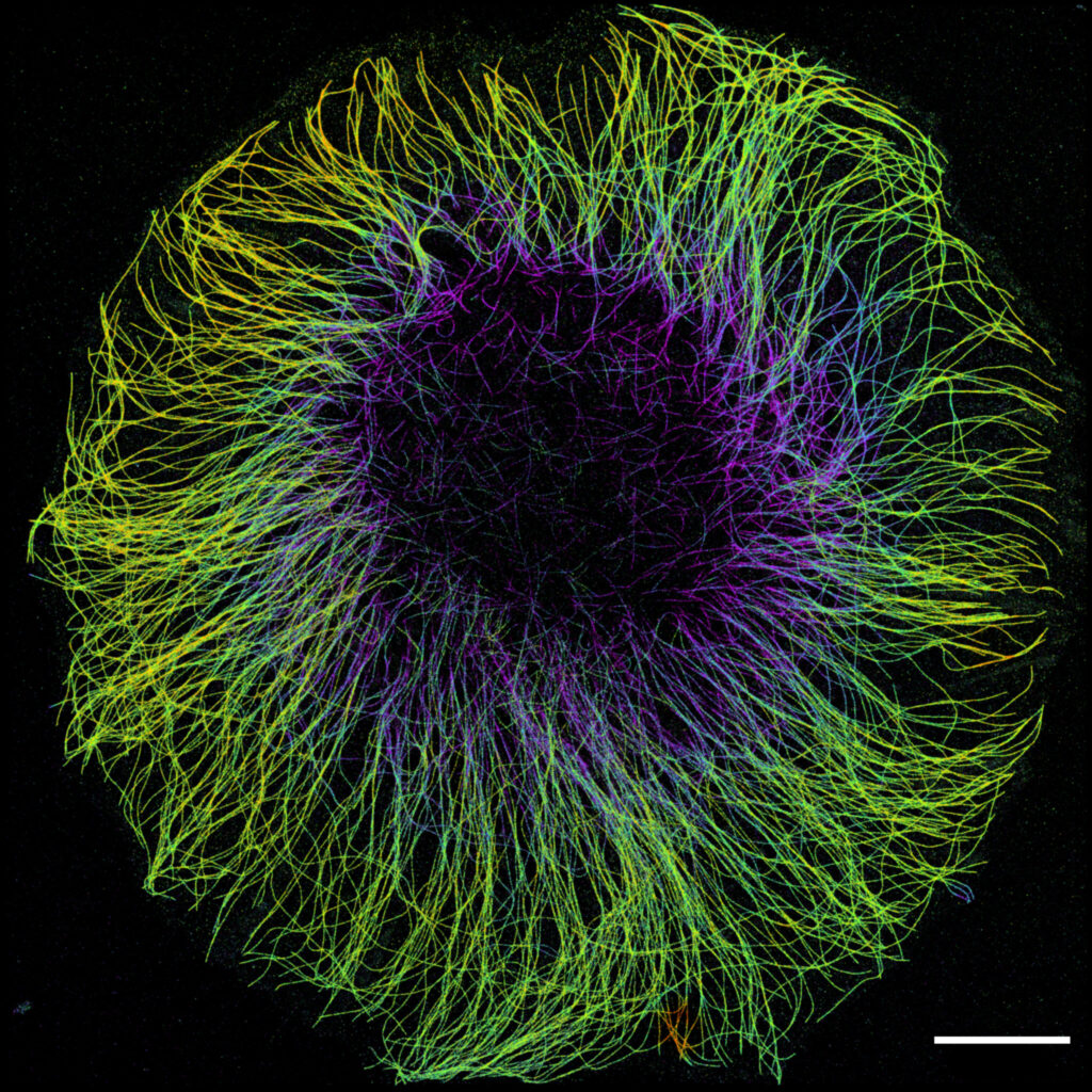
1st Place: Laurent LE, Lévêque-Fort Team, Institut des Sciences Moléculaires d’Orsay
“In the blink of an eye”
COS7 fixed cell. Alpha-tubulin labeled with DNA-PAINT and imaged with Atto 647N. Axial information is obtained by virtual-SAF measurement known as DONALD.
SMLM Fluorescence Microscopy with DNA-PAINT with DONALD detection
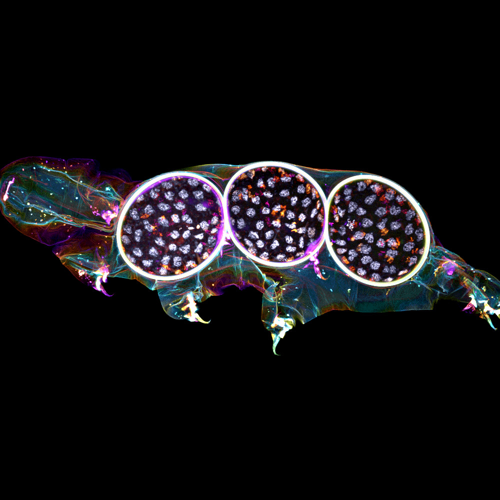
2nd Place:Gonzalo QUIROGA-ARTIGAS, Team Contrôle cytoplasmique de la stabilité du génome, Centre de recherche en Biologie Cellulaire de Montpellier
“Tardigrade embryos protected by mother’s molt”
Tardigrades commonly align the time of molting with egg laying. In this image we observe a tardigrade molt covering three developing embryos (DNA in white). The microscopy technology applied was confocal microscopy, and the research aimed to investigate the synchronization of embryo development in tardigrades.
Confocal microscopy
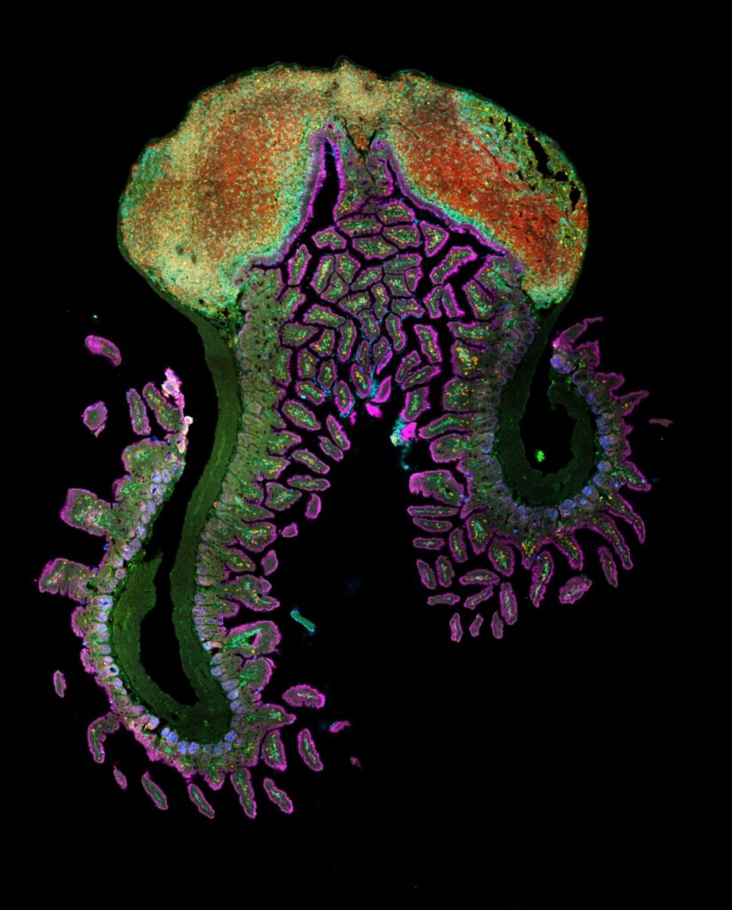
3rd Place:Hugues LELOUARD, Gorvel team, Centre d’Immunologie de Marseille Luminy
“Intestinal octopus”
Small intestine section from a LyzM-eGFP mouse containing one Peyer’s patch and stained for proliferative cells (Ki-67, yellow), Paneth cells (UEA-I, blue), epithelial cells (EpCAM, magenta), naive B cells (IgD, red), T cells (CD3, orange), helper T cells/macrophages (CD4, cyan), phagocytes (CD11c, turquoise), monocyte-derived phagocytes (GFP, green).
10-color spectral confocal microscopy
Congratulations to the winners!
Explore all the images submitted here:
As stated in the Terms & Conditions of the contest, foreign participants non-affiliated to a French institution are featured in the gallery, but were not evaluated as part of the contest.
As the 2023 edition of the France-BioImaging Image Contest admissions is still running, we wanted to highlight our previous winners and their projects. Here is a quick throwback to our 2022 winners.
Before getting to the heart of the matter, we want to remind you that you still have time (before November 10th) to submit your best images and try to win your registration fees for one 2024 microscopy-related event! Please make sure you upload your images on the following link:
Last year, we enjoyed the winning images submitted for their artistic take and their quality. Thanks to Carole SIRET, Magalie BENARD and Frédéric FERCOQ for their beautiful images!
1st Place: Carole SIRET, Van de Pavert Team, Centre d'Immunologie de Marseille-Luminy
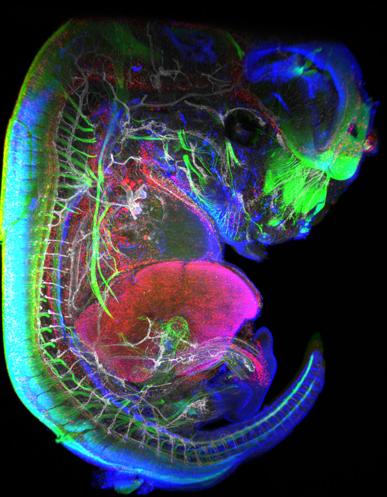
"Little Monster"
The embryonic formation of lymph nodes, small organs essential for the immune response, is now known. Using light sheet microscopy, scientists were able to determine the dynamics at work in this 13.5-day-old mouse embryo. In blue, the lymphoid cells (LTi), derived from the haematogenous endothelium, a specific tissue of the embryo. They pass into the liver where they proliferate before migrating through the body to give rise to lymph nodes. The 3D information obtained thus makes it possible to follow the interactions of lymph nodes with their environment, in particular with nerve cells, in green, and blood vessels, in white. The lymphatic endothelial cells and some macrophages are visible in red.
Lightsheet Microscopy
Carole Siret is a Research engineer, expert in Lightsheet microscopy, at the Centre d’Immunology Marseille Luminy (CIML) since 2018. She is working in Dr Serge van de Pavert team where they study immune system development. They are particularly interested in the lymph nodes (LN) formation during mouse embryogenesis.
The image she submitted is a projection from a lightsheet acquisition on the UMII (Miltenyi). This image illustrates an E13.5 mouse embryo stained for neurons, LTi (Tissue inducer cells which are the precursor cells for the lymph node), lymphatic and blood vessels. This acquisition was done in the context of the study of the role of Cxcl12 in embryonic LN formation. From previous work it is clear that Cxcl13 and Ccl21 are not expressed present near blood vessels, but it likely that some chemokines, possibly Cxcl12, could be expressed on the endothelial cells. We focus on Cxcl12 since this chemokine has shown to be important for the attraction of several hematopoietic cells. Although it was shown that the receptor for Cxcl12, Cxcr4, is expressed by the mature hematopoietic inducer cells, it is not clear whether it also expressed by the progenitor hematopoietic inducer cells. Next to the possible attraction of hematopoietic cells towards the lymph node anlagen, Cxcl12 is involved in the attraction of nerve fibers. Therefore, the possible role of Cxcl12 could be to both attract hematopoietic cells as well as nerve fibers to initiate a region which is permissive to form lymph nodes.
Thanks to the France-Bioimaging Image Contest, Carole participated to the SFI Congress, where, this year, it was a special joint conference both between the Société Française d’Immunologie (SFI) and the Deutsche Gesellschaft für Immunologie (DGfI). It was a great opportunity to exchange with people at the cutting edge of the immunology field.
2nd Place:Magalie BENARD, Plateforme de Recherche en IMAgerie CEllulaire de Normandie (PRIMACEN), Research infrastructure HeRacLeS, Inserm US 51, CNRS UAR 2026,
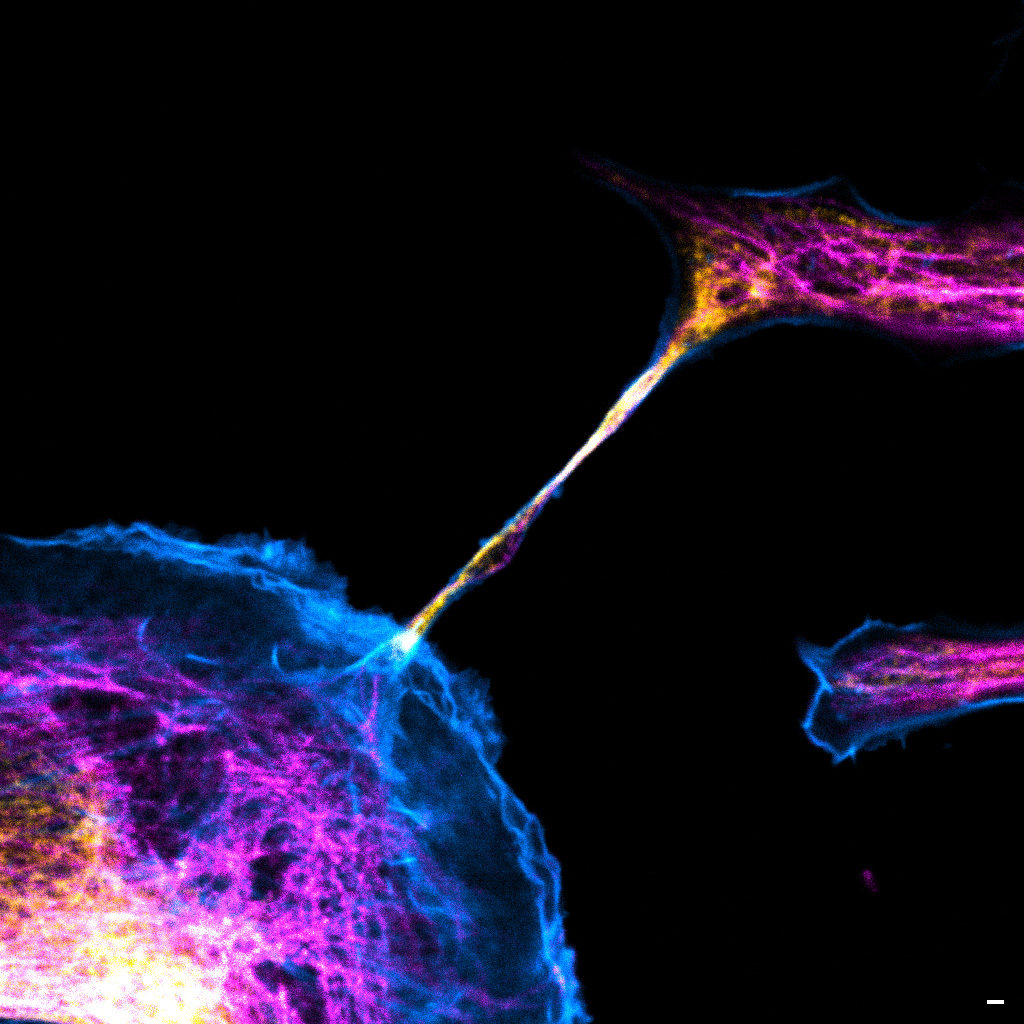
"The communication link with others"
Image of a cellular interconnection between two human tumor cells whose cytoskeleton has been labeled with anti-tubulin (ATTO-647N), anti-vimentin (AlexaFluor594) antibodies and with Phalloidin probe (AlexaFluor488). Scale bar 1µm.
Confocal microscopy
Magalie Bénard is a Research Engineer and the Technical Manager at the Cellular Imaging Facilty PRIMACEN (Plate-forme de Recherche en IMAgerie CEllulaire de Normandie).
The image she submitted is a confocal image representing a cellular interconnection tunneling nanotube (TNTs) between two human tumour cells. In a cancer case, some cells are able to express spontaneously TNTs with cytoskeleton protein composition corresponding to specific role of this communication mechanism. In the winning image, the TNT is composed of tubulin (magenta), actin (cyan) and vimentin (yellow) proteins. Called TNT1, this nanotube allows the transfer of intracellular elements such as RNA, proteins or organelles. Moreover, due to the thinness of TNTs, their photo-sensitivity and their fragility, live-cell imaging is technically challenging with regards to potentially damaging methods. Magalie and her team have developed an adapted method to observe TNTs in living cell with high resolution imaging (STED) enhanced by FLIM by using red and near infrared probes.
France-Bioimaging sponsored her participation to the ELMI (European Light Microscopy Initiative Meeting June 6-9, 2023) congress. During this event, she had the chance to present her project through a poster. This congress also offered a great opportunity to have an overview and the last updates on state-of-the-art imaging techniques.
3rd Place:Frédéric FERCOQ, Parasites et Protistes Libres (PPL), Museum National d'Histoire Naturelle
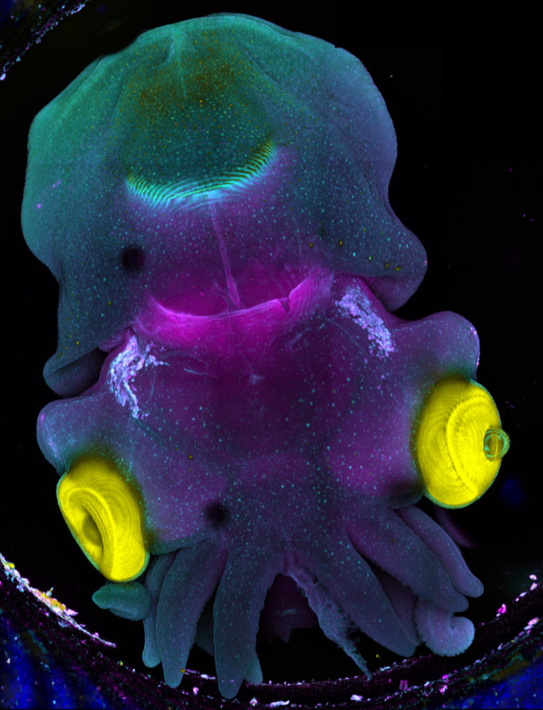
"Sepia"
Stage 25 cuttlefish embryo (Sepia officinalis) observed under a confocal microscope. The cuttlefish was cleared and the tissue autofluorescence was captured.
This image was produced in collaboration with Laure BONNAUD-PONTICELLI and Luis MOLINA from the BOREA laboratory.
Confocal microscopy
Frédéric Fercoq is a postdoc scientist in the Parasitology laboratory of the Muséum National d’Histoire Naturelle (MNHN) in Paris. My main interest is on how myeloid cells participate to the control of parasitic infections, but sometimes at the price of collateral tissue damage. This project involves a lot of microscopy of immune cells, parasites and host tissues to analyse the complex interactions taking place at the site of infection.
The image he submitted has nothing to do with his main project! As he has the chance to work on very different topics and models, this image was acquired as a proof of concept for imaging full embryos of the cuttlefish Sepia officinalis for Frédéric's colleagues Laure BONNAUD-PONTICELLI and Luis MOLINA (BOREA laboratory, MNHN). They work on the nervous system of cephalopod and on the influence of environmental factors during its development. They are now optimizing fluorescent staining for neuronal markers to test the effect of light on the nervous system in situ.
France-Bioimaging sponsored his participation to the FOM (Focus on Microscopy) 2023 congress in Porto. He had the chance to be granted the opportunity to both present his current project through a poster and to give an oral presentation. He was also amazed by the new avenues opened up by the cutting-edge imaging techniques presented throughout the conference.
The France-BioImaging Image Contest is back for its 5th edition!
This image contest is open to all within the imaging community: core facility staff and users, R&D labs teams and co-workers, students… Submit your best microscopy images for a chance to showcase your skills, research and creativity to the French bioimaging community and beyond, allowing people to see the visual appeal of the life sciences. Images from the contest will be featured on France-BioImaging communication tools, online and in print.
France-BioImaging and all the French community aims to develop and promote innovative imaging technologies and methods. But microscopy images can also take an artistic, creative look and make the invisible world beautiful.
We are all eager to see your work !
Prizes
1 to 3 images will be awarded depending on the quantity and quality of the entries submitted. France-BioImaging will cover the registration fees for one 2024 microscopy related event of the winners’ choice (FOM, ELMI, EMC, COMULIS conference, etc.).
Important: Only French or foreign participants affiliated to a French institution can enter the contest. Foreign participants non-affiliated to a French institution can submit images and will be featured in the gallery, but will not be evaluated as part of the contest.
Submission deadline: Friday, November 10th, 2023, 23h59 UTC+2.
France BioImaging and all the French community aims to develop and promote innovative imaging technologies and methods. But microscopy images can also take an artistic, creative look and make the invisible world beautiful, allowing people to see the visual appeal of the life sciences.
We enjoyed the diversity of the images submitted with many different microscopy techniques, models and applications represented. A big thank you to all the participants!
The National Coordination Team and the Executive Board are proud to announce the winners of the FBI Image Contest 2022:
1st Place: Carole SIRET, Van de Pavert Team, Centre d’Immunologie de Marseille-Luminy
“Little Monster”
The embryonic formation of lymph nodes, small organs essential for the immune response, is now known. Using light sheet microscopy, scientists were able to determine the dynamics at work in this 13.5-day-old mouse embryo. In blue, the lymphoid cells (LTi), derived from the haematogenous endothelium, a specific tissue of the embryo. They pass into the liver where they proliferate before migrating through the body to give rise to lymph nodes. The 3D information obtained thus makes it possible to follow the interactions of lymph nodes with their environment, in particular with nerve cells, in green, and blood vessels, in white. The lymphatic endothelial cells and some macrophages are visible in red.
Lightsheet Microscopy
2nd Place:Magalie BENARD, Plateforme de Recherche en IMAgerie CEllulaire de Normandie (PRIMACEN), Research infrastructure HeRacLeS, Inserm US 51, CNRS UAR 2026,
“The communication link with others”
Image of a cellular interconnection between two human tumor cells whose cytoskeleton has been labeled with anti-tubulin (ATTO-647N), anti-vimentin (AlexaFluor594) antibodies and with Phalloidin probe (AlexaFluor488). Scale bar 1µm.
Confocal microscopy
3rd Place:Frédéric FERCOQ, Parasites et Protistes Libres (PPL), Museum National d’Histoire Naturelle
“Sepia”
Stage 25 cuttlefish embryo (Sepia officinalis) observed under a confocal microscope. The cuttlefish was cleared and the tissue autofluorescence was captured.
This image was produced in collaboration with Laure BONNAUD-PONTICELLI and Luis MOLINA from the BOREA laboratory.
Confocal microscopy
Congratulations to the winners!
Explore all the images submitted here:
As stated in the Terms & Conditions of the contest, foreign participants non-affiliated to a French institution are featured in the gallery, but were not evaluated as part of the contest.
As the 2022 edition of the France-BioImaging Image Contest admissions is coming to an end, we wanted to highlight our previous winners and their projects. Here is a quick throwback to our 2021 winners.
Before getting to the heart of the matter, we want to remind you that you still have time (before November 11th) to submit your best images and try to win your registration fees for one 2023 microscopy-related event! Please make sure you upload your images on the following link:
Last year, we enjoyed the winning images submitted for their artistic take and their quality. Thanks to Léna Meneux, Eunice HoYee Chan, Camille Boutin et Nicolas Brouilly for their beautiful images!
1st place:Léna Meneux, Eye Team, Institut des Neurosciences de Montpellier
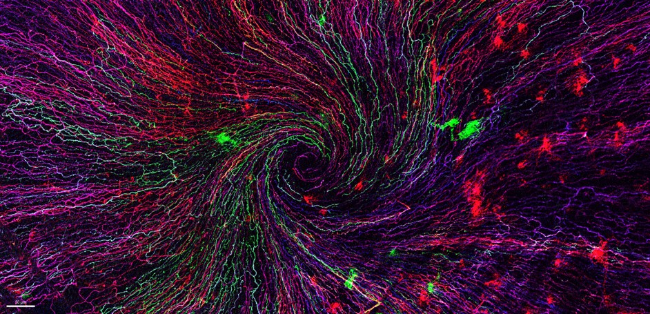
"The eye of the storm"
Sensory fibers of a mouse cornea imaged with a confocal microscope. The corneal nervesconverge toward the centre forming a vortex. This particular transgenic mouse model allows stochastic expression of fluorescent proteins, unravelling the heterogeneity of the fiber origines inside the corneal epithelium. Acknowledgements to Karine Loulier for the mouse model and Laetitia Hudececk for her help during the acquisition.
In the Institut des Neurosciences de Montpellier since 2020, Léna is a PhD student working in the team Eye lead by Dr. Frédéric Michon. This team is investigating the mechanisms related to the preservation and the integrity of the anterior part of the eye, including the lacrimal gland, the tears and the cornea. Léna’s project focuses on the cellular and molecular effects of the corneal innervation on the corneal homeostasis. The project goes further as they aim at highlighting new targets able to prevent and/or repair corneal damage.
The image she submitted for the 2021 France-BioImaging Image Contest (The eye of the storm) represents the sensory fibers of a mouse cornea. This innervation follows a typical pattern where all the nerves converge toward the centre forming a vortex. This particular transgenic mouse model allows random expression of fluorescent proteins, unravelling the heterogeneity of the fibers’ origin inside the corneal epithelium. As cornea is the most innervated tissue in the whole body, this model shows the differences between fibers. In pathological context, for example wound injury, it is thus possible to follow a specific fiber during the healing process.
France-Bioimaging sponsored her participation to the FOM (Focus on Microscopy)2022 congress where she presented her project through a poster. Even though the congress was online, it gave her the opportunity to share her results with experts and as a consequence, to gather advice on her ongoing experiments.
2nd place:Eunice HoYee Chan, Muscle Dynamics Team, Developmental Biology Institute of Marseille (IBDM)
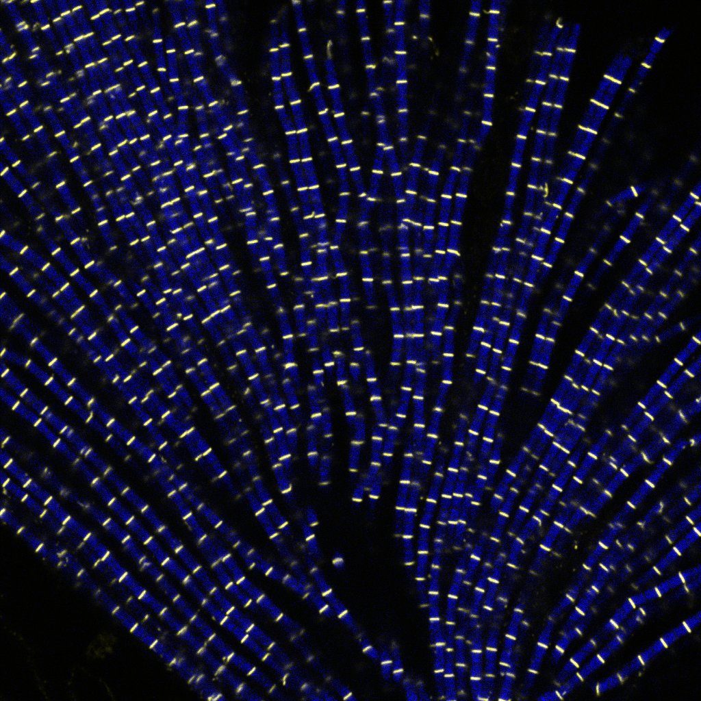
"Sarcomeric bouquet"
Myofibrils isolated from Drosophila indirect flight muscle labelled with titin (yellow) and actin (blue). Image captured from confocal microscope. We are studying the role of titin protein in muscle mechanics and organisation during development.
Research engineer in Frank Schnorrer's team at Institut de Biologie du Développement de Marseille (IBDM), Eunice focuses her research on Drosophila muscle dynamic and organisation during development using advanced biophysical and imaging techniques.
The image she submitted named “Sarcomeric bouquet" was from one of her very first muscle myofibrils isolation experiment. She dissected flight muscles from flies and labelled the individualised myofibrils with Llama nanobodies conjugated with different epitopes. Those labelled myofibrils were then subjected to various imaging methods including standard confocal microscopy, super resolution microscopy and cryo electron-tomogram. Using these novel labelling tools and imaging techniques, her team could study the dynamic and organisation of muscles during development in details.
France-BioImaging sponsored her registration to the 49th European Muscle Conference in Prague (22-26 September 2022). As she is new to the muscle field, this conference offered a great opportunity to have a broad view on different kind of state-of-the-art imaging techniques. Besides, she gave a presentation during the conference, highlighting her work and initiating discussion.
3rd Place:Camille Boutin, Biology of multiciliated cells Team, Developmental Biology Institute of Marseille (IBDM) &Nicolas Brouilly, PICsL Imaging facility, Electron Microscopy department
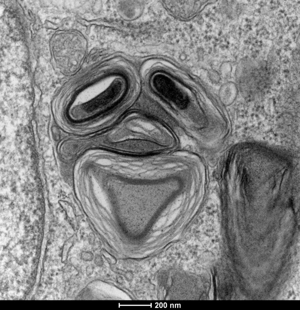
"Clown"
Lamellar structure in a differentiating multiciliated cell observed by transmission electron microscopy with a Tecnai G2 200kV FEI.
Camille is a researcher in Laurent Kodjabachian’s group at the Institut de Biologie du Développement de Marseille (IBDM). She develops projects as a principal investigator on the compartmentalization and sizing of multiciliated cells. With this in mind, she routinely uses confocal and super-resolution microscopy but also scanning and transmission electron microscopy and tomography.
Nicolas is in charge of the Electron Microscopy Unit of the Plateforme d’imagerie commune du site de Luminy (PICsL). In addition to the routine sample preparation and 2D TEM imaging, this imaging facility offers, to internal and external users, advanced sample preparation (cryo-methods, immunolabelling...) and advanced imaging (tomography, CLEM, serial blockface…).
To understand the production of multiple centrioles in multiciliate cells, they focused on the deuterosome, a membrane-less organelle that has been described 50 years ago but whose composition, organisation and function remain unknown to this day. In this context they have developed an inducible multiciliated cells line. This image was taken during the initial characterisation of this cell line by transmission electron microscopy.
Thanks to the France-Bioimaging Image Contest, Nicolas participated to the COST COMULIS Conference that was held by the Cyprus Institute in Nikosia. It was a great opportunity to exchange with the people at the cutting edge of the multi-modal imaging field. The program covered subjects such as the sample preparation for multi-modal imaging, image analysis and integrated industrial partners.
The France-BioImaging Image Contest is back for its 4th edition!
This image contest is open to all within the imaging community: core facility staff and users, R&D labs teams and co-workers, students… Submit your best microscopy images for a chance to showcase your skills, research and creativity to the French bioimaging community and beyond, allowing people to see the visual appeal of the life sciences. Images from the contest will be featured on France-BioImaging communication tools, online and in print.
France-BioImaging and all the French community aims to develop and promote innovative imaging technologies and methods. But microscopy images can also take an artistic, creative look and make the invisible world beautiful.
We are all eager to see your work !
Prizes
1 to 3 images will be awarded depending on the quantity and quality of the entries submitted. France-BioImaging will cover the registration fees for one 2023 microscopy related event of the winners’ choice (FOM, ELMI, EMC, COMULIS conference, etc.).
Important: Only French or foreign participants affiliated to a French institution can enter the contest. Foreign participants non-affiliated to a French institution can submit images and will be featured in the gallery, but will not be evaluated as part of the contest.
Submission deadline: Friday, November 11th, 2022, 23h59 UTC+2.
France BioImaging and all the French community aims to develop and promote innovative imaging technologies and methods. But microscopy images can also take an artistic, creative look and make the invisible world beautiful, allowing people to see the visual appeal of the life sciences.
We enjoyed the diversity of the images submitted with many different microscopy techniques, models and applications represented. A big thank you to all the participants!
The National Coordination Team and the Executive Board are proud to announce the winners of the FBI Image Contest 2021:
1st Place: Léna Meneux, Eye Team, Institut des Neurosciences de Montpellier
“The eye of the storm”
Sensory fibers of a mouse cornea imaged with a confocal microscope. The corneal nervesconverge toward the centre forming a vortex. This particular transgenic mouse model allows stochastic expression of fluorescent proteins, unravelling the heterogeneity of the fiber origines inside the corneal epithelium. Acknowledgements to Karine Loulier for the mouse model and Laetitia Hudececk for her help during the acquisition.
Confocal microscopy
2nd Place:Eunice HoYee Chan, Muscle Dynamics Team, Developmental Biology Institute of Marseille (IBDM)
“Sarcomeric bouquet”
Myofibrils isolated from Drosophila indirect flight muscle labelled with titin (yellow) and actin (blue). Image captured from confocal microscope. We are studying the role of titin protein in muscle mechanics and organisation during development.
Confocal LSM880
3rd Place:Camille Boutin, Biology of multiciliated cells Team, Developmental Biology Institute of Marseille (IBDM) &Nicolas Brouilly, PICsL Imaging facility, Electron Microscopy department
“Clown”
Lamellar structure in a differentiating multiciliated cell observed by transmission electron microscopy with a Tecnai G2 200kV FEI.
Transmission Electron Microscopy, Tecnai G2 200kV FEI
Congratulations to the winners!
Explore all the images submitted here:
As stated in the Terms & Conditions of the contest, foreign participants non-affiliated to a French institution are featured in the gallery, but were not evaluated as part of the contest.
The France BioImaging Image Contest is back for its 3rd edition!
This image contest is open to all within the imaging community: core facility staff and users, R&D labs teams and co-workers, students… Submit your best microscopy images for a chance to showcase your skills, research and creativity to the French bioimaging community and beyond, allowing people to see the visual appeal of the life sciences. Images from the contest will be featured on France BioImaging communication tools, online and in print.
France BioImaging and all the French community aims to develop and promote innovative imaging technologies and methods. But microscopy images can also take an artistic, creative look and make the invisible world beautiful.
We are all eager to see your work !
Prizes
1 to 3 images will be awarded depending on the quantity and quality of the entries submitted. France BioImaging will cover the registration fees for one 2022 microscopy related event of the winners’ choice (FOM, ELMI, EMC, COMULIS conference, etc.).
Important: Only French or foreign participants affiliated to a French institution can enter the contest. Foreign participants non-affiliated to a French institution can submit images and will be featured in the gallery, but will not be evaluated as part of the contest.
Submission deadline: Friday, October 15th, 2021, 23h59 UTC+2.
We use cookies on our website to give you the most relevant experience by remembering your preferences and repeat visits. By clicking “Accept All”, you consent to the use of ALL the cookies. However, you may visit "Cookie Settings" to provide a controlled consent.
This website uses cookies to improve your experience while you navigate through the website. Out of these, the cookies that are categorized as necessary are stored on your browser as they are essential for the working of basic functionalities of the website. We also use third-party cookies that help us analyze and understand how you use this website. These cookies will be stored in your browser only with your consent. You also have the option to opt-out of these cookies. But opting out of some of these cookies may affect your browsing experience.
Necessary cookies are absolutely essential for the website to function properly. These cookies ensure basic functionalities and security features of the website, anonymously.
Cookie
Duration
Description
cookielawinfo-checkbox-analytics
11 months
This cookie is set by GDPR Cookie Consent plugin. The cookie is used to store the user consent for the cookies in the category "Analytics".
cookielawinfo-checkbox-functional
11 months
The cookie is set by GDPR cookie consent to record the user consent for the cookies in the category "Functional".
cookielawinfo-checkbox-necessary
11 months
This cookie is set by GDPR Cookie Consent plugin. The cookies is used to store the user consent for the cookies in the category "Necessary".
cookielawinfo-checkbox-others
11 months
This cookie is set by GDPR Cookie Consent plugin. The cookie is used to store the user consent for the cookies in the category "Other.
cookielawinfo-checkbox-performance
11 months
This cookie is set by GDPR Cookie Consent plugin. The cookie is used to store the user consent for the cookies in the category "Performance".
viewed_cookie_policy
11 months
The cookie is set by the GDPR Cookie Consent plugin and is used to store whether or not user has consented to the use of cookies. It does not store any personal data.
Functional cookies help to perform certain functionalities like sharing the content of the website on social media platforms, collect feedbacks, and other third-party features.
Performance cookies are used to understand and analyze the key performance indexes of the website which helps in delivering a better user experience for the visitors.
Analytical cookies are used to understand how visitors interact with the website. These cookies help provide information on metrics the number of visitors, bounce rate, traffic source, etc.
Advertisement cookies are used to provide visitors with relevant ads and marketing campaigns. These cookies track visitors across websites and collect information to provide customized ads.